. 2022 Jan 12;14(1):2275-2290.
doi: 10.1021/acsami.1c19241. Epub 2021 Dec 21.

Surface Properties of Colloidal Particles Affect Colloidal Self-Assembly in Evaporating Self-Lubricating Ternary Droplets

Affiliations

Surface Properties of Colloidal Particles Affect Colloidal Self-Assembly in Evaporating Self-Lubricating Ternary Droplets

Olga Koshkina et al. ACS Appl Mater Interfaces. .

Abstract

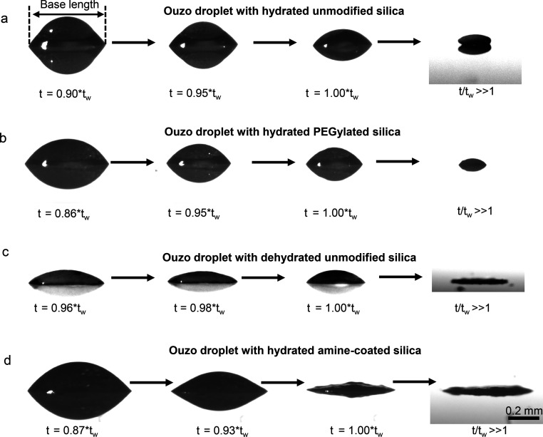
In this work, we unravel the role of surface properties of colloidal particles on the formation of supraparticles (clusters of colloidal particles) in a colloidal Ouzo droplet. Self-lubricating colloidal Ouzo droplets are an efficient and simple approach to form supraparticles, overcoming the challenge of the coffee stain effect in situ. Supraparticles are an efficient route to high-performance materials in various fields, from catalysis to carriers for therapeutics. Yet, the role of the surface of colloidal particles in the formation of supraparticles using Ouzo droplets remains unknown. Therefore, we used silica particles as a model system and compared sterically stabilized versus electrostatically stabilized silica particles─positively and negatively charged. Additionally, we studied the effect of hydration. Hydrated negatively charged silica particles and sterically stabilized silica particles form supraparticles. Conversely, dehydrated negatively charged silica particles and positively charged amine-coated particles form flat film-like deposits. Notably, the assembly process is different for all the four types of particles. The surface modifications alter (a) the contact line motion of the Ouzo droplet and (b) the particle-oil and particle-substrate interactions. These alterations modify the particle accumulation at the various interfaces, which ultimately determines the shape of the final deposit. Thus, by modulating the surface properties of the colloidal particles, we can tune the shape of the final deposit, from a spheroidal supraparticle to a flat deposit. In the future, this approach can be used to tailor the supraparticles for applications such as optics and catalysis, where the shape affects the functionality.

Keywords: Ouzo effect; colloidal self-assembly; colloidal stabilization; evaporation induced self-assembly; self-lubrication; silica particles; supraparticles.


Conflict of interest statement

The authors declare no competing financial interest.

Figures

Figure 1
Figure 1
Surface modification of silica particles. Stöber silica particles were covalently modified with (6-aminohexyl)-aminopropyltrimethoxysilane or PEG-triethoxysilane, leading to positively charged amine-modified or sterically stabilized PEGylated particles, respectively. After the synthesis, all particles were purified and dried at 60 °C. To subsequently study the effect of hydration, particles were resuspended and incubated in water (hydrated particles). Alternatively, particles were resuspended in ethanol prior to preparation of the Ouzo-mixtures (dehydrated particles). Transmission electron micrograph of negatively charged silica particles is shown. Scale bar 500 nm.
Figure 2
Figure 2
Surface modification of the colloidal particles affects the shapes of the final deposits obtained from evaporating Ouzo droplets. (a–d) Side view shadowgraphy images of the final deposit obtained after evaporation of droplets that were loaded with different silica particles, as mentioned at the top of the panel. (e–h) Top view images of the final deposits, corresponding to the side view images. Hydrated unmodified silica particles and PEGylated silica particles form supraparticles while the dehydrated unmodified silica particles and amine-coated silica particles form flat film-like deposits. Scale bar 0.1 mm.
Figure 3
Figure 3
Side-view shadowgraph images of colloidal Ouzo droplets, at different time instances, showing the drop evaporation and steps leading to final deposit formation for (a) hydrated unmodified silica, (b) hydrated PEGylated silica, (c) dehydrated unmodified silica and (d) hydrated amine-coated silica. Hydrated unmodified silica particles and PEGylated silica form supraparticles, while dehydrated unmodified silica and amine-coated silica particles form flat deposits. Scale bar 0.2 mm.
Figure 4
Figure 4
(a) Evolution of the droplet volume with time t normalized to time tw. There is no appreciable difference in the volume evolution between droplets with different particles. (b) The base length shows different trends in the final phase of the evaporation process (zoomed region), depending on the type of silica particles. Refer to SI Section 3 for additional plots and repeat measurements. tw is estimated from the variation in volume and base length over time. tw corresponds to the time when the plots of both volume vs time and base length vs time reach a plateau. At t = tw, most of the water has evaporated but some trace amount of water could be left behind, which cannot be measured.
Figure 5
Figure 5
Effects of surface modifications on particle–oil interactions. The overlay of fluorescence signals from the particles (red) and oil (yellow) is shown. (a, e) Hydrated silica particles (NP) do not interact with oil microdroplets (MD). In contrast, PEGylation (b, f), dehydration (c, g), and surface modification with amine (d, h) lead to the adsorption of the particles (NP) onto the surface of oil microdroplets (MD). Thus, the surface modifications and dehydration lead to formation of structures similar to Pickering emulsions. The composition of the mixtures was the following (by weight): t-anethole/particles/water/ethanol: 0.020/0.0016/0.55/0.43. Overview images (a–d): Scale bar 20 μm. Zoomed in images (e–f): Scale bar 10 μm. See also Figures S9–S14 and figures in supplementary file Z1 for larger overview images, images for the Ouzo mixtures containing dehydrated amine-coated silica and dehydrated PEGylated silica, and for repetition of the experiments.
Figure 6
Figure 6
Reflection channel of confocal microscopy shows the difference in the oil microdroplets on the substrate for the different silica particles. (b), (d), and (h) are the zoomed in images of the square marked regions in (a), (c), and (g). For the case of dehydrated unmodified silica particles, (f) shows an image taken at a higher magnification of another droplet, to clearly resolve the small oil microdroplets. All images are taken at time t/tw ≈ 0.35, except (f) which is taken at t/tw ≈ 0.32. Scale bar 0.4 mm for (a), (c), (e), and (g). Scale bar 50 μm for zoomed in images. In the case of the droplet containing dehydrated unmodified silica particles (e and f), a large number of very small oil microdroplets (diameter ∼5 μm) are nucleated on the substrate, in stark contrast to the other cases.
Figure 7
Figure 7
Confocal microscopy reveals similarities and differences in the formation of supraparticles in droplets containing hydrated unmodified silica (a and c) and hydrated PEG coated silica (b and d). Overlay of emission signals of perylene (yellow, showing oil) and rhodamine (red, showing silica particles) at different stages of the evaporation. (a and b) Side-view or vertical cross section, reconstructed from layer-wise scanning (Scale bar 100 μm); (c and d) bottom-view of a horizontal plane 0–10 μm from the glass substrate (Scale bar 0.2 mm). The contrast of the images has been enhanced to show all the relevant features in the images. (e) In Ouzo droplet containing PEG coated silica particles, Pickering microdroplets on the substrate merge with the outer shell, making the outer shell radially asymmetric. Images in (e) are the zoomed image of the dotted rectangular region in (d)-(ii). Scale bar 50 μm. The number at the bottom of each image shows the time in relation to the time tw when almost all water has evaporated, but oil is still surrounding the silica deposit. Both hydrated unmodified silica and PEG coated silica form supraparticles, even though the outer shell of PEG coated silica has a film-like appearance.
Figure 8
Figure 8
Confocal microscopy shows the formation of flat film-like structures in droplets containing dehydrated unmodified silica (a and c) and hydrated amine-coated silica (b and d). (a–d) Overlay of emission signals of perylene (yellow, showing oil) and rhodamine (red, silica particles) at different stages of the evaporation – (a and b) side-view or vertical cross section, reconstructed from layer-wise scanning (Scale bar 100 μm); (c and d) bottom-view of a horizontal plane 0–10 μm from the glass substrate (Scale bar 0.2 mm). The contrast of the images has been enhanced to show all the relevant features in the images. The number at the bottom shows the time in relation to the time tw when water has evaporated almost completely, but oil is still surrounding the silica deposit. (e–g) Pickering microdroplets observed in an evaporating Ouzo droplet containing dehydrated unmodified silica particles, at t = 0.64tw. (f) and (g) are zoomed in images of the dotted rectangular region in (e). (g) Fluorescence emission signals only from perylene to show oil microdroplets close to the outer shell. Scale bar 50 μm. The evolution of outer shell at the air–water interface and close to the substrate is different for dehydrated unmodified silica particles and amine-coated silica particles. Nevertheless, both particles form flat deposits at the end of the droplet evaporation process.
Figure 9
Figure 9
Schematic showing the mechanism of final deposits formation with the different silica particles. (a) Hydrated unmodified silica particles and (b) PEGylated silica particles form supraparticles while (c) dehydrated unmodified silica and (d) amine-coated silica particles form flat deposits. The differences in the shape of the outer shell leads to differences in the shape of the final deposit.

Similar articles

Cited by

References

    1. Wintzheimer S.; Granath T.; Oppmann M.; Kister T.; Thai T.; Kraus T.; Vogel N.; Mandel K. Supraparticles: Functionality from Uniform Structural Motifs. ACS Nano 2018, 12 (6), 5093–5120. 10.1021/acsnano.8b00873. - DOI - PubMed
    1. Si K. J.; Chen Y.; Shi Q.; Cheng W. Nanoparticle Superlattices: The Roles of Soft Ligands. Adv. Sci. 2018, 5 (1), 1700179.10.1002/advs.201700179. - DOI - PMC - PubMed
    1. Yu Y.; Yang X.; Liu M.; Nishikawa M.; Tei T.; Miyako E. Amphipathic Nanodiamond Supraparticles for Anticancer Drug Loading and Delivery. ACS Appl. Mater. Interfaces 2019, 11 (21), 18978–18987. 10.1021/acsami.9b04792. - DOI - PubMed
    1. Zhang Z.; Rahmat J. N.; Mahendran R.; Zhang Y. Controllable Assembly of Upconversion Nanoparticles Enhanced Tumor Cell Penetration and Killing Efficiency. Adv. Sci. 2020, 7 (24), 2001831.10.1002/advs.202001831. - DOI - PMC - PubMed
    1. Liu J.; Xiao M.; Li C.; Li H.; Wu Z.; Zhu Q.; Tang R.; Xu A. B.; He L. Rugby-Ball-Like Photonic Crystal Supraparticles with Non-Close-Packed Structures and Multiple Magneto-Optical Responses. J. Mater. Chem. C 2019, 7 (47), 15042–15048. 10.1039/C9TC05438C. - DOI