. 2022 Apr;303(1):162-170.
doi: 10.1148/radiol.211222. Epub 2021 Dec 21.

Normal Growth, Sexual Dimorphism, and Lateral Asymmetries at Fetal Brain MRI

Affiliations

Normal Growth, Sexual Dimorphism, and Lateral Asymmetries at Fetal Brain MRI

Fedel Machado-Rivas et al. Radiology. 2022 Apr.

Abstract

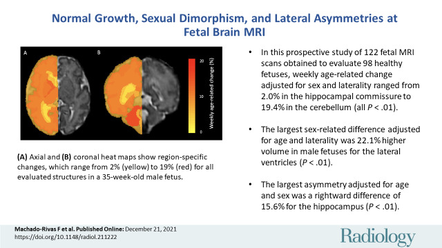
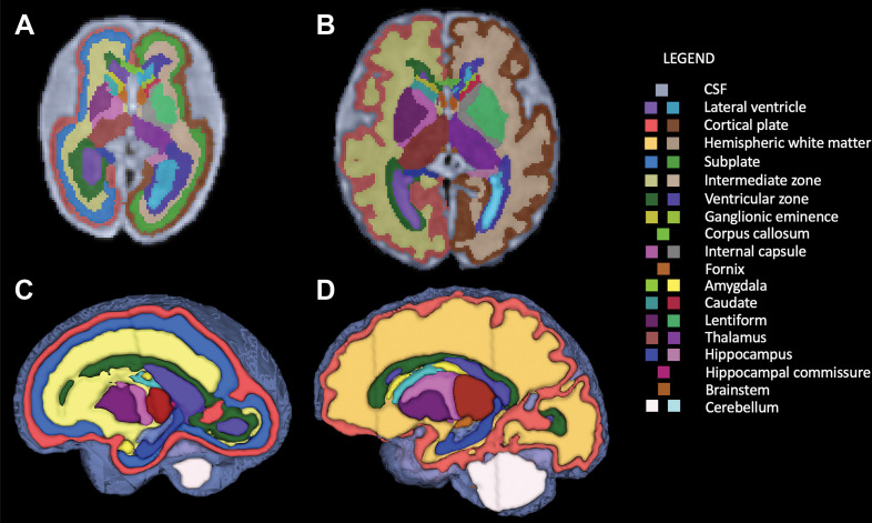
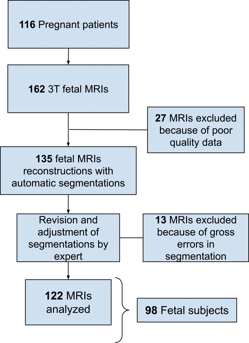
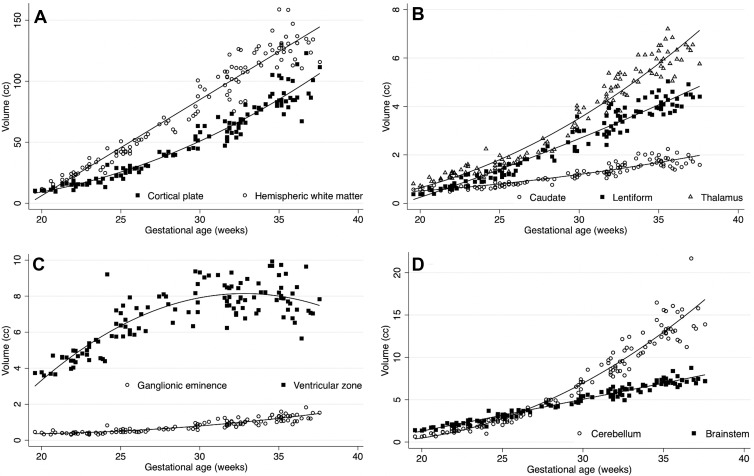
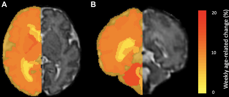
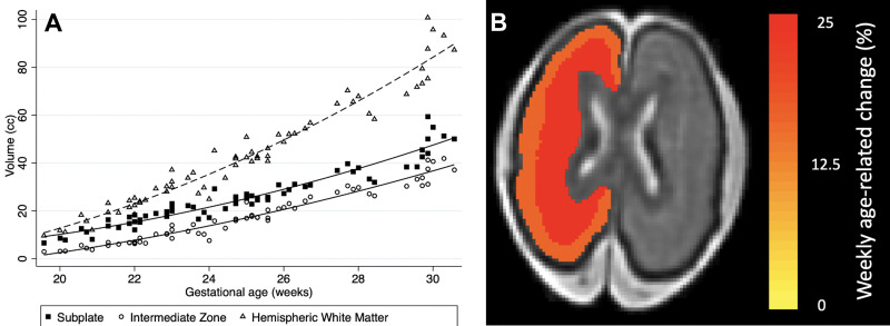
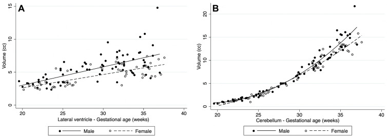
Background Tools in image reconstruction, motion correction, and segmentation have enabled the accurate volumetric characterization of fetal brain growth at MRI. Purpose To evaluate the volumetric growth of intracranial structures in healthy fetuses, accounting for gestational age (GA), sex, and laterality with use of a spatiotemporal MRI atlas of fetal brain development. Materials and Methods T2-weighted 3.0-T half-Fourier acquired single-shot turbo spin-echo sequence MRI was performed in healthy fetuses from prospectively recruited pregnant volunteers from March 2013 to May 2019. A previously validated section-to-volume reconstruction algorithm was used to generate intensity-normalized superresolution three-dimensional volumes that were registered to a fetal brain MRI atlas with 28 anatomic regions of interest. Atlas-based segmentation was performed and manually refined. Labels included the bilateral hippocampus, amygdala, caudate nucleus, lentiform nucleus, thalamus, lateral ventricle, cerebellum, cortical plate, hemispheric white matter, internal capsule, ganglionic eminence, ventricular zone, corpus callosum, brainstem, hippocampal commissure, and extra-axial cerebrospinal fluid. For fetuses younger than 31 weeks of GA, the subplate and intermediate zones were delineated. A linear regression analysis was used to determine weekly age-related change adjusted for sex and laterality. Results The final analytic sample consisted of 122 MRI scans in 98 fetuses (mean GA, 29 weeks ± 5 [range, 20-38 weeks]). All structures had significant volume growth with increasing GA (P < .001). Weekly age-related change for individual structures in the brain parenchyma ranged from 2.0% (95% CI: 0.9, 3.1; P < .001) in the hippocampal commissure to 19.4% (95% CI: 18.7, 20.1; P < .001) in the cerebellum. The largest sex-related differences were 22.1% higher volume in male fetuses for the lateral ventricles (95% CI: 10.9, 34.4; P < .001). There was rightward volumetric asymmetry of 15.6% for the hippocampus (95% CI: 14.2, 17.2; P < .001) and leftward volumetric asymmetry of 8.1% for the lateral ventricles (95% CI: 3.7, 12.2; P < .001). Conclusion With use of a spatiotemporal MRI atlas, volumetric growth of the fetal brain showed complex trajectories dependent on structure, gestational age, sex, and laterality. © RSNA, 2021 Online supplemental material is available for this article. See also the editorial by Rollins in this issue.


Conflict of interest statement

Disclosures of conflicts of interest: F.M.R. No relevant relationships. J.G. No relevant relationships. J.J.C. No relevant relationships. C.V.A. No relevant relationships. O.A. Participation on the data safety monitoring board or advisory board of Boston Children’s Hospital and Harvard Medical School. S.K.W. No relevant relationships. A.G. No relevant relationships. C.J. No relevant relationships.

Figures

None
Graphical abstract
Segmentation of fetal brain structures. Twenty-eight individual
structures were segmented and refined for all fetuses, and for fetuses less
than 31 weeks of gestational age, the fetal white matter transient zones
were additionally segmented. Image shows two-dimensional axial fetal
segmentations for (A) a 25-week-old fetus and (B) a 36-week-old fetus, as
well as (C, D) the three-dimensional volumetric segmentations (C corresponds
with A, and D with B). A color bar is shown with labels for all
structures.
Figure 1:
Segmentation of fetal brain structures. Twenty-eight individual structures were segmented and refined for all fetuses, and for fetuses less than 31 weeks of gestational age, the fetal white matter transient zones were additionally segmented. Image shows two-dimensional axial fetal segmentations for (A) a 25-week-old fetus and (B) a 36-week-old fetus, as well as (C, D) the three-dimensional volumetric segmentations (C corresponds with A, and D with B). A color bar is shown with labels for all structures.
Flowchart of our study sample shows inclusion and exclusion. There was
a total of 116 pregnant patients who were imaged, resulting in 162 fetal MRI
examinations. Twenty-seven MRI examinations were excluded because of
poor-quality data, resulting in 135 MRI examinations that were reconstructed
and segmented. Then, 13 MRI examinations were excluded because of gross
errors in segmentation, resulting in 122 MRI examinations analyzed
(corresponding to 98 fetuses).
Figure 2:
Flowchart of our study sample shows inclusion and exclusion. There was a total of 116 pregnant patients who were imaged, resulting in 162 fetal MRI examinations. Twenty-seven MRI examinations were excluded because of poor-quality data, resulting in 135 MRI examinations that were reconstructed and segmented. Then, 13 MRI examinations were excluded because of gross errors in segmentation, resulting in 122 MRI examinations analyzed (corresponding to 98 fetuses).
Plots show volumetric age-related change. Age-related change had high
variability between the structures evaluated. (A) Cortical plate and
hemispheric white matter; (B) the deep gray matter structures: caudate
nucleus, lentiform nucleus, and thalamus; (C) the proliferative
compartments: ganglionic eminence and ventricular zone; and (D) the
cerebellum and brainstem.
Figure 3:
Plots show volumetric age-related change. Age-related change had high variability between the structures evaluated. (A) Cortical plate and hemispheric white matter; (B) the deep gray matter structures: caudate nucleus, lentiform nucleus, and thalamus; (C) the proliferative compartments: ganglionic eminence and ventricular zone; and (D) the cerebellum and brainstem.
Heat maps show volumetric age-related change. Weekly age-related
percentage change is overlaid in a 35-week-old male fetus. (A) Axial and (B)
coronal heat maps show region-specific changes, which range from 2% (yellow)
to 19% (red) for all evaluated structures.
Figure 4:
Heat maps show volumetric age-related change. Weekly age-related percentage change is overlaid in a 35-week-old male fetus. (A) Axial and (B) coronal heat maps show region-specific changes, which range from 2% (yellow) to 19% (red) for all evaluated structures.
Subanalysis of the fetal hemispheric white matter transient zones in
fetuses younger than 31 weeks of gestational age. (A) Plot shows volumetric
age-related change for the fetal white matter transient zones (subplate and
intermediate zone). (B) Heat map depicts weekly age-related change of the
subplate (25.2%) and the intermediate zone (15.7%) overlaid in a 25-week-old
female fetus.
Figure 5:
Subanalysis of the fetal hemispheric white matter transient zones in fetuses younger than 31 weeks of gestational age. (A) Plot shows volumetric age-related change for the fetal white matter transient zones (subplate and intermediate zone). (B) Heat map depicts weekly age-related change of the subplate (25.2%) and the intermediate zone (15.7%) overlaid in a 25-week-old female fetus.
Plots show volumetric differences by sex. In structures with sexual
dimorphism, male fetuses had higher volumes on average: (A) shows rates for
the lateral ventricles and (B) for the cerebellum.
Figure 6:
Plots show volumetric differences by sex. In structures with sexual dimorphism, male fetuses had higher volumes on average: (A) shows rates for the lateral ventricles and (B) for the cerebellum.

Comment in

References

    1. Kainz B , Steinberger M , Wein W , et al. . Fast volume reconstruction from motion corrupted stacks of 2D slices . IEEE Trans Med Imaging 2015. ; 34 ( 9 ): 1901 – 1913 . - PMC - PubMed
    1. Gholipour A , Rollins CK , Velasco-Annis C , et al. . A normative spatiotemporal MRI atlas of the fetal brain for automatic segmentation and analysis of early brain growth . Sci Rep 2017. ; 7 ( 1 ): 476 . - PMC - PubMed
    1. Kyriakopoulou V , Vatansever D , Davidson A , et al. . Normative biometry of the fetal brain using magnetic resonance imaging . Brain Struct Funct 2017. ; 222 ( 5 ): 2295 – 2307 . - PMC - PubMed
    1. Andescavage NN , du Plessis A , McCarter R , et al. . Complex trajectories of brain development in the healthy human fetus . Cereb Cortex 2017. ; 27 ( 11 ): 5274 – 5283 . - PMC - PubMed
    1. Clouchoux C , Guizard N , Evans AC , du Plessis AJ , Limperopoulos C . Normative fetal brain growth by quantitative in vivo magnetic resonance imaging . Am J Obstet Gynecol 2012. ; 206 ( 2 ): 173.e1 – 173.e8 . - PubMed

Publication types