Observational Study
. 2022 Apr;303(1):184-190.
doi: 10.1148/radiol.2021204616. Epub 2021 Dec 21.

Mucus Plugs in Asthma at CT Associated with Regional Ventilation Defects at 3He MRI

Affiliations
Observational Study

Mucus Plugs in Asthma at CT Associated with Regional Ventilation Defects at 3He MRI

David G Mummy et al. Radiology. 2022 Apr.

Abstract

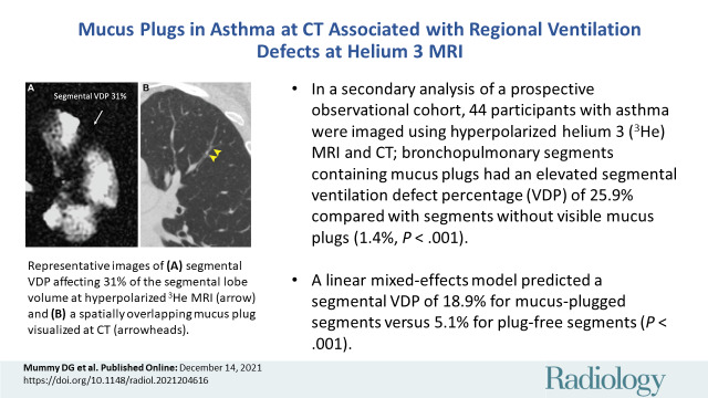
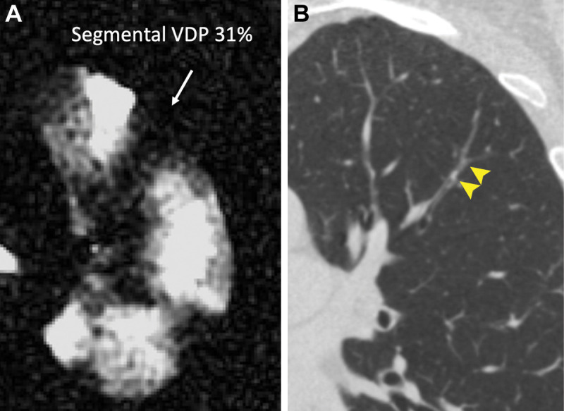
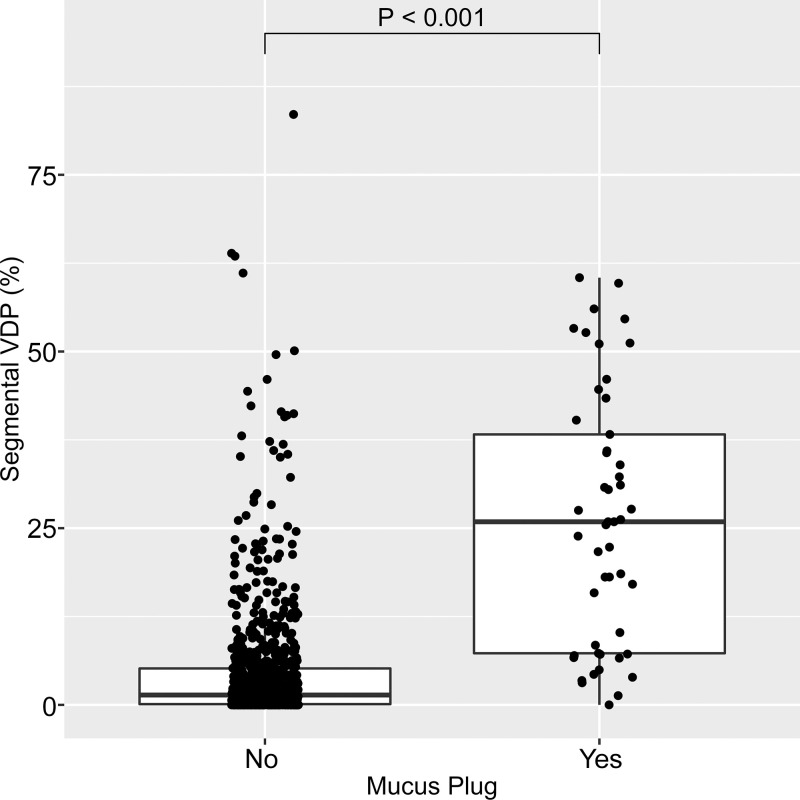
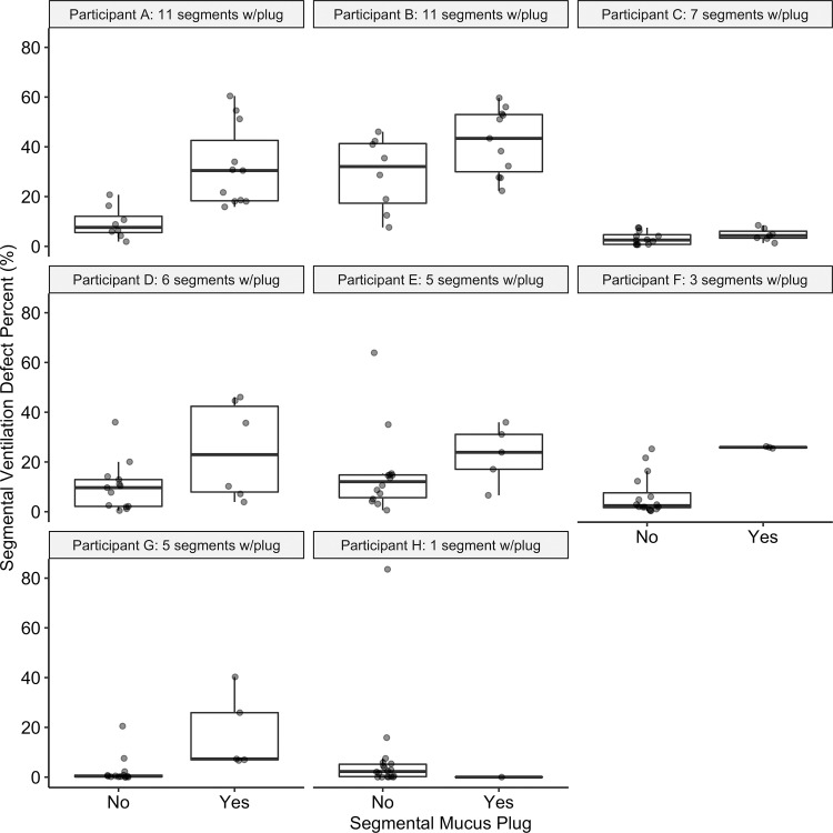
Background Airway mucus plugs in asthma are associated with exacerbation frequency, increased eosinophilia, and reduced lung function. The relationship between mucus plugs and spatially overlapping ventilation abnormalities observed at hyperpolarized gas MRI has not been assessed quantitatively. Purpose To assess regional associations between CT mucus plugs scored by individual bronchopulmonary segment and corresponding measurements of segmental ventilation defect percentage (VDP) at hyperpolarized helium 3 (3He) MRI. Materials and Methods In this secondary analysis of a Health Insurance Portability and Accountability Act-compliant prospective observational cohort, participants in the Severe Asthma Research Program (SARP) III (NCT01760915) between December 2012 and August 2015 underwent hyperpolarized 3He MRI to determine segmental VDP. Segmental mucus plugs at CT were scored by two readers, with segments scored as plugged only if both readers agreed independently. A linear mixed-effects model controlling for interpatient variability was then used to assess differences in VDP in plugged versus plug-free segments. Results Forty-four participants with asthma were assessed (mean age ± standard deviation, 47 years ± 15; 29 women): 19 with mild-to-moderate asthma and 25 with severe asthma. Mucus plugs were observed in 49 total bronchopulmonary segments across eight of 44 patients. Segments containing mucus plugs had a median segmental VDP of 25.9% (25th-75th percentile, 7.3%-38.3%) versus 1.4% (25th-75th percentile, 0.1%-5.2%; P < .001) in plug-free segments. Similarly, the model estimated a segmental VDP of 18.9% (95% CI: 15.7, 22.2) for mucus-plugged segments versus 5.1% (95% CI: 3.3, 7.0) for plug-free segments (P < .001). Participants with one or more mucus plugs had a median whole-lung VDP of 11.1% (25th-75th percentile, 7.1%-18.9%) versus 3.1% (25th-75th percentile, 1.1%-4.4%) in those without plugs (P < .001). Conclusion Airway mucus plugging at CT was associated with reduced ventilation in the same bronchopulmonary segment at hyperpolarized helium 3 MRI, suggesting that mucus plugging may be an important cause of ventilation defects in asthma. © RSNA, 2021 Online supplemental material is available for this article.


Conflict of interest statement

Disclosures of conflicts of interest: D.G.M. Scientific consultant for Polarean Imaging. E.M.D. No relevant relationships. K.J.C. Was consultant for Imbio; employed by Imbio. M.D.E. No relevant relationships. B.M.E. No relevant relationships. J.D.N. Consultant and medical advisor for VIDA; employed by VIDA and National Institutes of Health (NIH); grants/grants pending with NIH; payment for lectures including service on speakers bureaus, payment for development of educational presentations, and travel/accommodations/meeting expenses unrelated to activities listed from VIDA; patents (planned, pending, or issued) with and stock/stock options in VIDA; payment for manuscript preparation and royalties from Elsevier. D.S.G. No relevant relationships. S.K.N. Patent with Wisconsin Alumni Research Foundation. M.L.S. Patent with John Fahy, UCSF; Deputy Editor of Radiology. R.L.S. No relevant relationships. N.N.J. Institution supported by grant from AstraZeneca; consultant for Pulmocide and GlaxoSmithKline. L.C.D. Consultant for AstraZeneca, Sanofi. J.V.F. Consulting fee or honorarium from Gossamer, Arrowhead, Ionis Pharmaceuticals, and Connect Biopharma. S.B.F. Consultant for Sanofi/Regeneron, Polarean, Caladrius; payment for lectures including service on speakers bureaus for Sanofi/Regeneron.

Figures

None
Graphical abstract
Flowchart of study sample and image processing steps with number and
percentage of total at each stage. All imaging was performed after
administration of a bronchodilator. Segmental mucus plugs were scored by a
team of radiologists as part of a larger study that required 3-year
follow-up CT. Thus, patients who were lost to follow-up because they moved
out of the area or otherwise withdrew from the study (n = 5) were not scored
and were excluded from our final sample. SARPIII = Severe Asthma Research
Program III, VDP = ventilation defect percentage.
Figure 1:
Flowchart of study sample and image processing steps with number and percentage of total at each stage. All imaging was performed after administration of a bronchodilator. Segmental mucus plugs were scored by a team of radiologists as part of a larger study that required 3-year follow-up CT. Thus, patients who were lost to follow-up because they moved out of the area or otherwise withdrew from the study (n = 5) were not scored and were excluded from our final sample. SARPIII = Severe Asthma Research Program III, VDP = ventilation defect percentage.
Representative images of (A) segmental ventilation defect percentage
(VDP) affecting 31% of the segmental lobe volume at hyperpolarized helium 3
MRI (arrow) and (B) a spatially overlapping mucus plug visualized at CT
(arrowheads).
Figure 2:
Representative images of (A) segmental ventilation defect percentage (VDP) affecting 31% of the segmental lobe volume at hyperpolarized helium 3 MRI (arrow) and (B) a spatially overlapping mucus plug visualized at CT (arrowheads).
Box plot shows segmental ventilation defect percentage (VDP) in
segments with no mucus plug (left) and with at least one mucus plug (right).
Horizontal lines represent the median, 25th percentile, and 75th percentile
values. Each dot represents one individual bronchopulmonary segment. Of the
825 total bronchopulmonary segments evaluated, 49 (5.9%) were identified as
having a mucus plug. Segments with mucus plugs had a median segmental VDP of
25.9% (25th–75th percentile, 7.3%–38.3%) versus 1.4%
(25th–75th percentile, 0.1%–5.2%) in segments without a mucus
plug (P < .001).
Figure 3:
Box plot shows segmental ventilation defect percentage (VDP) in segments with no mucus plug (left) and with at least one mucus plug (right). Horizontal lines represent the median, 25th percentile, and 75th percentile values. Each dot represents one individual bronchopulmonary segment. Of the 825 total bronchopulmonary segments evaluated, 49 (5.9%) were identified as having a mucus plug. Segments with mucus plugs had a median segmental VDP of 25.9% (25th–75th percentile, 7.3%–38.3%) versus 1.4% (25th–75th percentile, 0.1%–5.2%) in segments without a mucus plug (P < .001).
Plots show segmental ventilation defect percentage (VDP) in plugged
versus nonplugged segments in each of the eight individual participants
(participants A–H) with identified mucus plugs. Horizontal lines
represent the median, 25th percentile, and 75th percentile values. Each dot
represents one individual bronchopulmonary segment. The number of plugs
identified in each participant ranged from 1 to 11, as indicated in each
subplot title, with a median value of 5.5 plugged segments. Median segmental
VDP in plugged segments was higher than that in the unplugged segments in
all but one participant (participant H, with only one mucus plug
identified). In specific participants, there is considerable overlap in
values for plugged versus unplugged segments, as in participant B, for whom
nearly all segments have substantial segmental VDP, and in participant C,
for whom all segments have relatively low segmental VDP.
Figure 4:
Plots show segmental ventilation defect percentage (VDP) in plugged versus nonplugged segments in each of the eight individual participants (participants A–H) with identified mucus plugs. Horizontal lines represent the median, 25th percentile, and 75th percentile values. Each dot represents one individual bronchopulmonary segment. The number of plugs identified in each participant ranged from 1 to 11, as indicated in each subplot title, with a median value of 5.5 plugged segments. Median segmental VDP in plugged segments was higher than that in the unplugged segments in all but one participant (participant H, with only one mucus plug identified). In specific participants, there is considerable overlap in values for plugged versus unplugged segments, as in participant B, for whom nearly all segments have substantial segmental VDP, and in participant C, for whom all segments have relatively low segmental VDP.

Similar articles

Cited by

References

    1. Downie SR , Salome CM , Verbanck S , Thompson B , Berend N , King GG . Ventilation heterogeneity is a major determinant of airway hyperresponsiveness in asthma, independent of airway inflammation . Thorax 2007. ; 62 ( 8 ): 684 – 689 . - PMC - PubMed
    1. Teague WG , Tustison NJ , Altes TA . Ventilation heterogeneity in asthma . J Asthma 2014. ; 51 ( 7 ): 677 – 684 . - PubMed
    1. Mummy DG , Kruger SJ , Zha W , et al. . Ventilation defect percent in helium-3 magnetic resonance imaging as a biomarker of severe outcomes in asthma . J Allergy Clin Immunol 2018. ; 141 ( 3 ): 1140 – 1141.e4 . - PMC - PubMed
    1. Mummy DG , Carey KJ , Evans MD , et al. . Ventilation defects on hyperpolarized helium-3 MRI in asthma are predictive of 2-year exacerbation frequency . J Allergy Clin Immunol 2020. ; 146 ( 4 ): 831 – 839.e6 . - PMC - PubMed
    1. Svenningsen S , Eddy RL , Lim HF , Cox PG , Nair P , Parraga G . Sputum eosinophilia and magnetic resonance imaging ventilation heterogeneity in severe asthma . Am J Respir Crit Care Med 2018. ; 197 ( 7 ): 876 – 884 . - PubMed

Publication types