. 2022 Feb;298(2):101515.
doi: 10.1016/j.jbc.2021.101515. Epub 2021 Dec 18.

Inhibition of Jumonji demethylases reprograms severe dilated cardiomyopathy and prolongs survival

Affiliations

Inhibition of Jumonji demethylases reprograms severe dilated cardiomyopathy and prolongs survival

Tram Anh Tran et al. J Biol Chem. 2022 Feb.

Abstract

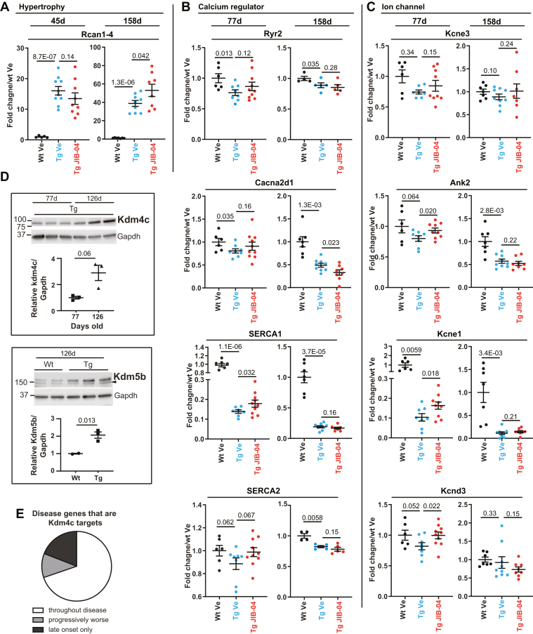
Hypertrophic/dilated cardiomyopathy, often a prequel to heart failure, is accompanied by maladaptive transcriptional changes that contribute to arrythmias and contractile misfunction. Transgenic mice constitutively expressing high levels of calcineurin are known to develop extreme heart hypertrophy, which progresses to dilated cardiomyopathy, and to die several weeks after birth. Here, we characterized aberrant transcriptional and epigenetic pathways in this mouse model and established a pharmacological approach to treat established cardiomyopathy. We found that H3K4me3 (trimethyl histone 3 lysine 4) and H3K9me3 (trimethyl histone 3 lysine 9) Jumonji histone demethylases are markedly increased at the protein level and show enhanced enzymatic activity in diseased hearts. These epigenetic regulators continued to increase with time, further affecting cardiac gene expression. Our findings parallel the lower H3K4me3 and H3K9me3 levels seen in human patients. Inhibition of Jumonji demethylase activities in vivo results in lower histone demethylase enzymatic function in the heart and higher histone methylation levels and leads to partial reduction of heart size, reversal of maladaptive transcriptional programs, improved heart function, and prolonged survival. At the molecular level, target genes of transcription factor myocyte enhancer factor 2 are specifically regulated in response to pharmacological or genetic inhibition of Jumonji demethylases. Similar transcriptional reversal of disease-associated genes is seen in a second disease model based on cardiac mechanical overload. Our findings validate pharmacological inhibitors of Jumonji demethylases as potential therapeutics for the treatment of cardiomyopathies across disease models and provide evidence of the reversal of maladaptive transcriptional reprogramming leading to partial restoration of cardiac function. In addition, this study defines pathways of therapeutic resistance upregulated with disease progression.

Keywords: JIB-04; Jumonji enzymes; calcineurin; cardiomyopathy; small-molecule inhibitor; transcriptional reprogramming.


Conflict of interest statement

Conflict of interest The authors declare that they have no conflicts of interest with the contents of this article.

Figures

Figure 1
Figure 1
Jumonji KDMexpression and activity are increased in CnA transgenic (Tg) hearts.A, Jumonji proteins increase in Tg hearts. Western blots of H3K4me3 and H3K9me3 Jumonji KDM demethylases in hearts from Wt versus CnATg mice. Protein extracts for Western blots of Kdm5a (whole cell), Kdm2b (whole cell), and Kdm4c (nuclear extract) are lysates from ventricle powders of Wt and Tg mice, between 35 and 42 days of age. B, quantification of Jumonji protein levels. Left panels, H3K4me3 demethylases. Kdm5a: Wt n = 6, Tg n = 6; Kdm2b: Wt n = 5, Tg n = 6. Right panel, H3K9me3 demethylase. Kdm4c: Wt n = 4, Tg n = 4. Data are average ± SEM. p Values are from Mann–Whitney's test. C, H3K4me3 and H3K9me3 Jumonji demethylase activity in heart nuclear extracts (left ventricle) from Wt versus CnATg mice between 84 and 103 days of age on exogenous trimethylated substrates. Left panel, H3K4me3 demethylase activity assays; Wt n = 4, Tg n = 4. Right panel, H3K9me3 demethylase activity assays, Wt n = 3, Tg n = 3. Data are average ± SEM. p Values are from t test, two-tailed, unequal variance. B and C,p ≤ 0.05 and ∗∗p ≤ 0.01. CnA, calcineurin A; H3K4me3, trimethylation of histone 3 lysine 4; H3K9me3, trimethylation of histone 9 lysine 4; KDM, histone lysine demethylase.
Figure 2
Figure 2
Pharmacological inhibition of Jumonji KDMs in the heart improves cardiac function and prolongs survival.A, H3K4me3 and H3K9me3 demethylase activity in nuclear extracts from two pairs of transgenic (Tg) mice treated with vehicle (Ve) or JIB-04 for 2 weeks (orange symbols) and two pairs treated for 7 weeks (green symbols), all starting at 4 weeks of age. p Values are t test, two-tailed, and paired. B, percent changes of Wt or Tg hearts treated with JIB-04 for 6 weeks, starting from 5 weeks of age, normalized to Ve control of the same genotype. Wt n = 10, Tg n = 12, p value is t test, two-tailed, and of unequal variance. C, echocardiography data of mice treated with JIB-04 in the same age range as mice in panel A (orange symbols). Stroke volume and cardiac output are shown. p Values are from t test, two-tailed, and of unequal variance. Wt Ve n = 12, Tg Ve n = 9, Tg JIB-04 = 14. D, percent survival of Tg mice treated with Ve or JIB-04, starting from 4 weeks of age. Ve n = 25, JIB-04 n = 19. p Value is Gehan–Breslow–Wilcoxon test. 75% median and 25% survival are shown by horizontal lines. E, body weight changes of Tg animals treated with Ve or JIB-04 over the duration of the survival experiment shown in D. F, quantification of heart rate and ST interval from ECG measurements of Wt mice treated with Ve and of CnATg mice treated with Ve or JIB-04 starting at 4 weeks of age. Ages of mice used for ECG range from 88 to 149 days old. Wt Ve n = 7, Tg Ve n = 5, and Tg JIB-04 n = 6. p Values are from t test, two-tailed, and of unequal variance. G, echocardiography parameters of Wt Ve-treated, Tg Ve-treated, and Tg JIB-04-treated mice in the same age range used for ECG measurements in F. Animals in F and G were treated for 9–13 weeks. Wt Ve n = 2, Tg Ve n = 4, and Tg JIB-04 n = 3. p Values are t test, one-tailed, and of unequal variance. A, B, C, E, F, and G, data are average ± SEM. ∗p ≤ 0.05, ∗∗p ≤ 0.01, ∗∗∗p ≤ 0.001, or p values are shown. H3K4me3, trimethylation of histone 3 lysine 4; H3K9me3, trimethylation of histone 9 lysine 4; KDM, histone lysine demethylase.
Figure 3
Figure 3
Jumonji enzyme inhibition reprograms gene expression in the heart toward normal myogenesis and contractile function.A, D, G, and J, GSEA curves of disease models showing the enrichment of genes in Hallmark myogenesis, Chen LVAD support of failing heart, muscle contraction genes, and MEF2 motifs, respectively. B, E, H, and K, GSEA curves showing that JIB-04 treatment depletes disease-enriched gene sets. C, F, I, and L, Venn diagrams showing genes that are upregulated in disease and reversed by JIB-04 treatment in either CnATg or TAC models. Genes used to make Venn diagram in I for muscle contraction are the sum of following GSEA terms: GO actin-mediated cell contraction, GO contractile fiber, GO muscle contraction, and GO heart process. Genes used to make Venn diagram in L for MEF2 motifs are the sum of genes with the following motifs: MEF2_02, MEF2_03, MEF2_Q6_01, CTAWWWATA_RSRFC4_Q2, and RSRFC4_Q2, in a region spanning up to 4 kb around the transcription start sites according to GSEA. CnA, calcineurin A; GO, Gene Ontology; GSEA, gene set enrichment analysis; LVAD, left ventricular assist device; MEF2, myocyte enhancer factor 2; TAC, transaortic constriction.
Figure 4
Figure 4
KDM transgenic (Tg) mouse hearts show similar deregulation of transcriptional pathways as TAC and MHC-CnA animals,and KDM KO in the heart partly mimics JIB-04 treatment.A, GSEA enrichment curve of Kdm3a Tg/Wt of Hallmark myogenesis. B, GSEA depletion curve of Kdm4a heart-specific (hs) KO/Wt of gene sest “Chen LVAD support of failing heart”. C, GSEA enrichment curves of Kdm4a Tg/Wt of muscle contraction genes, including GO muscle contraction and GO contractile fiber. D, GSEA enrichment curves of Kdm4a general (g) KO/Wt for fatty acid metabolism, including reactome fatty acid metabolism and Hallmark fatty acid metabolism. E, GSEA enrichment curves of Kdm4a Tg/Wt of MEF2 motifs, including MEF2_Q6_01 and RSRFC4_Q2. F, overlap of JIB-04-upregulated and KDM-regulated genes. Upper panel, Venn diagram of JIB-04-upregulated and Kdm3a- or Kdm4a KO-upregulated genes (JIB-04/Ve ≥1.5, Kdm3a or Kdm4a KO/Wt ≥1.3). Lower panel, Venn diagram of JIB-04-upregulated and Kdm3a- or Kdm4a Tg-downregulated genes (JIB-04/Ve ≥1.5; Kdm3a or Kdm4a Tg/Wt ≤0.77). G, overlap of JIB-04-downregulated and KDM-regulated genes. Upper panel, Venn diagram of JIB-04-downregulated and Kdm3a- or Kdm4a KO-downregulated genes (JIB-04/Ve ≤0.67, Kdm3a or Kdm4a KO/Wt ≤0.77). Lower panel, Venn diagram of JIB-04-downregulated and Kdm3a- or Kdm4a Tg-upregulated genes (JIB-04/Ve ≤0.67; Kdm3a or Kdm4a Tg/Wt ≥1.3). CnA, calcineurin A; GSEA, gene set enrichment analysis; KDM, histone lysine demethylase; LVAD, left ventricular assist device; MEF2, myocyte enhancer factor 2; MHC, myosin heavy chain; TAC, transaortic constriction.
Figure 5
Figure 5
Reduced transcriptional responsiveness of CnATg animals to Jumonji inhibitor coincides with increased levels of Jumonji proteins in older animals during disease progression. Relative mRNA levels in response to JIB-04 treatment of hypertrophy (A), calcium regulator (B), and ion channel genes (C) at early (45 or 77 days old) versus late (125–158 days old) time points of CnATg mice. In all cases, RNA was extracted from left ventricle from mice treated starting at 4 to 5 weeks of age and measured by quantitative RT–PCR except for older animal time points for Ryr2 and SERCA2 (RNA-Seq). About 45-day-old mice: Wt vehicle (Ve) n = 5, Tg Ve n = 10, Tg JIB-04 n = 9; 77-day-old mice: Wt Ve n = 5–6, Tg Ve n = 7–8, Tg JIB-04 n = 9–10; 158-day-old mice: Wt Ve n = 7, Tg Ve n = 8–9, Tg JIB-04 n = 7–8; Ryr2 and SERCA2 158 days: Wt Ve n = 4, Tg Ve n = 5, Tg JIB-04 n = 5. D, Western blot analysis and quantification of Kdm4c (upper panel) and Kdm5b (lower panel). Mice used in Kdm4c Western blot are Tg mice at 77 (n = 3) or 126 (n = 3) days old. Mice used in Kdm5b Western blot are Wt or Tg, as indicated, at 126 days old. Wt n = 2; Tg n = 3. Data are average ± SEM, p values are from t test, one-tailed, and of unequal variance. Arrowhead: nonspecific band. Samples are from ventricle powders. E, pie chart of disease genes that are Kdm4c targets modulated throughout the disease, progressively modulated, or modulated at late time points only when Kdm4c protein is further increased. See text for details and Table S9 for a list of genes and their expression over time in Tg hearts. CnA, calcineurin A; SERCA2, sarcoplasmic/endoplasmic reticulum calcium ATPase 2; Tg, transgenic.

Similar articles

Cited by

References

    1. Dorn G.W., 2nd, Force T. Protein kinase cascades in the regulation of cardiac hypertrophy. J. Clin. Invest. 2005;115:527–537. - PMC - PubMed
    1. Frey N., Olson E.N. Cardiac hypertrophy: The good, the bad, and the ugly. Annu. Rev. Physiol. 2003;65:45–79. - PubMed
    1. Maillet M., van Berlo J.H., Molkentin J.D. Molecular basis of physiological heart growth: Fundamental concepts and new players. Nat. Rev. Mol. Cell Biol. 2013;14:38–48. - PMC - PubMed
    1. Cao D.J., Wang Z.V., Battiprolu P.K., Jiang N., Morales C.R., Kong Y., Rothermel B.A., Gillette T.G., Hill J.A. Histone deacetylase (HDAC) inhibitors attenuate cardiac hypertrophy by suppressing autophagy. Proc. Natl. Acad. Sci. U. S. A. 2011;108:4123–4128. - PMC - PubMed
    1. El-Nachef D., Oyama K., Wu Y.Y., Freeman M., Zhang Y., MacLellan W.R. Repressive histone methylation regulates cardiac myocyte cell cycle exit. J. Mol. Cell Cardiol. 2018;121:1–12. - PMC - PubMed

Publication types

MeSH terms