. 2022 Mar 2;30(3):1227-1238.
doi: 10.1016/j.ymthe.2021.12.013. Epub 2021 Dec 18.

Tim-3 regulates sepsis-induced immunosuppression by inhibiting the NF-κB signaling pathway in CD4 T cells

Affiliations

Tim-3 regulates sepsis-induced immunosuppression by inhibiting the NF-κB signaling pathway in CD4 T cells

Siyuan Huang et al. Mol Ther. .

Abstract

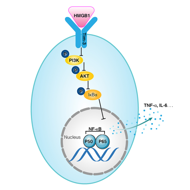
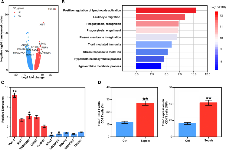
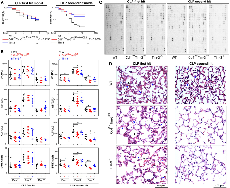
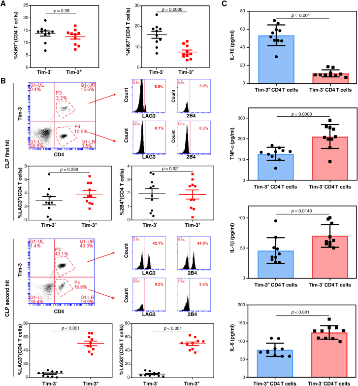
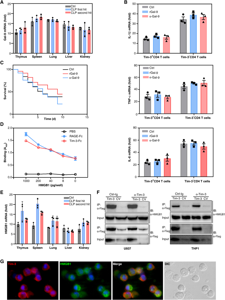
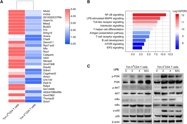
Immunosuppression in response to severe sepsis remains a serious human health concern. Evidence of sepsis-induced immunosuppression includes impaired T lymphocyte function, T lymphocyte depletion or exhaustion, increased susceptibility to opportunistic nosocomial infection, and imbalanced cytokine secretion. CD4 T cells play a critical role in cellular and humoral immune responses during sepsis. Here, using an RNA sequencing assay, we found that the expression of T cell-containing immunoglobulin and mucin domain-3 (Tim-3) on CD4 T cells in sepsis-induced immunosuppression patients was significantly elevated. Furthermore, the percentage of Tim-3+ CD4 T cells from sepsis patients was correlated with the mortality of sepsis-induced immunosuppression. Conditional deletion of Tim-3 in CD4 T cells and systemic Tim-3 deletion both reduced mortality in response to sepsis in mice by preserving organ function. Tim-3+ CD4 T cells exhibited reduced proliferative ability and elevated expression of inhibitory markers compared with Tim-3-CD4 T cells. Colocalization analyses indicated that HMGB1 was a ligand that binds to Tim-3 on CD4 T cells and that its binding inhibited the NF-κB signaling pathway in Tim-3+ CD4 T cells during sepsis-induced immunosuppression. Together, our findings reveal the mechanism of Tim-3 in regulating sepsis-induced immunosuppression and provide a novel therapeutic target for this condition.

Keywords: CD4 T cells; T cell-containing immunoglobulin and mucin domain-3 (Tim-3); biomarker; sepsis-induced immunosuppression.


Conflict of interest statement

Declaration of interests All authors declare that they have no competing financial interests.

Figures

None
Graphical abstract
Figure 1
Figure 1
The proportion of Tim-3+ CD4 T cells is increased in CD4 T cells from septic immunosuppression patients (A) Volcano plot of CD4 T cells from healthy volunteers and septic immunosuppression patients. The top five upregulated genes were Tim-3, XIST, TMEM38B, LIMS2, and IL10RB. The top five downregulated genes were ADA2, LDLRAD4, PRMT6, MMACHC, and TENM1. (B) Enriched gene ontology (GO) functions of upregulated genes in CD4 T cells. Upregulated genes in CD4 T cells of septic immunosuppression patients were enriched in immune-related pathways, especially lymphocyte- and leukocyte-related pathways, as well as several metabolic pathways. (C) Verification of the transcription levels of the top five upregulated and top five downregulated genes in CD4 T cells between sepsis patients and healthy volunteers (means ± SD; n = 30; ∗p < 0.05 and ∗∗p < 0.01 versus healthy control group, one-way ANOVA). (D) Tim-3 expression on CD4 T cells in 30 sepsis patients and 30 healthy volunteers by flow cytometry. The percentage of Tim-3-positive CD4 T cells (means ± SD; n = 30; ∗∗p < 0.01 versus healthy control group, one-way ANOVA). Tim-3 expression on CD4 T cells is presented as the mean fluorescence intensity (means ± SD; n = 30; ∗∗p < 0.01 versus healthy control group, one-way ANOVA).
Figure 2
Figure 2
Tim-3 knockout reduces sepsis mortality by preserving organ function (A) Survival curves of WT, Cd4CreTim-3fL/fL, and Tim-3−/− mice in the CLP first hit and second hit models of sepsis. p = 0.0062 compared with WT and Cd4CreTim-3fL/fL and p = 0.008 compared with WT and Tim-3−/− mouse in CLP second hit mouse of sepsis (n = 30 mice per group; Kaplan–Meier survival analysis). (B) Serum enzyme activity in the heart, liver, and kidney of CLP first and second hit mouse post 1, 3, and 7 days (n = 5; ∗p < 0.05 and ∗∗p < 0.01 versus WT, one-way ANOVA). (C) Secreted cytokines in WT, Cd4CreTim-3fL/fL, and Tim-3−/− mouse peripheral blood were profiled using a Proteome Profiler™ Array Mouse XL Cytokine Array Kit. (D) Hematoxylin and eosin staining of WT, Cd4CreTim-3fL/fL, and Tim-3−/− mouse lungs: alveolar septum thickening (blue arrow), leukocyte infiltration (blue arrowhead), alveolar congestion and edema (black arrow). Magnification, 200×; Scale bars, 100 μm.
Figure 3
Figure 3
Tim-3 is correlated with a more severe exhaustion state of CD4 T cells during septic immunosuppression (A) The proliferation ability of Tim-3+ CD4 T cells and Tim-3 CD4 T cells after CLP first hit (left) and second hit (right) is shown as the percentage of Ki67+ cells. Data are representative of three independent experiments with 10 mice per group in each experiment (p = 0.0006 in CLP second hit mouse of sepsis, one-way ANOVA). (B) Expression of LAG3 and 2B4 on Tim-3+ CD4 T cells and Tim-3 CD4 T cells after CLP first hit and second hit models (n = 10; p < 0.01 in CLP second hit mouse of sepsis, one-way ANOVA). (C) Release of IL-10, TNF-α, IL-1β, and IL-6 in Tim-3+ CD4 T cells and Tim-3 CD4 T cells in response to 100 ng/mL LPS stimulation in vitro for 24 h (p = 0.0008 for TNF-α, p = 0.0143 for IL-1β, p < 0.01 for IL-10 and IL-6, one-way ANOVA).
Figure 4
Figure 4
HMGB1 is a ligand for Tim-3 on CD4 T cells in septic immunity (A) RT-PCR quantification of Gal-9 mRNA in the thymus, spleen, lung, liver, and kidney of CLP first hit and second hit model mice. The results are presented relative to the expression of GAPDH (n = 3; means ± SD; one-way ANOVA). (B) RT-PCR analysis of IL-1β, TNF-α, and IL-6 mRNA without or with rGal-9 or α-Gal-9 treatment for 12 h. The results are presented relative to GAPDH expression (n = 3; means ± SD; one-way ANOVA). (C) Administration of rGal-9 or α-Gal-9 to mice prevented CLP second hit-induced death (n = 20 mice per group; Kaplan–Meier survival analysis). (D) Binding of biotin-labeled rHMGB1 to plates coated with PBS or fusions of Fc-RAGE (RAGE-Fc) or Fc-Tim-3 measured by colorimetric analysis and presented as absorbance at 450 nm. (E) RT-PCR quantification of HMGB1 mRNA in the thymus, spleen, lung, liver, and kidney of CLP first hit and second hit mouse models. The results are presented relative to the expression of GAPDH (n = 3; means ± SD; p < 0.05 thymus versus lung, liver, or kidney in CLP second hit model; p < 0.05 spleen versus lung, liver, or kidney in CLP second hit model by one-way ANOVA). (F) Immunoprecipitation of U937 or THP1 cells transfected with vector encoding Flag-tagged Tim-3 or control vector and stimulated for 2 h with HMGB1 in the presence of control immunoglobulin (Ig) or mAb to Tim-3, followed by IP with mAb M2 to the FLAG tag and immunoblot analysis with anti-HMGB1 or anti-Flag. (G) Immunofluorescence image of Tim-3 (red) in Tim-3+ CD4 T cells colocalized with recombinant HMGB1 (green). Magnification, 600×.
Figure 5
Figure 5
Expression of Tim-3 on CD4 T cells regulates septic immunosuppression through the NF-κB/TNF-α pathway (A) Heatmap of the top 30 upregulated genes in Tim-3 CD4 T cells compared to Tim-3+ CD4 T cells isolated from CLP second hit mice. (B) Enriched gene ontology functions of upregulated genes in Tim-3 CD4 T cells. Upregulated genes were enriched in NF-κB signaling, LPS-stimulated MAPK, and Toll-like receptor signaling. (C) Western blot analysis of the indicated protein expression in Tim-3+/Tim-3 CD4 T cells in response to treatment with 100 ng/mL LPS for 0, 2, 4, or 6 h.

References

    1. Rudd K.E., Johnson S.C., Agesa K.M., Shackelford K.A., Tsoi D., Kievlan D.R., Colombara D.V., Ikuta K.S., Kissoon N., Finfer S., et al. Global, regional, and national sepsis incidence and mortality, 1990-2017: analysis for the Global Burden of Disease Study. Lancet. 2020;395:200–211. - PMC - PubMed
    1. Mira J.C., Gentile L.F., Mathias B.J., Efron P.A., Brakenridge S.C., Mohr A.M., Moore F.A., Moldawer L.L. Sepsis pathophysiology, chronic critical illness, and persistent inflammation-immunosuppression and catabolism syndrome. Crit. Care Med. 2017;45:253–262. - PMC - PubMed
    1. Van Der Poll T., Van De Veerdonk F.L., Scicluna B.P., Netea M.G. The immunopathology of sepsis and potential therapeutic targets. Nat. Rev. Immunol. 2017;17:407–420. - PubMed
    1. Fattahi F., Ward P.A. Understanding immunosuppression after sepsis. Immunity. 2017;47:3–5. - PubMed
    1. Gotts J.E., Matthay M.A. Sepsis: pathophysiology and clinical management. BMJ. 2016;353:i1585. - PubMed

Publication types

Substances