. 2022 Feb;602(7896):328-335.
doi: 10.1038/s41586-021-04340-2. Epub 2021 Dec 21.

Activation mechanism of PINK1

Affiliations

Activation mechanism of PINK1

Zhong Yan Gan et al. Nature. 2022 Feb.

Erratum in

  • Publisher Correction: Activation mechanism of PINK1.
    Gan ZY, Callegari S, Cobbold SA, Cotton TR, Mlodzianoski MJ, Schubert AF, Geoghegan ND, Rogers KL, Leis A, Dewson G, Glukhova A, Komander D. Gan ZY, et al. Nature. 2022 Mar;603(7903):E33. doi: 10.1038/s41586-022-04591-7. Nature. 2022. PMID: 35293391 Free PMC article. No abstract available.

Abstract

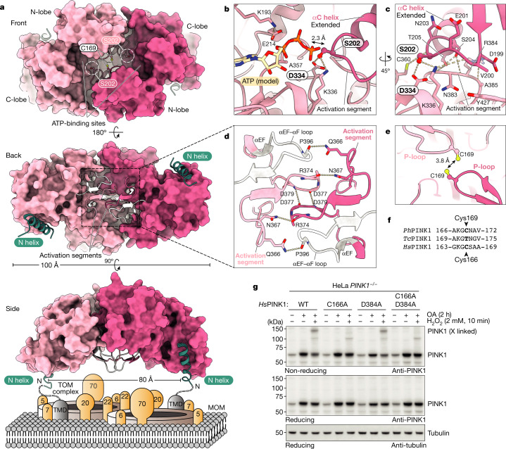
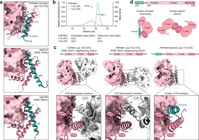
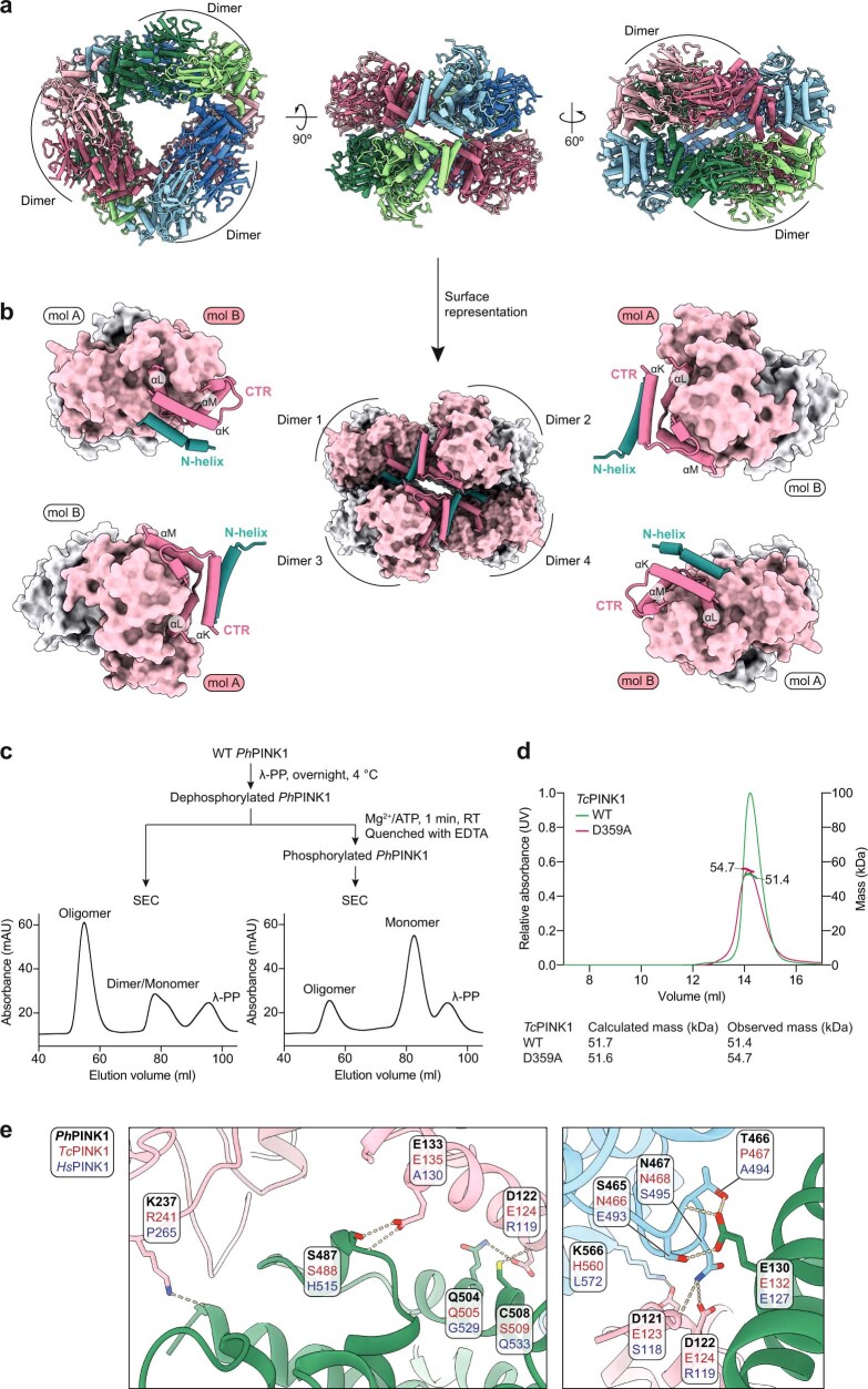
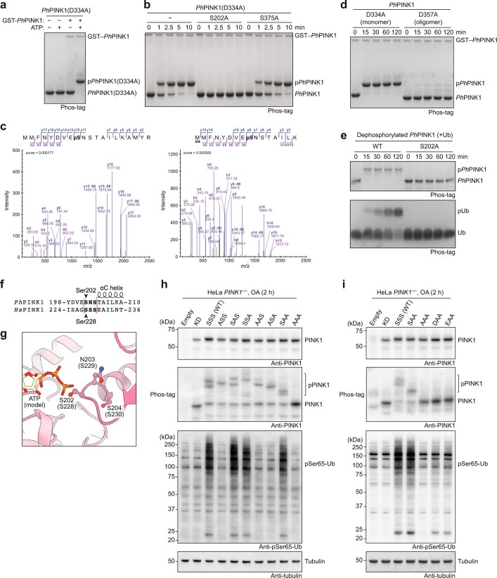
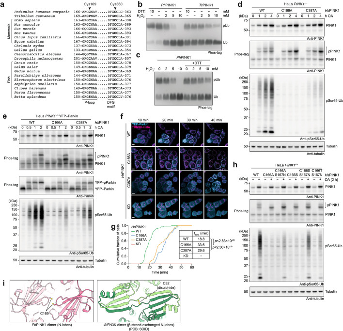
Mutations in the protein kinase PINK1 lead to defects in mitophagy and cause autosomal recessive early onset Parkinson's disease1,2. PINK1 has many unique features that enable it to phosphorylate ubiquitin and the ubiquitin-like domain of Parkin3-9. Structural analysis of PINK1 from diverse insect species10-12 with and without ubiquitin provided snapshots of distinct structural states yet did not explain how PINK1 is activated. Here we elucidate the activation mechanism of PINK1 using crystallography and cryo-electron microscopy (cryo-EM). A crystal structure of unphosphorylated Pediculus humanus corporis (Ph; human body louse) PINK1 resolves an N-terminal helix, revealing the orientation of unphosphorylated yet active PINK1 on the mitochondria. We further provide a cryo-EM structure of a symmetric PhPINK1 dimer trapped during the process of trans-autophosphorylation, as well as a cryo-EM structure of phosphorylated PhPINK1 undergoing a conformational change to an active ubiquitin kinase state. Structures and phosphorylation studies further identify a role for regulatory PINK1 oxidation. Together, our research delineates the complete activation mechanism of PINK1, illuminates how PINK1 interacts with the mitochondrial outer membrane and reveals how PINK1 activity may be modulated by mitochondrial reactive oxygen species.


Conflict of interest statement

D.K. serves on the Scientific Advisory Board of BioTheryX Inc. The other authors declare no competing interests.

Figures

Fig. 1
Fig. 1. Crystal structure of the cytosolic portion of PhPINK1.
a, PhPINK1 constructs that were used previously (Protein Data Bank (PDB): 6EQI) and structurally characterized in this study. The mitochondrial-targeting sequence (MTS), outer mitochondrial membrane localization signal (OMS), TMD, N-helix and linker, insertion-2 (i2), insertion-3 (i3) and the CTR are indicated. b, The crystal structure of unphosphorylated PhPINK1(D334A) (amino acids 115–575), with an extended αC helix and disordered insertion-3. The N-helix (teal) packs against the CTR and directly follows the predicted TMD that interacts with the TOM complex (not to scale) (Extended Data Figs. 1 and 2 and Supplementary Table 1). aa, amino acids. c, EOPD mutations in the N-helix and CTR affect the interface. Mutations according to refs. ,. d, The previous structure of the PhPINK1–Ub TVLN complex (PDB: 6EQI; Ub TVLN is shown in grey, without the nanobody), with a kinked αC helix, phosphorylated (p) Ser202 and ordered insertion-3.
Fig. 2
Fig. 2. Oligomerization of a kinase inactive PhPINK1 enables cryo-EM.
a, SEC–MALS analysis of PhPINK1 (amino acids 115–575) variants (Fig. 1a). Absorbance was measured at a wavelength of 280 nm. Theoretical and observed molecular mass values are indicated. Each protein displayed identical behaviour in at least three SEC runs, and SEC–MALS experiments were performed twice. b, Cryo-EM density map for the PhPINK1(D357A) dodecamer at 2.48 Å, indicating monomers in different colours and dimers in shades of the same colour. Left, top view with three-fold symmetry. Right, side view with two-fold symmetry (Extended Data Fig. 3 and Supplementary Table 2).
Fig. 3
Fig. 3. The PhPINK1 dimer before trans-autophosphorylation.
a, The structure of the PhPINK1(D357A) dimer in surface representation. Top, front view of the dimer with empty ATP-binding sites. The Ser202-containing loop (Ser202 circled in white) reaches into the acceptor site of the opposing kinase domain. Middle, back view of the dimer, with complementary activation segments (cartoon, coloured). Bottom, side view of the dimer showing the N-helices, indicating how it may sit on the MOM interacting with a TOM complex. A molecular model of the PhPINK1 dimer manually docked onto the TOM complex is shown in Extended Data Fig. 5d. be, Detailed views in stick representation. The dotted lines indicate hydrogen bonds. b, ATP-binding and trans-autophosphorylation interactions. ATP was modelled from PDB 2PHK (ref. ) as before. c, Coordination of the Ser202-containing loop. d, Dimer interactions through activation segments and αEF–αF loops. e, The close proximity of P-loop Cys169 residues during dimer formation (Extended Data Fig. 5e). f, Conservation of PhPINK1 Cys169 in HsPINK1 (Cys166) but not TcPINK1 (Thr172). g, Formation of a disulphide-linked HsPINK1 dimer in HeLa cells that were treated with H2O2. HsPINK1 was expressed in HeLa PINK1−/− cells and stabilized with OA treatment (Methods). H2O2 treatment leads to a disulphide-linked HsPINK1 dimer band visualized on a non-reducing gel, which is absent with C166A mutation, suggesting HsPINK1 also dimerizes through Cys166. A putative dimer-trapping mutation in HsPINK1, D384A (D357A in PhPINK1; compare with Fig. 2a), does not further stabilize the dimer. The experiments were performed in biological triplicate with identical results. The uncropped blots are provided in Supplementary Fig. 1.
Fig. 4
Fig. 4. The cryo-EM structure of Ser202-phosphorylated PhPINK1 reveals a conformational change.
a, Flowchart for the generation of the phosphorylated and partially cross-linked WT PhPINK1 dodecamer. RT, room temperature. b, Profile of the final SEC run. c, Phos-tag analysis of the PhPINK1 species. The final oligomer fraction comprises homogenously phosphorylated PhPINK1 and was used for cryo-EM analysis. The experimental workflow shown in ac was performed once in this exact configuration. The uncropped gel is provided in Supplementary Fig. 1. d, Cryo-EM density map of the Ser202-phosphorylated WT PhPINK1 dimer at 3.07 Å. A break in symmetry in the N-lobe is visible (Methods, Extended Data Fig. 7 and Supplementary Table 2). e, EM density for the N-lobe of molecule A shows an extended αC helix (orange) with the phosphorylated Ser202 at the tip. f, The EM density for the N-lobe of molecule B shows a less-ordered state of the αC helix seemingly in transition. g, h, 3D variability analysis enabled the clustering of distinct states of the N-lobe in molecule B. g, In the first cluster, the αC helix (orange) is kinked, and extra density can be modelled by a poly-Ala model of insertion-3 (yellow). h, The second cluster resembles molecule A with an extended αC helix and disordered insertion-3.
Fig. 5
Fig. 5. Regulation of PINK1 by oxidation and the model of PINK1 activation.
a, Cys169—which is involved in dimerization (Fig. 3)—and Cys360 line the ATP-binding pocket of PhPINK1 and are also close to the substrate ubiquitin (PDB: 6EQI). ATP was modelled as in Fig. 3b. b, Phos-tag analysis of PhPINK1-mediated ubiquitin phosphorylation, with increasing concentrations of H2O2. Mutations of Cys169 and Cys360 render PhPINK1 less active but also unresponsive to H2O2. The experiments were performed in biological triplicate with identical results. The uncropped gel is provided in Supplementary Fig. 1. c, Conservation of Cys169 and its context in PhPINK1, HsPINK1 and salmon (Salmo salar) PINK1 (Extended Data Fig. 9a). d, The workflow for the experiment in e. Details are provided in the Methods. Heavy membranes isolated from OA-treated HeLa PINK1−/− cells expressing WT HsPINK1 or HsPINK1(C166S/S167N) (SN, mutating the P-loop to a fish-like sequence to generate active Cys166-lacking HsPINK1) were pretreated with increasing concentrations of H2O2 (as indicated in e) to oxidize membrane-associated active HsPINK1, before incubation with recombinant ubiquitin and ATP. DTT, dithiothreitol. e, Western blotting of the samples generated in d, indicating reversible inactivation of HsPINK1 by H2O2, which was not observed using the Cys166-lacking SN mutant. The experiments were performed in biological triplicate. Uncropped blots are provided in Supplementary Fig. 1. f, Quantification of pSer65-Ub band intensities from experiments in e and its repeats (n = 3; Supplementary Fig. 2). Intensities were adjusted on the basis of PINK1 levels (reducing gel) and normalized to the 0 mM H2O2 condition for each HsPINK1 variant. Band intensities were quantified using ImageLab (Bio-Rad, v.6.1). g, The model for PINK1 activation on the surface of depolarized mitochondria. We expect that αC helix kinking (1) precedes ordering of insertion-3 (2) to form the ubiquitin-binding site. ROS, reactive oxygen species. Source data
Extended Data Fig. 1
Extended Data Fig. 1. Crystal structures of unphosphorylated PINK1.
a, Constructs used in crystal structures of TcPINK1 (PDB: 5OAT and 5YJ9). Phosphomimetics substituted for Ser205 (equivalent to Ser202 in PhPINK1) and other residues, although one structure (PDB: 5OAT) still showed phosphorylation at Ser207 (equivalent to Ser204 in PhPINK1, and Ser230 in HsPINK1, see below). Constructs were further engineered as indicated and described,. b, Depiction of TcPINK1 crystal structures, with key features indicated. c, 2|Fo|–|Fc| electron density contoured at 1.5σ, covering the PhPINK1(D334A) molecule in the asymmetric unit. Right, detail for regions of interest including the αC helix and Ser202-containing loop and the N-helix. d, Our crystal structure of unphosphorylated PhPINK1(D334A) (compare Fig. 1) in the same orientation as in b, for comparison. e, Two different zoomed views of the ATP-binding site of unphosphorylated PhPINK1, with an ATP molecule modelled in from PDB 2PHK (ref. ) as before. Left, highlighting key features common to most active kinases, including the P-loop (grey), αC helix (orange) and activation segment (pink). The DFG and HRD motifs (dotted outlines) which include Asp357 involved in coordination of Mg2+, and the substrate-binding Asp334 involved in phosphoryl transfer (and mutated to Ala in the crystal structure) are indicated. Glu214 and Lys193 form a crucial salt bridge to coordinate ATP. Right, assembled catalytic (C) and regulatory (R) spines in red and blue, respectively, indicative of an active kinase conformation. f, Crystal contact involving N-helix residue Trp129, which binds to the N-lobe of a symmetry related molecule. ASU, asymmetric unit.
Extended Data Fig. 2
Extended Data Fig. 2. The N-helix and its role in keeping PINK1 monomeric.
a, The pseudokinases SgK223 (PDB: 5VE6) and SgK269 (PDB: 6BHC) were structurally characterised only recently,, and contain a CTR similar to PINK1 that is complemented by an N-helix, in a highly analogous fashion now shown for PINK1. While conceptually similar, the organization of the N-helix on the CTR is however distinct. SgK223 and SgK269 utilize a cross-architecture to provide a dimerization interface, whereas the orientation in PINK1 is parallel to the αK helix of the CTR. b, SEC–MALS analysis of PhPINK1 with (amino acids 115–575) or without (amino acids 143–575) the N-helix. The shorter PhPINK1 construct tends to form less well-defined dimers, whereas the longer variant is a monomer. Experiments were performed three times with identical results. c, Previous TcPINK1 and PhPINK1 structures dimerized in the crystal lattice through the CTR, reflecting an available interaction surface since the N-helix was missing. TcPINK1 structures dimerized identically (left, only shown for PDB 5OAT). The relative orientation of PhPINK1 molecules in PDB 6EQI was different to TcPINK1 (middle). Right, PhPINK1(D334A) structure including the N-helix, for comparison. d, Schematic of the N-helix–CTR interaction, and situation in previous PINK1 structures, as identified in structures and in SEC–MALS.
Extended Data Fig. 3
Extended Data Fig. 3. PhPINK1(D357A) oligomerization enables EM studies.
a, Elution profile of the PhPINK1(D357A) mutant during purification by SEC. Data shown is from a representative experiment of three runs. b, Thermal denaturation studies of purified PhPINK1 (amino acids 115–575) variants. The crystallized D334A mutant has a melting curve profile and melting temperature similar to WT PhPINK1. PhPINK1(D357A) shows an unusual profile with a high secondary melting temperature. Technical duplicates were measured in three independent experiments. Average melting temperatures are indicated. c, Negative stain EM analysis of PhPINK1(D357A) revealed a highly ordered oligomer suitable for cryo-EM studies. Minimal processing was performed in RELION (v.3.1), and a subset of the resulting 2D classes is depicted as insets. Scale bar, 50 nm. Negative staining for this sample was performed once, before advancing to cryo-EM analysis. d, Flowchart for cryo-EM analysis as described in Methods. e, 2D classification of particles reveal a 2D-crystalline arrangement of PhPINK1(D357A) oligomers in some areas of the grid. f, Final cryo-EM density maps (coloured by local resolution) for the PhPINK1(D357A) dodecamer (left) at 2.48 Å and the extracted dimer (right) at 2.35 Å. g, Examples of map quality for the final 2.35 Å density covering the PhPINK1(D357A) dimer. Source data
Extended Data Fig. 4
Extended Data Fig. 4. Further analysis of the PhPINK1 oligomeric state.
a, Model built into the PhPINK1(D357A) dodecamer cryo-EM density as in Fig. 2b. b, Side view of the oligomer showing how the N-helix–CTR area facilitates contacts to connect four dimers. c, We were concerned that PhPINK1 oligomerization may only happen with a specific mutant protein and tested whether the oligomer would also form with unphosphorylated WT PhPINK1. WT PhPINK1 is phosphorylated at multiple sites when purified from E. coli (see), and was hence dephosphorylated by λ-PP and repurified by SEC as indicated (see Methods). Like the inactive D357A mutant, a prominent oligomer, as well as a dimer/monomer equilibrium is apparent on SEC. Alternatively, the WT protein was first dephosphorylated by λ-PP, then rephosphorylated by adding Mg2+/ATP for 1 min, and the kinase and λ-PP were inactivated by EDTA (see Methods). Phosphorylation destabilized the oligomer to a predominant monomeric species, explaining why WT PhPINK1 does not form the oligomer, and suggesting that autophosphorylation resolves the oligomer and dimer (see below). These experiments were part of the purification process and were performed three times with identical results. d, SEC–MALS analysis of TcPINK1 variants, to show that corresponding TcPINK1 constructs (amino acids 117–570) do not show oligomeric behaviour. TcPINK1 purifications revealing monomeric behaviour were performed at least twice, and a SEC–MALS experiment was performed once. e, Residues mediating hydrogen bonds (dotted lines) in the oligomer interfaces. Lack of conservation of oligomer contacts between PhPINK1, TcPINK1 and HsPINK1 likely explain why TcPINK1 does not form an oligomer, and why we do not expect an identical oligomer in HsPINK1.
Extended Data Fig. 5
Extended Data Fig. 5. Structural and biochemical analysis of the PhPINK1 dimer.
a, Comparison between PhPINK1 autophosphorylation and ubiquitin phosphorylation resolved previously (PDB: 6EQI), with relevant details in the active site (top row) and in the activation segment (bottom row). The right panel shows substrate disposition in relation to a modelled ATP molecule as in Fig. 3b. The dimeric PhPINK1 autophosphorylation complex appears to place Ser202 in an ideal phospho-accepting position. b, Comparison of activation segment structures in all published PINK1 structures, revealing high structural similarity. Ser375 (PhPINK1)/Ser377 (TcPINK1) is shown in ball-and-stick representation; this residue was mutated to Asp in one prior structure (see Extended Data Fig. 1a). c, PhPINK1 Ser375 corresponds to Ser402 in HsPINK1, which is a reported phosphorylation site,, but is located within the activation segment and out of reach of the substrate-binding site within dimeric PhPINK1. Our structure does not reveal how autophosphorylation at this residue could be facilitated in cis or trans (left), nor how phosphorylation through e.g. an upstream kinase would contribute to PINK1 activity or function, since phosphorylation would likely disrupt the dimer (right). ATP was modelled as in Fig. 3b. d, Manual docking of dimeric PhPINK1 onto a cryo-EM structure of dimeric human TOM complex (PDB: 7CK6). The PhPINK1 dimer was oriented with its two N-helices (spanning ~80 Å) aligning with the two TOM7 subunits of the TOM complex dimer. TOM7 has been reported as essential for PINK1 stabilization on the TOM complex,. Note that some TOM components have considerable cytosolic domains that would need to be accommodated in addition to a PINK1 dimer. e, Unidentified density in the 2.35 Å cryo-EM map connects Cys169 in the dimer. f, Time course of PhPINK1 and TcPINK1 disulphide formation upon treatment with 2 mM H2O2, resolved on a non-reducing SDS–PAGE gel. The dimer/oligomer stabilizing PhPINK1 D357A mutation (Fig. 3) enables fast disulphide formation that is averted by an additional C169A mutation. TcPINK1 WT or D359A (equivalent to PhPINK1(D357A)) mutant do not show fast cross-linking behaviour observed in PhPINK1. Engineering of an additional Thr172 to Cys mutation (TcPINK1(T172C/D359A)) results in rapid emergence of cross-linked TcPINK1 dimers upon oxidation. Experiments were performed in biological triplicate with identical results. See Supplementary Fig. 1 for uncropped gels. g, EOPD mutations in the activation segment and αEF–αF loop. Mutations according to. h, HsPINK1 EOPD mutants listed in g were expressed in HeLa PINK1−/− cells, stabilized with OA and treated with H2O2 to assess PINK1 dimerization, autophosphorylation and ubiquitin phosphorylation activity (see Methods). The control D384A mutant (mutation of the DFG motif, equivalent to D357A in PhPINK1) was included as an inactive mutant. As anticipated, it is able to dimerize (oxidative cross-link formed) but unable to autophosphorylate or generate phosphorylated ubiquitin. Oxidative dimerization enables separation of pure catalytic mutants and dimerization deficient mutants. Experiments were performed in biological triplicate with identical results. See Supplementary Fig. 1 for uncropped blots.
Extended Data Fig. 6
Extended Data Fig. 6. Analysis of autophosphorylation sites in PhPINK1 and HsPINK1.
a, Phos-tag analysis of inactive, monomeric PhPINK1(D334A), which does not autophosphorylate when incubated with ATP. Phosphorylation by WT GST-tagged PhPINK1 for 2 h leads to a shift of the entire protein consistent with a single phosphorylation site, and a small amount (1–2%) shifts to a higher species indicating additional autophosphorylation events after prolonged incubation. A representative gel of three independent experiments is shown. See Supplementary Fig. 1 for uncropped gel. b, As in a but shown in a time course experiment. Mutating Ser202 to Ala abrogates the observed gel shift, indicating that Ser202 is the sole site of phosphorylation. Mutation of Ser375 (equivalent to HsPINK1 Ser402) to Ala does not impede phosphorylation. A representative gel of three independent experiments is shown. See Supplementary Fig. 1 for uncropped gel. c, Mass spectrometry confirms the detection of a phosphate at Ser202 of PhPINK1(D334A) after phosphorylation with WT GST–PhPINK1. See Methods. d, Time course experiment as in b, with monomeric PhPINK1(D334A) or oligomeric PhPINK1(D357A). While the monomer can be phosphorylated by WT GST–PhPINK1, the oligomer is not efficiently phosphorylated since Ser202 is buried in the stable dimer/oligomer structure. This experiment was performed twice with identical results. See Supplementary Fig. 1 for uncropped gel. e, Time course experiment using dephosphorylated WT PhPINK1 (amino acids 119–575, which we found to be oligomerization impaired) and PhPINK1 (amino acids 119–575) S202A, and with ubiquitin in the reaction. See Methods for details. Phosphorylation of PhPINK1 at Ser202 is essential for ubiquitin phosphorylation, as both phosphorylation events are completely abrogated if Ser202 cannot be phosphorylated. Representative gels of three independent experiments are shown. See Supplementary Fig. 1 for uncropped gels. f–i, Analysis of autophosphorylation sites in HsPINK1. f, In HsPINK1, the Ser202 equivalent residue is Ser228, which is a known, important autophosphorylation site. In PhPINK1, the Ser202 site is followed by Asn203 and Ser204, and Ser204 can be phosphorylated when PhPINK1 is expressed in E. coli. Also, the Ser204 equivalent residue in TcPINK1 (Ser207) was phosphorylated in a previous crystal structure (Extended Data Fig. 1a). In HsPINK1, Ser228 is followed by Ser229 and Ser230, and we investigated whether phosphorylation of these residues occurs and whether it contributes to HsPINK1 activity. (g) Structural detail of the Ser202-containing loop in the active site of the PhPINK1 autophosphorylation dimer. Equivalent HsPINK1 residues are in brackets. ATP was modelled as in Fig. 3b. Only Ser202 is in the phospho-acceptor site, but the other residues may occupy the site and be phosphorylated subsequently, if the end of the αC helix slightly unravels; since we expect conformational changes in this region (see below), this seemed feasible. h, i, HsPINK1 variants with combinations in the Ser228-containing loop as indicated (empty, vector control; KD, kinase dead, a triple mutant K219A, D362A (HRD motif), D384A (DFG motif); SSS (WT) refers to the WT sequence with Ser228, Ser229, Ser230; ASS refers to Ala228, Ser229, Ser230; etc.) were expressed in HeLa PINK1−/− cells, treated with OA for 2 h, and subjected to western blotting (see Methods). h, A Phos-tag gel probed with anti-PINK1 antibody reveals that KD and AAA mutants remain unphosphorylated, while the WT HsPINK1 (SSS) shows multiple phosphorylation states, indicating more than one phosphorylation event in this loop. All three Ser residues appear to be phosphorylatable. These results were correlated with appearance of Ser65-phosphorylated ubiquitin in the same cell lysates, revealed by a ubiquitin Ser65-phosphospecific antibody. WT HsPINK1 leads to a strong signal for phosphorylated ubiquitin, while Ala mutation in Ser228 does not lead to a phosphorylated ubiquitin signal. In contrast, if only Ser228 is present (e.g. SAA mutant) the phosphorylated ubiquitin signal is indistinguishable from that of WT HsPINK1. i, As in h, but with further substitution of Ser228. Phosphomimetic residues Asp228 or Glu228, followed by Ala229 and Ala230, lead to a small increase in phosphorylated ubiquitin, in the absence of autophosphorylation. The results in h and i indicate that also in HsPINK1, Ser228 phosphorylation is essential to turn PINK1 into a ubiquitin kinase, and a phosphomimetic is a weak substitute. Other residues in this area may also become phosphorylated but do not trigger ubiquitin phosphorylation. Experiments shown in h and i were performed in biological triplicate with identical results. See Supplementary Fig. 1 for uncropped blots.
Extended Data Fig. 7
Extended Data Fig. 7. Structure of a cross-linked, phosphorylated PhPINK1 reveals asymmetry and conformational changes in the N-lobe.
a, Final phosphorylated and cross-linked PhPINK1 oligomer on a non-reducing SDS–PAGE gel. See Supplementary Fig. 1 for uncropped gel. b, Flowchart for cryo-EM analysis, as in Extended Data Fig. 3. c, Local resolution maps and resolution calculations. d, Map quality for selected regions. e, EM density for the phosphorylated Ser202 of molecule A, in the substrate-binding site of molecule B.
Extended Data Fig. 8
Extended Data Fig. 8. Assessment of PhPINK1 dimer stability by measuring oligomer formation.
a, Our studies had revealed that disruption of the kinase–substrate interaction had a detrimental effect for dimer formation. We wondered whether the underlying dimerization mechanism would facilitate the reported effect that each copy of PINK1 is phosphorylated during mitophagy,,. Based on our biochemical data (Fig. 2a and Extended Data Fig. 4c) we conceptualized PINK1 dimer interactions as a function of the number of intact Ser202–Asp334 contacts. Ser202 phosphorylation would resolve the Ser202–Asp334 contacts individually, leading to one contact point after the first phosphorylation event, and zero contact points when both PINK1 molecules are phosphorylated. It was unclear whether the dimer is stable after the first phosphorylation event, when only one Ser202–Asp334 contact point exists within the dimer. b, We explored whether we would be able to generate PhPINK1 oligomers with an unphosphorylated PhPINK1(D334A) mutant and Ser202-phosphorylated PhPINK1 (phospho-PhPINK1), as indicated, with the knowledge that homo-oligomerization is disfavoured (zero Ser202–Asp334 contact points, highlighted in red). Formation of a hetero-oligomer between unphosphorylated PhPINK1(D334A) and phospho-PhPINK1 would indicate that the dimer is stable with just one Ser202–Asp334 contact (highlighted in green). c, SEC analysis of the mutants confirms that unphosphorylated PhPINK1(D334A) or phospho-PhPINK1 do not form oligomers, and hence, zero Ser202–Asp334 contact points are not sufficient. In contrast, enabling one contact point, as per the green scenario in b where variants are mixed, enables oligomerization of PhPINK1. Each individual protein showed identical results in every purification run (= 3), and the mixing experiments were performed in biological triplicate. d, Phos-tag analysis confirms that the oligomer species is a 1:1 mixture of unphosphorylated PhPINK1(D334A) and phospho-PhPINK1. Experiments were performed in biological triplicate. See Supplementary Fig. 1 for uncropped gel. Together, these experiments show that the dynamic equilibrium favours dimer formation until each copy of PINK1 has been phosphorylated, ensuring full phosphorylation.
Extended Data Fig. 9
Extended Data Fig. 9. Regulation of PINK1 activity by oxidation.
a, Extended sequence alignment indicating that Cys169 and Cys360 are well conserved in PINK1, and invariant in mammalian PINK1. Cys169 is a Thr in TcPINK1, and a Ser in many fish species. b, Comparison of PhPINK1 and TcPINK1, for their ability to be regulated by oxidation, shown in Phos-tag ubiquitin phosphorylation assays (see Fig. 5b). While PhPINK1 activity is abrogated with H2O2, TcPINK1 with Thr172 in the P-loop, remains active. The observed reduction in TcPINK1 activity could be a result of oxidation of the conserved Cys362 (Cys360 in PhPINK1) in the active site. Experiments were performed in biological triplicate. See Supplementary Fig. 1 for uncropped gel. c, Inhibition of PhPINK1 ubiquitin phosphorylation activity can be reversed with DTT, suggesting reversible regulatory oxidation. See Methods. Experiments were performed in biological triplicate. See Supplementary Fig. 1 for uncropped gel. d, Time course assessment of HsPINK1 Cys–Ala mutants transiently expressed in HeLa PINK1−/− cells. OA treatment leads to accumulation of HsPINK1 and slightly altered autophosphorylation for both C166A and C387A. The HsPINK1(C166A) mutant was almost completely deficient in ubiquitin phosphorylation, while HsPINK1(C387A) showed highly reduced but still detectable phosphorylated ubiquitin levels. Experiments were performed in biological triplicate. See Supplementary Fig. 1 for uncropped blots. e, Time course of HsPINK1 mutants as in d, but using stable HsPINK1 expression in the presence of YFP–Parkin. Presence of YFP–Parkin seemingly increases levels of phosphorylated ubiquitin for all HsPINK1 variants as compared to d, yet overall, phosphorylated ubiquitin and phosphorylated Parkin levels remain strongly diminished with HsPINK1(C166A), and to a lesser degree with HsPINK1(C387A). Experiments were performed in biological triplicate. See Supplementary Fig. 1 for uncropped blots. f, Translocation of YFP–Parkin (cyan) to mitochondria (magenta, TOM20–Halo) in HeLa PINK1−/− stably expressing HsPINK1 Cys–Ala variants upon OA treatment, imaged using lattice light sheet microscopy. Maximum intensity projections are shown for four different timepoints. YFP–Parkin translocation is delayed in cells expressing either HsPINK1(C166A) or HsPINK1(C387A) relative to WT hHsPINK1. A kinase dead (KD) HsPINK1 variant was included as a control. Images are representative of three independent experiments. Scale bar is 10 μm. See Supplementary Video 1. g, Quantification of YFP–Parkin translocation in f. The cumulative fraction of cells exhibiting YFP–Parkin translocation is shown over time. Approximately 70 cells were counted per cell line. Each curve was fitted to determine the time for 50% of the cells to feature translocation. Significant differences between curves were determined using a two-sample Kolmogorov–Smirnov test. A MATLAB script and Source Data are available as Supplementary Material. Exact p values are: WT–C166A: p < 2.83 × 10−20; WT–C387A: p < 2.36 × 10−18. h, HsPINK1 mutants expressed in HeLa PINK1−/− cells and treated with OA for 2 h. Activity of HsPINK1(C166A) and HsPINK1(C166S) can be partially restored by an additional S167N mutation, mimicking the sequence observed in many fish species. A TcPINK1-like sequence introduced into HsPINK1, C166T/S167N, is less active than the HsPINK1(C166S/S167N) mutant. The additional Asn in the P-loop of the HsPINK1(C166S/S167N) mutant recovers activity of HsPINK1(C166S), suggesting that both residues are also important in ubiquitin/Ubl substrate interactions, and not merely involved in dimerization. Experiments were performed in biological triplicate. See Supplementary Fig. 1 for uncropped blots. i, A redox active switch in fructosamine-3-kinases (PDB: 6OID) is conceptually similar, utilizing a Cys at an identical position (Cys32) for Cys-mediated cross-linking and regulation of kinase activity by oxidation, however the overall orientation of kinase domains is dissimilar. Source data
Extended Data Fig. 10
Extended Data Fig. 10. Analysis of the HsPINK1 model as predicted by AlphaFold2,.
(a, b) Predicting the structure of HsPINK1 via AlphaFold2 (a, left), resulted in a model remarkably similar to PhPINK1 from the PhPINK1–Ub TVLN complex (b, right), and features a kinked αC helix, ordered insertion-3, and an extended N-helix that binds and extends from the CTR directly into the membrane. Consistently, predicting a complex between HsPINK1 with the ubiquitin-like (Ubl) domain of human Parkin (a, right), places the Ubl domain at HsPINK1 analogously to the PhPINK1–Ub TVLN complex (b, right). Both predictions are dissimilar to unphosphorylated PhPINK1 (b, left) and TcPINK1 (Extended Data Fig. 1). Insets show the detail of the N-lobe with a kinked αC helix and an ordered insertion-3. The prediction is somewhat surprising, as it suggests HsPINK1 to be an active ubiquitin kinase even without Ser228 phosphorylation, which contradicts biochemical analysis. Since AlphaFold2 does not yet predict the impact of post-translational modifications, we interpret the prediction such that it is possible and even likely, that HsPINK1 can adopt a ubiquitin-phosphorylation-competent conformation consistent with our previous PhPINK1–Ub TVLN complex structure. c, d, We next used AlphaFold2 to predict a dimer of HsPINK1. c, Strikingly, AlphaFold2 predicts a symmetric dimer with a dimer interface identical to the one shown for PhPINK1 (compare with d and Fig. 3). In fact, we have already validated this arrangement of HsPINK1 molecules via our Cys166 cross-linking experiments in Fig. 3g. However, in the predicted dimer of HsPINK1, the αC helix is kinked and Ser228 does not contact the second molecule. This is different from our conclusions in Extended Data Fig. 8, but again may be a result of not incorporating the effect of Ser228 phosphorylation. We therefore anticipate that unphosphorylated HsPINK1 can also adopt a conformation with an extended αC helix that places Ser228 into the active site of the dimeric molecule to facilitate autophosphorylation prior to forming the depicted conformation. AlphaFold2 predictions hence support the notion that the activation model proposed in Fig. 5g applies to HsPINK1.

References

    1. Valente EM, et al. Hereditary early-onset Parkinson’s disease caused by mutations in PINK1. Science. 2004;304:1158–1160. - PubMed
    1. Pickrell AM, Youle RJ. The roles of PINK1, Parkin, and mitochondrial fidelity in Parkinson’s disease. Neuron. 2015;85:257–273. - PMC - PubMed
    1. Kondapalli C, et al. PINK1 is activated by mitochondrial membrane potential depolarization and stimulates Parkin E3 ligase activity by phosphorylating serine 65. Open Biol. 2012;2:120080. - PMC - PubMed
    1. Kazlauskaite A, et al. Parkin is activated by PINK1-dependent phosphorylation of ubiquitin at Ser65. Biochem. J. 2014;460:127–139. - PMC - PubMed
    1. Koyano F, et al. Ubiquitin is phosphorylated by PINK1 to activate parkin. Nature. 2014;510:162–166. - PubMed

Publication types