. 2021 Dec 21;11(1):24166.
doi: 10.1038/s41598-021-03123-z.

Involvement of the zebrafish trrap gene in craniofacial development

Affiliations

Involvement of the zebrafish trrap gene in craniofacial development

Taichi Suzuki et al. Sci Rep. .

Abstract

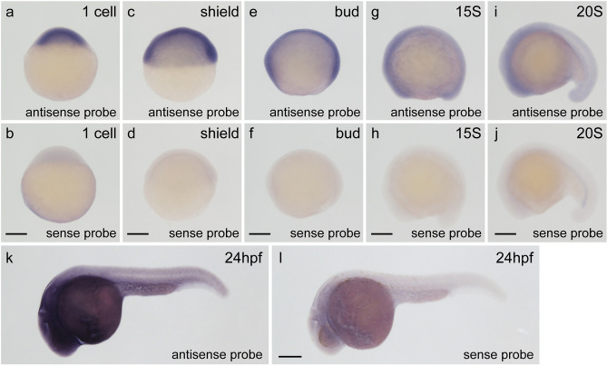
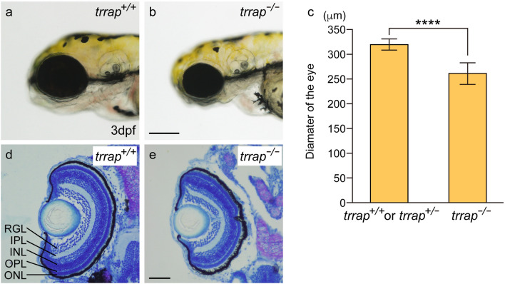
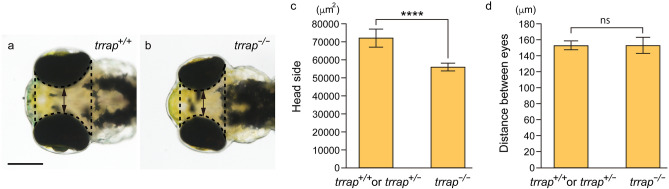
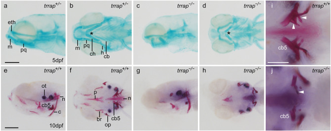
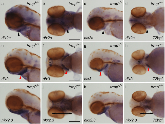
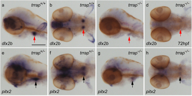
Trrap (transformation/transcription domain-associated protein) is a component shared by several histone acetyltransferase (HAT) complexes and participates in transcriptional regulation and DNA repair; however, the developmental functions of Trrap in vertebrates are not fully understood. Recently, it has been reported that human patients with genetic mutations in the TRRAP gene show various symptoms, including facial dysmorphisms, microcephaly and global developmental delay. To investigate the physiological functions of Trrap, we established trrap gene-knockout zebrafish and examined loss-of-function phenotypes in the mutants. The trrap zebrafish mutants exhibited smaller eyes and heads than the wild-type zebrafish. The size of the ventral pharyngeal arches was reduced and the mineralization of teeth was impaired in the trrap mutants. Whole-mount in situ hybridization analysis revealed that dlx3 expression was narrowly restricted in the developing ventral pharyngeal arches, while dlx2b expression was diminished in the trrap mutants. These results suggest that trrap zebrafish mutants are useful model organisms for a human disorder associated with genetic mutations in the human TRRAP gene.


Conflict of interest statement

The authors declare no competing interests.

Figures

Figure 1
Figure 1
Expression of the trrap gene during zebrafish embryogenesis. (a,b) One-cell stage. (c,d) Shield stage. (e,f) Bud stage. (g,h) Fifteen-somite (15S) stage. (i,j) Twenty-somite (20S) stage. (k,l) Twenty-four hours post-fertilization (hpf). All pictures are lateral views. Dorsal is right (c). Anterior is left (el). The expression of the trrap gene was examined by whole-mount in situ hybridization (WISH) using an antisense trrap DIG probe (a,c,e,g,i,k) or a sense trrap DIG probe (b,d,f,h,j,l). Scale bar, 200 μm.
Figure 2
Figure 2
Small eyes in the trrap-zebrafish mutant. (a) Wild-type fish (trrap+/+) at 3 dpf. (b) trrap mutants (trrap−/−) at 3 dpf. Scale bar, 200 μm. (c) The eye diameters in larvae containing the wild-type allele (trrap+/+: n = 2; trrap+/−: n = 6) and larvae containing trrap mutant alleles (trrap−/−: n = 8) were measured. The error bars indicate the standard deviation. Asterisks indicate statistical significance between the wild-type and the mutant zebrafish. ****P < 0.0001. (d,e) Cross-sections of the wild-type (trrap+/+) (d) and the trrap-mutant (trrap−/−) zebrafish (e) at 3 dpf were stained with toluidine blue (0.1%). The eye diameter was reduced in the trrap mutants, whereas the laminated retinas consisting of three layers (RGL, INL and ONL) and two plexiform layers (IPL and OPL) developed normal in the wild-type and mutant zebrafish. RGL retinal ganglion cell layer, IPL inner plexiform layer, INL inner nuclear layer, OPL outer plexiform layer, ONL outer nuclear layer. Genomic DNA was isolated from individual caudal fins, and genotyping was performed by genomic PCR. Scale bar, 50 μm.
Figure 3
Figure 3
Small heads in the trrap-zebrafish mutant zebrafish. (a) Wild-type fish (trrap+/+) at 3 dpf. (b) trrap mutants (trrap−/−) at 3 dpf. Scale bar, 200 μm. (c) The sizes of the head excluding the eyes (dashed line) in the wild-type (trrap+/+: n = 1; trrap+/−: n = 7) and trrap mutant (trrap−/−: n = 8) zebrafish were measured. The error bars indicate the standard deviation. Asterisks indicate statistical significance between the wild-type and mutant zebrafish. ****P < 0.0001. (d) The distance between the eyes (double arrow) in the wild-type (trrap+/+: n = 1; trrap+/−: n = 7) and trrap mutant (trrap−/−: n = 8) zebrafish were measured. The error bars indicate the standard deviation; ns, not significant.
Figure 4
Figure 4
Morphological defects in the pharyngeal arches and teeth of the trrap mutants. (ad) Alcian blue staining of head cartilage at 5 dpf. (a,b) Wild-type fish (trrap+/−). (c,d) trrap mutants (trrap−/−). (a,c) Lateral view. (b,d) Ventral view. The angle of the paired ceratohyals (indicated with an asterisk) in the trrap mutants was larger than that in the wild-type fish. eth ethmoid plate, m Meckel’s cartilage, pq palatoquadrate, ch ceratohyal, h hyosymplectic, cb ceratobranchials. Scale bar, 200 μm. Genomic DNA was isolated from individual fins, and genotyping of individual larvae was performed by genomic PCR. (ej) Alizarin red staining of cranial bones at 10 dpf. (e,f,i) Wild-type fish (trrap+/+). (g,h,j) trrap mutant (trrap−/−). (e,g) Lateral view. (f,hj) Ventral view. The white arrowheads indicate mineralized teeth (i), whereas tooth mineralization was diminished in the trrap mutants (j). ot otolith, n notochord, cb5 ceratobranchial 5, c cleithrum, p parasphenoid, br branchiostegal rays, op opercle. Scale bar, 200 μm (eh). Scale bar, 100 μm (i,j). Genomic DNA was isolated from individual fins, and genotyping of individual larvae was performed by genomic PCR.
Figure 5
Figure 5
Differential expression of pharyngeal arch genes in the trrap mutants. Whole-mount in situ hybridization (WISH) analysis for dlx2a (ad), dlx3 (eh), and nkx2.3 (il) at 72 hpf. (a,c,e,g,i,k) Lateral views. (b,d,f,h,j,l) Ventral views. All pictures show the anterior aspect to the left. The expression of dlx2a (black arrowheads), dlx3 (red arrowheads) and nkx2.3 (double arrows) in the ventral pharyngeal arches was narrowed and restricted in the trrap mutants. The asterisks indicate the position of the mouth. After images were taken, genomic DNA was isolated from individual larvae, and genotyping was performed by genomic PCR. Scale bar, 200 μm.
Figure 6
Figure 6
Differential expression of tooth marker genes in the trrap mutants. WISH analysis for dlx2b (ad) and pitx2 (eh) at 72 hpf. (a,c,e,g) Lateral views. (b,d,f,h) Ventral views. All pictures show the anterior aspect to the left. The expression of dlx2b (red arrow) and pitx2 (black arrow) in the developing teeth was weak in the mutants. After images were taken, genomic DNA was isolated from individual larvae, and genotyping was performed by genomic PCR. Scale bar, 200 μm.

References

    1. Murr R, Vaissière T, Sawan C, Shukla V, Herceg Z. Orchestration of chromatin-based processes: Mind the TRRAP. Oncogene. 2007;26:5358–5372. doi: 10.1038/sj.onc.1210605. - DOI - PubMed
    1. Herceg Z, et al. Disruption of Trrap causes early embryonic lethality and defects in cell cycle progression. Nat. Genet. 2001;29:206–211. doi: 10.1038/ng725. - DOI - PubMed
    1. Tapias A, et al. Trrap-dependent histone acetylation specifically regulates cell-cycle gene transcription to control neural progenitor fate decisions. Cell Stem Cell. 2014;14:632–643. doi: 10.1016/j.stem.2014.04.001. - DOI - PubMed
    1. Sawan C, et al. Histone acetyltransferase cofactor Trrap maintains self-renewal and restricts differentiation of embryonic stem cells. Stem Cells. 2013;31:979–991. doi: 10.1002/stem.1341. - DOI - PubMed
    1. Cogné B, et al. Missense variants in the histone acetyltransferase complex component gene TRRAP cause autism and syndromic intellectual disability. Am. J. Hum. Genet. 2019;104:530–541. doi: 10.1016/j.ajhg.2019.01.010. - DOI - PMC - PubMed

Publication types

MeSH terms

Substances