. 2021 Nov 27;24(12):103524.
doi: 10.1016/j.isci.2021.103524. eCollection 2021 Dec 17.

Closantel is an allosteric inhibitor of human Taspase1

Affiliations

Closantel is an allosteric inhibitor of human Taspase1

Vanessa Luciano et al. iScience. .

Abstract

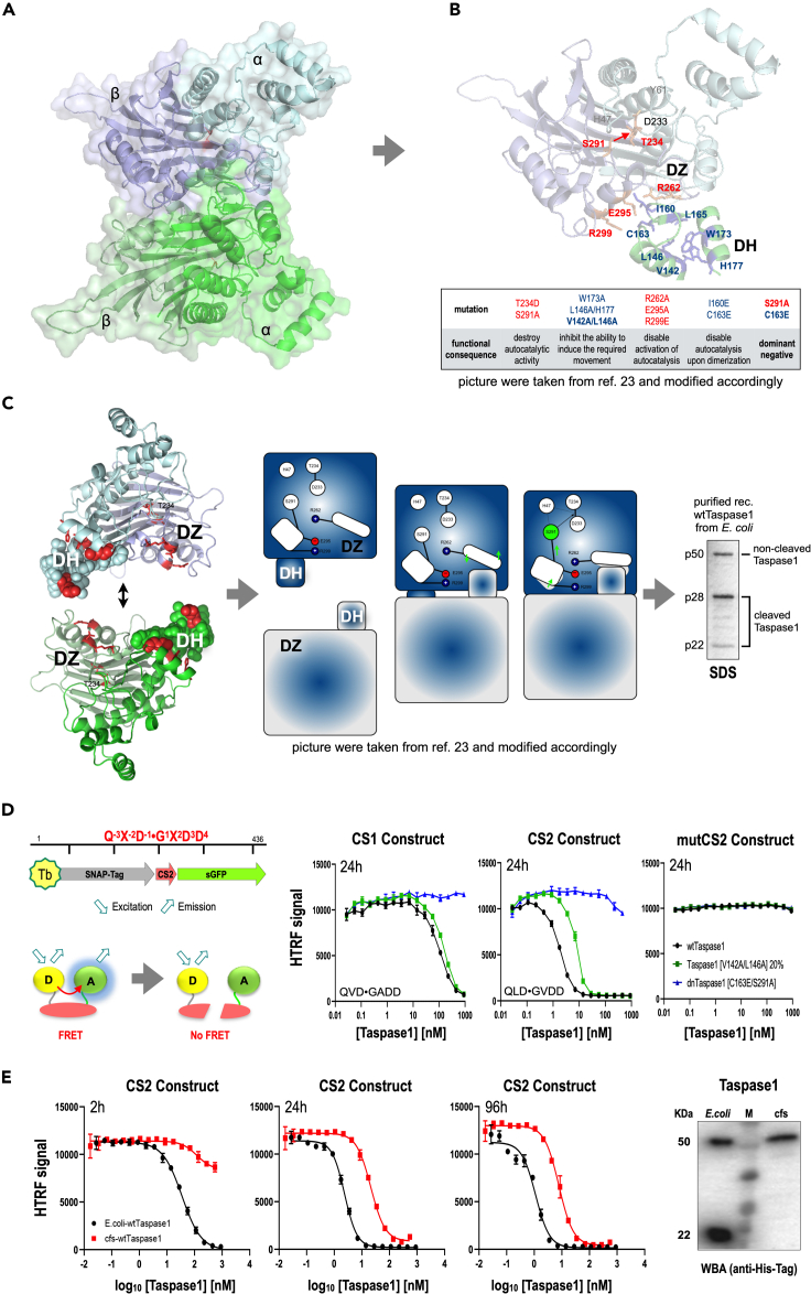
Dimerization of Taspase1 activates an intrinsic serine protease function that leads to the catalytic Thr234 residue, which allows to catalyze the consensus sequence Q-3X-2D-1⋅G1X2D3D4, present in Trithorax family members and TFIIA. Noteworthy, Taspase1 performs only a single hydrolytic step on substrate proteins, which makes it impossible to screen for inhibitors in a classical screening approach. Here, we report the development of an HTRF reporter assay that allowed the identification of an inhibitor, Closantel sodium, that inhibits Taspase1 in a noncovalent fashion (IC50 = 1.6 μM). The novel inhibitor interferes with the dimerization step and/or the intrinsic serine protease function of the proenzyme. Of interest, Taspase1 is required to activate the oncogenic functions of the leukemogenic AF4-MLL fusion protein and was shown in several studies to be overexpressed in many solid tumors. Therefore, the inhibitor may be useful for further validation of Taspase1 as a target for cancer therapy.

Keywords: Biochemistry; Biophysical chemistry; Structural biology.


Conflict of interest statement

The authors declare that they have no competing interests.

Figures

None
Graphical abstract
Figure 1
Figure 1
Molecular function of Taspase1 and setting up the HTRF assay (A) Molecular modeling of the Taspase1 homodimer with its α and β subunits (20). (B) Model of a single monomer of Taspase1 with the docking head of the second subunit (amino acids 140–180) and the critical amino acids that are all necessary to explain the intrinsic serine protease function (D233, T234, R262, S291, E295, and R299). (C) During the dimerization process, the two docking heads bind to the docking zone of the opposing Taspase1 monomer, respectively. The scheme in the middle explains the steps that lead to the positioning of the serine residue next to the D233/T234 peptide bond. On the right: Taspase1 isolated from E. coli, demonstrating that more than half of the Taspase1 molecules are already hydrolyzed into P28 and p22 (as visualized on Coomassie-stained SDS page). (D) Substrate protein. The SNAP-tag is labeled with Terbium cryptate (Tb-SNAP) that serves as the FRET donor. Superfolder GFP (sGFP) is utilized as FRET acceptor. Tb-SNAP and sGFP are linked to each other by the cleavage sites from the MLL protein (consensus sequence is indicated). FRET occurs with high efficiency when the substrate is not hydrolyzed. Cleavage causes separation of SNAP and sGFP, which results in drastic reduction of FRET. The reduction of sGFP fluorescence translates into HTRF being almost zero when being recorded on completely hydrolyzed substrate. Right panels: hydrolysis of the substrate protein with MLL CS1 and CS2, respectively. The cleavage site sequences are indicated. Three different Taspases were used: wild-type Taspase1, Taspase1 with V142A/L1476A that has about 20% catalytic activity, as well as dnTASP1 (C163E, S291A). 1 nM of CS1 or CS2 substrate protein were incubated for 24 h with Taspase1 variants at concentrations ranging from 0.027 nM to 900 nM (16 serial dilutions), respectively. Data are the mean ± SD; n = 3. (E) E. coli-produced already auto-activated Taspase1 (black) is compared with freshly cell-free-produced (cfs) Taspase1 (both wild type). Cleavage of CS2 was detected by HTRF after 2, 24, and 96 h. Data are the mean ± SD; n = 3. R2 = 0.82 for cfs after 2 h. For all over curves R2 was ≥0.98. This comparison revealed an augmented window for screening of potential inhibitors when using 30 nM cfs-Taspase1 and detection after 24 h. On the right: western blot experiment performed with E. coli- and cfs-Taspase1, indicating that cfs-Taspase1 is not yet auto-activated and hence fully suitable for screening of inhibitors with potentially different modes of inhibition.
Figure 2
Figure 2
Screening potential inhibitors and validating candidate drugs (A) Screening of FDA-approved drugs. The HTRF assay was used to screen a library with 1,200 FDA-approved drugs for their ability to inhibit cfs-Taspase1. The readout after 24 h incubation is presented; readout after 96 h is shown in Figure S1. About 25% uncut CS2 after 24 h was set as a threshold, and several candidates were identified. All identified Tetracycline derivatives were later identified as false positives showing autofluorescence in this assay. Two compounds were further investigated (Primaquine bisphosphate and Closantel sodium). Detailed information on both drugs is given below. (B) HTRF CS2 cleavage assay as dose response for verification of the two initial hits. At concentrations up to 12.5 μM, Primaquine (red) did not prevent cleavage and was hence classified as false positive. Closantel with or without sodium as counterion was validated as a novel potential Taspase1 inhibitor with an inhibitory activity in the low micromolar range. Data are the mean ± SD; n = 3. (C) CS2 cleavage assay for verification of inhibition and estimation of IC50 for Closantel sodium acting on cfs- and E. coli-Taspase1. Substrate protein (1 nM) was incubated for 24 h with 30 nM cfs- or 100 nM of E. coli-Taspase1 protein, respectively. Data are the mean ± SD; n = 3. R2 ≥ 0.986. For cfs-Taspase1 the assay was performed in parallel also with 8 nM CS2 substrate protein of which cleavage was also detected using western blot as an orthogonal assay, which also served as a control for 0% uncut versus 100% uncut CS2 used for referencing (Figure S2). Analysis of the HTRF assay revealed that inhibition of cfs-Taspase1 by Closantel sodium is characterized by an even lower IC50 and a much steeper hill slope in comparison to the inhibition of E. coli-Taspase1. (D) Same assay principle as employed in C, but instead of Closantel sodium here the E. coli-Taspase1 was titrated. CS2 substrate protein was set to 20 nM and treated for 24 h with Taspase1 ranging from 5 nM–5 μM or no Taspase1 as control. The inhibitory potential of Closantel sodium was tested at constant concentrations of 2, 6, and 12.5 μM with 5% DMSO in comparison to DMSO alone as control. Data are the mean ± SD; n = 4. After readout by HTRF, the corresponding quadruplicates were pooled, and cleavage of CS2 was detected by western blot; Figure S3. (E) Cleavage assay of cfs-Taspase1 in the absence or presence of different concentrations of Closantel sodium. cfs-Taspase1 was freshly prepared, immediately transferred into HTRF assay buffer, and then incubated for 12 h at RT with Closantel sodium at the indicted concentrations or 5% DMSO alone. As references cfs-Taspase1 DMSO controls were also obtained at time point 0, 12, and 24 h. The two samples on the left show E. coli-Taspase1 for comparison. Activation of cfs-Taspase1 is not taking place when Closantel is present at a concentration >1.6 μM, which corresponds to the IC50 value determined in the HTRF assay. (F) ITC experiments revealed that Closantel sodium is indeed attenuating the interaction between Taspase1 and CS2 substrate protein. CS2 (20 μM) was titrated into 1 μM E. coli-Taspase1 with or without 12.5 μM Closantel sodium being present. The baseline-corrected raw heat traces are presented as an overlay. Full evaluation is provided in Figure S5. (G) Testing Closantel sodium in living cells. Closantel sodium is inhibiting the hydrolysis of a substrate protein (GFP-CS2-BFP) as detected by western blot. A reduction in the intensity of the 26 kDa protein band, which is corresponding to GFP-CS2, is indicative for the inhibitory capacity of Closantel on co-transfected Taspase1. β-Actin served as internal loading control.

References

    1. Balkin D.M., Poranki M., Forester C.M., Dorsey M.J., Slavotinek A., Pomerantz J.H. TASP1 mutation in a female with craniofacial anomalies, anterior segment dysgenesis, congenital immunodeficiency and macrocytic anemia. Mol. Genet. Genomic Med. 2019;7:e818. - PMC - PubMed
    1. Brannigan J.A., Dodson G., Duggleby H.J., Moody P.C., Smith J.L., Tomchick D.R., Murzin A.G. A protein catalytic framework with an N-terminal nucleophile is capable of self-activation. Nature. 1995;378:416–419. doi: 10.1038/378416a0. - DOI - PubMed
    1. Bursen A., Moritz S., Gaussmann A., Moritz S., Dingermann T., Marschalek R. Interaction of AF4 wild-type and AF4⋅MLL fusion protein with SIAH proteins: indication for t(4;11) pathobiology? Oncogene. 2004;23:6237–6249. - PubMed
    1. Chen D.Y., Liu H., Takeda S., Tu H.C., Sasagawa S., Van Tine B.A., Lu D., Cheng E.H., Hsieh J.J. Taspase1 functions as a non-oncogene addiction protease that coordinates cancer cell proliferation and apoptosis. Cancer Res. 2010;70:5358–5367. - PMC - PubMed
    1. Chen D.Y., Lee Y., Van Tine B.A., Searleman A.C., Westergard T.D., Liu H., Tu H.C., Takeda S., Dong Y., Piwnica-Worms D.R., et al. A pharmacologic inhibitor of the protease Taspase1 effectively inhibits breast and brain tumor growth. Cancer Res. 2012;72:736–746. - PMC - PubMed

LinkOut - more resources