

Laser capture microdissection-capillary zone electrophoresis-tandem mass spectrometry (LCM-CZE-MS/MS) for spatially resolved top-down proteomics: a pilot study of zebrafish brain
- PMID: 34935839
- PMCID: PMC9066772
- DOI: 10.1039/d1mo00335f
Laser capture microdissection-capillary zone electrophoresis-tandem mass spectrometry (LCM-CZE-MS/MS) for spatially resolved top-down proteomics: a pilot study of zebrafish brain
Abstract
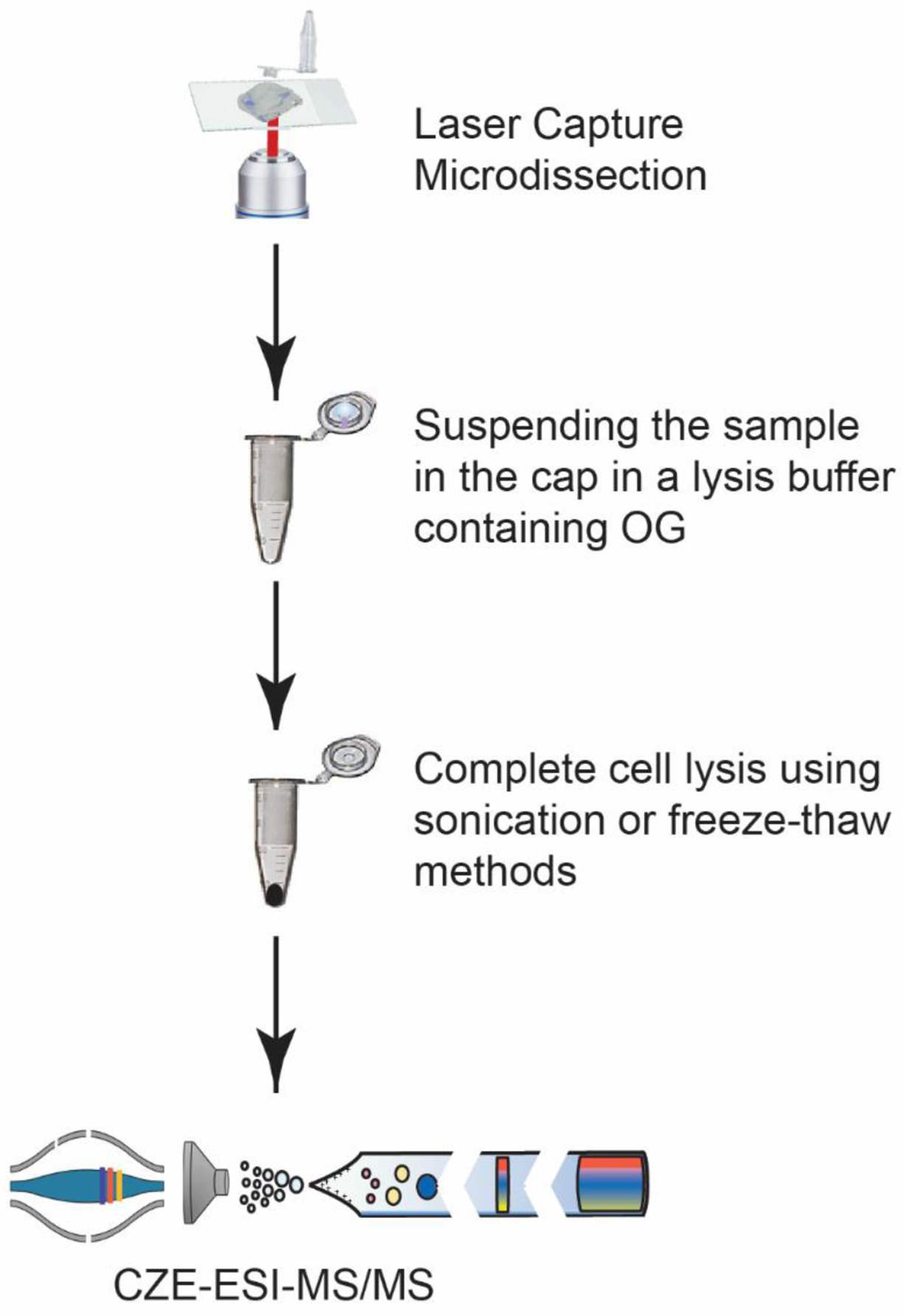
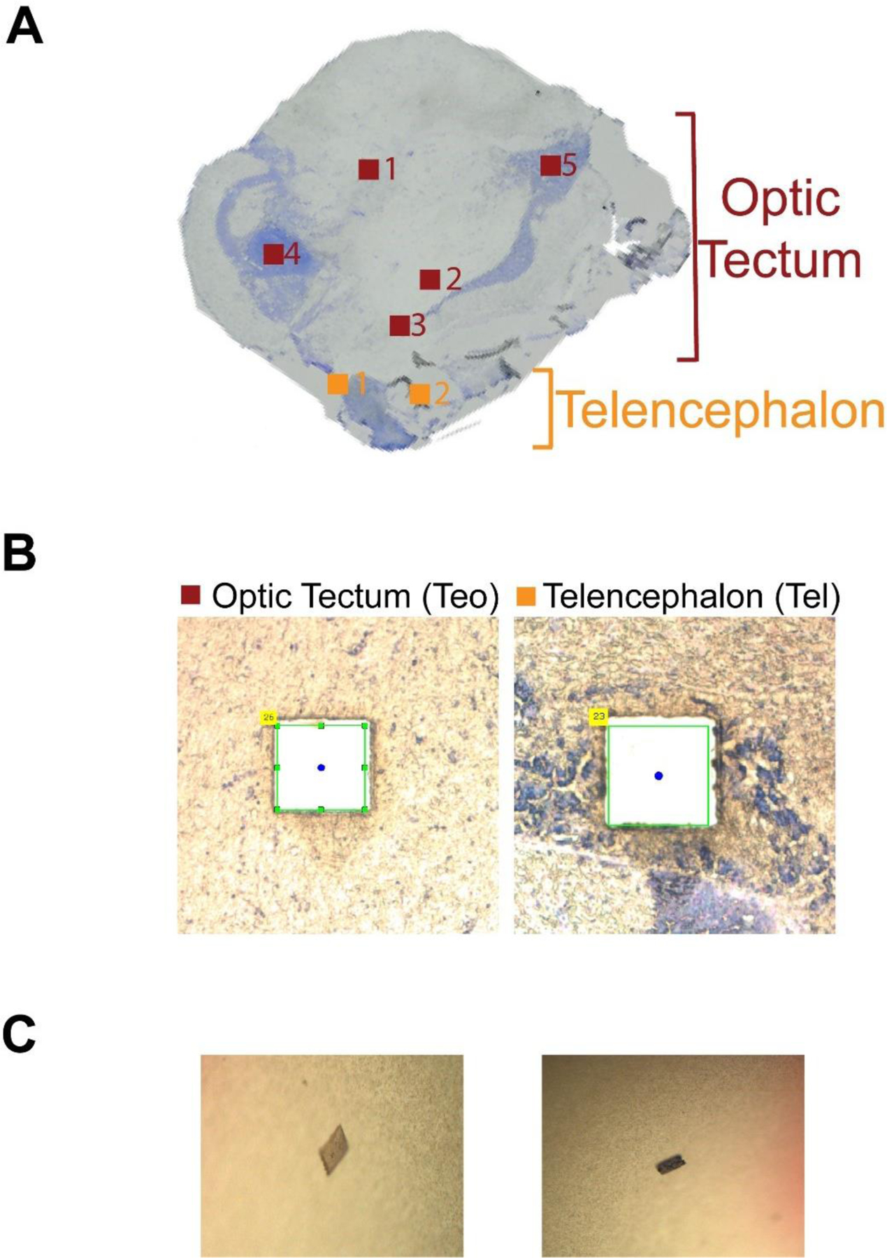
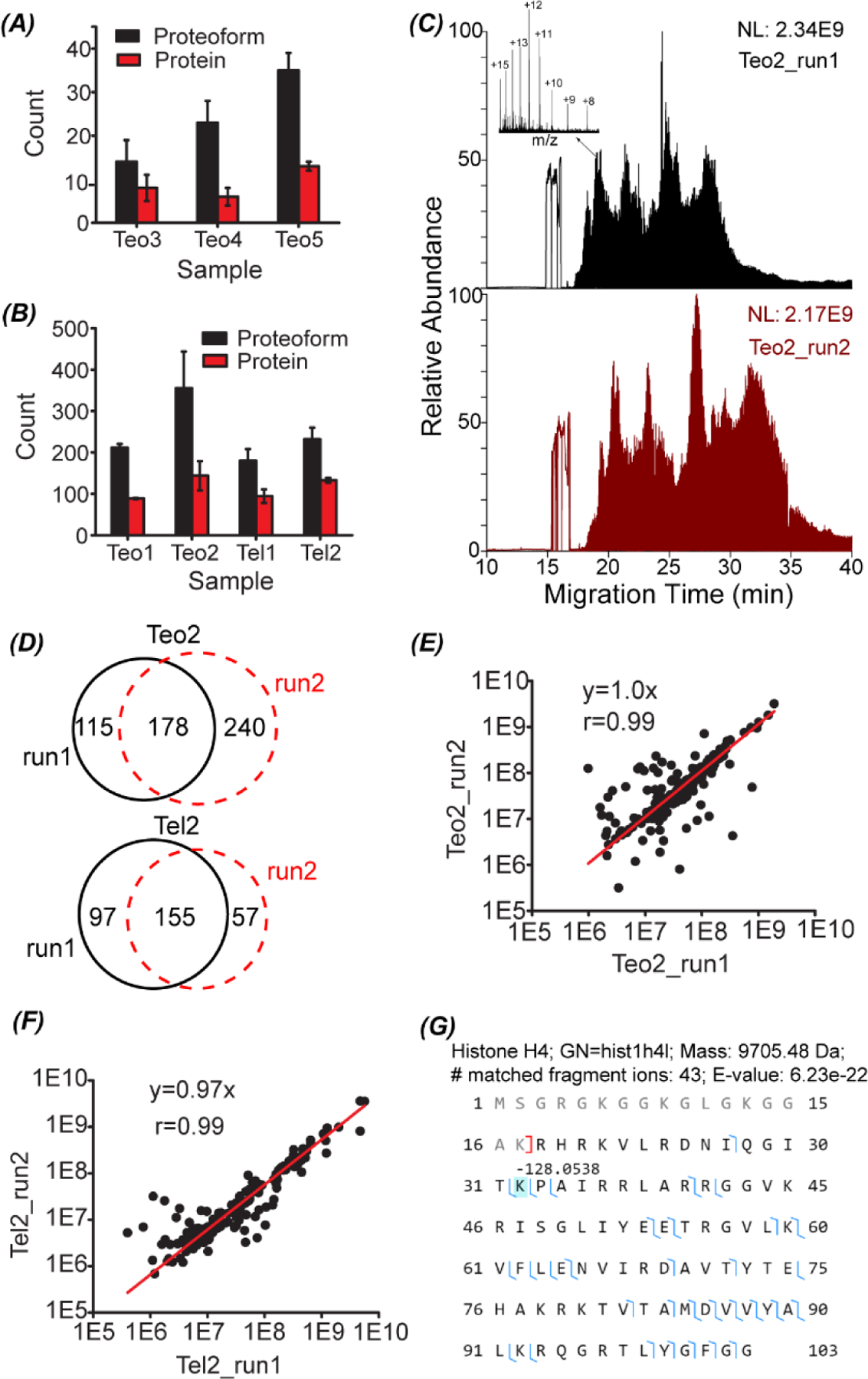
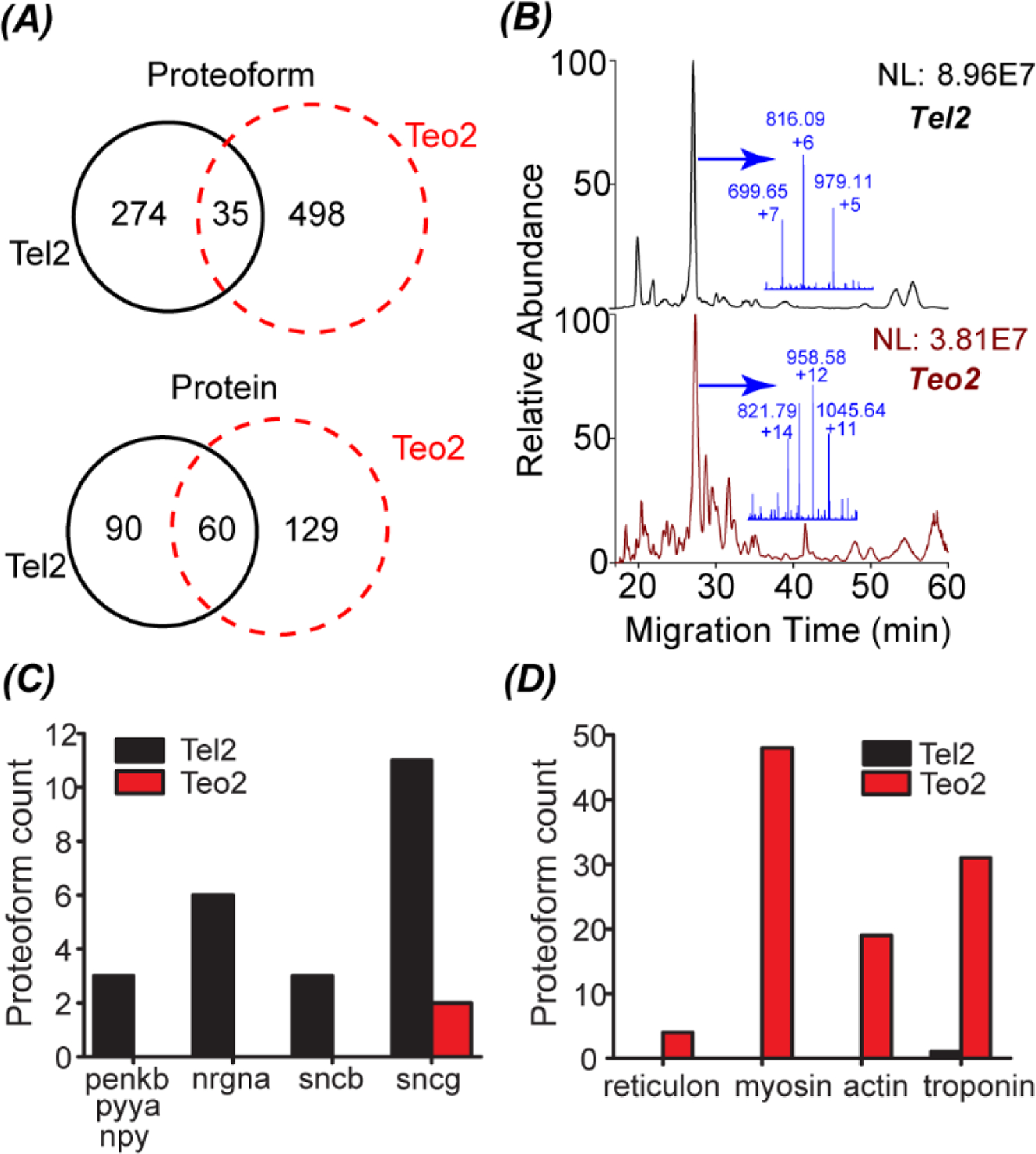
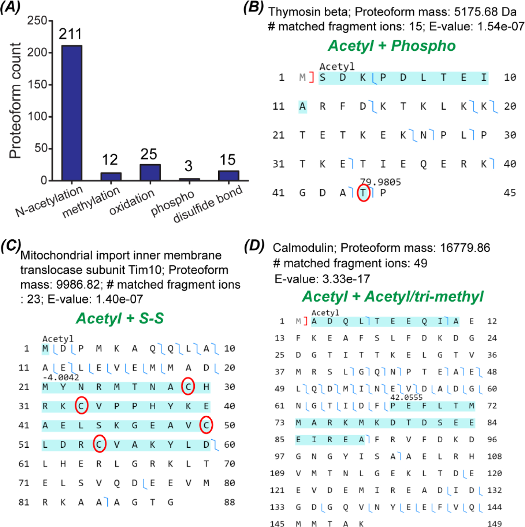
Mass spectrometry (MS)-based spatially resolved top-down proteomics (TDP) of tissues is crucial for understanding the roles played by microenvironmental heterogeneity in the biological functions of organs and for discovering new proteoform biomarkers of diseases. There are few published spatially resolved TDP studies. One of the challenges relates to the limited performance of TDP for the analysis of spatially isolated samples using, for example, laser capture microdissection (LCM) because those samples are usually mass-limited. We present the first pilot study of LCM-capillary zone electrophoresis (CZE)-MS/MS for spatially resolved TDP and used zebrafish brain as the sample. The LCM-CZE-MS/MS platform employed a non-ionic detergent and a freeze-thaw method for efficient proteoform extraction from LCM isolated brain sections followed by CZE-MS/MS without any sample cleanup step, ensuring high sensitivity. Over 400 proteoforms were identified in a CZE-MS/MS analysis of one LCM brain section via consuming the protein content of roughly 250 cells. We observed drastic differences in proteoform profiles between two LCM brain sections isolated from the optic tectum (Teo) and telencephalon (Tel) regions. Proteoforms of three proteins (npy, penkb, and pyya) having neuropeptide hormone activity were exclusively identified in the isolated Tel section. Proteoforms of reticulon, myosin, and troponin were almost exclusively identified in the isolated Teo section, and those proteins play essential roles in visual and motor activities. The proteoform profiles accurately reflected the main biological functions of the Teo and Tel regions of the brain. Additionally, hundreds of post-translationally modified proteoforms were identified.
Conflict of interest statement
Conflicts of interest
There are no conflicts of interest to declare.
Figures
References
-
- Zhu Y, Dou M, Piehowski PD, Liang Y, Wang F, Chu RK, Chrisler WB, Smith JN, Schwarz KC, Shen Y, Shukla AK, Moore RJ, Smith RD, Qian W-J and Kelly RT, Spatially Resolved Proteome Mapping of Laser Capture Microdissected Tissue with Automated Sample Transfer to Nanodroplets, Mol. Cell. Proteomics, 2018, 17(9), 1864–1874. - PMC - PubMed
-
- Clair G, Piehowski PD, Nicola T, Kitzmiller JA, Huang EL, Zink EM, Sontag RL, Orton DJ, Moore RJ, Carson JP, Smith RD, Whitsett JA, Corley RA, Ambalavanan N and Ansong C, Spatially-Resolved Proteomics: Rapid Quantitative Analysis of Laser Capture Microdissected Alveolar Tissue Samples, Sci. Rep, 2016, 6, 39223. - PMC - PubMed
Publication types
MeSH terms
Grants and funding
LinkOut - more resources
Full Text Sources
Miscellaneous