. 2021 Dec 24;7(52):eabk3273.
doi: 10.1126/sciadv.abk3273. Epub 2021 Dec 22.

Cryo-EM structure of the autoinhibited state of myosin-2

Affiliations

Cryo-EM structure of the autoinhibited state of myosin-2

Sarah M Heissler et al. Sci Adv. .

Abstract

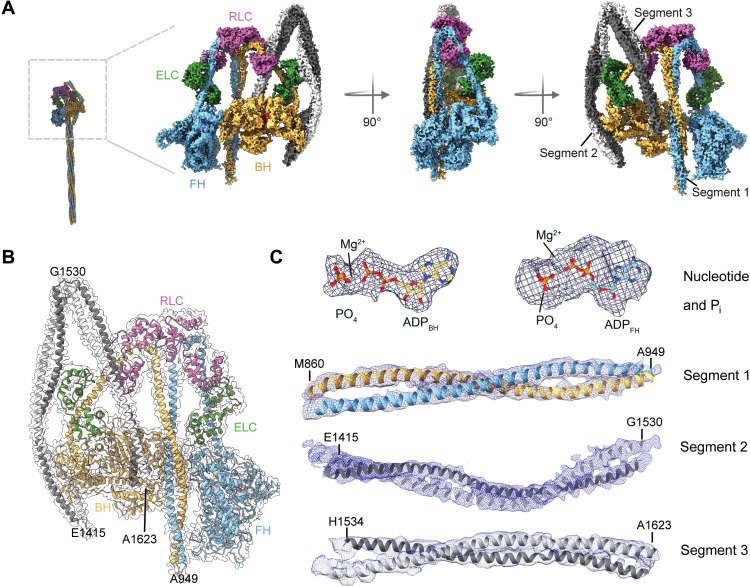
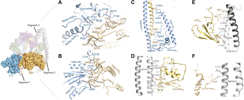
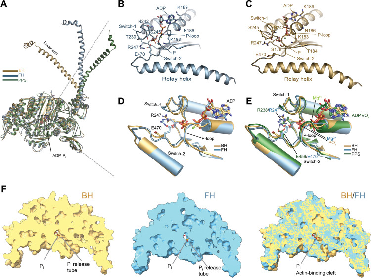
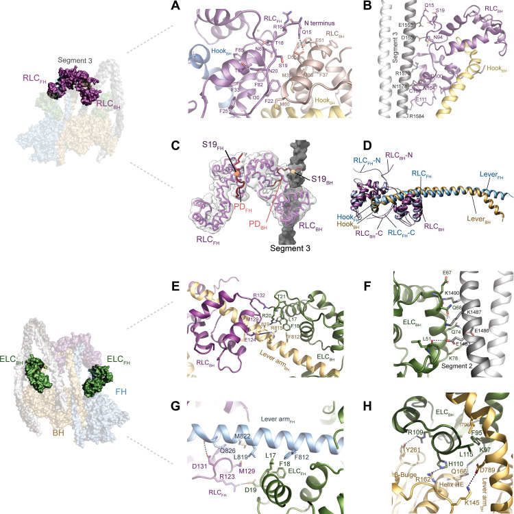
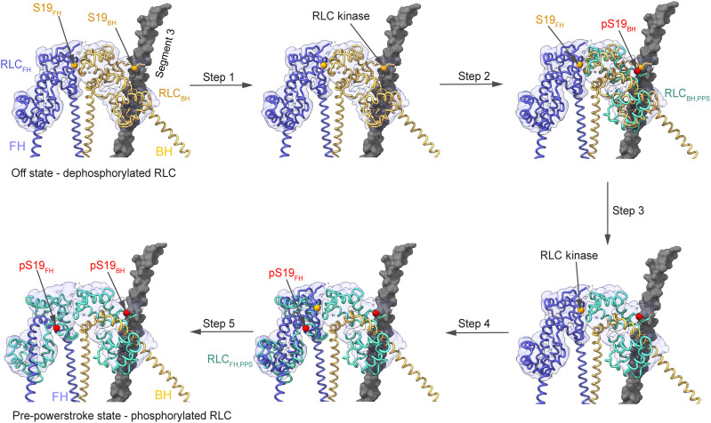
We solved the near-atomic resolution structure of smooth muscle myosin-2 in the autoinhibited state (10S) using single-particle cryo–electron microscopy. The 3.4-Å structure reveals the precise molecular architecture of 10S and the structural basis for myosin-2 regulation. We reveal the position of the phosphorylation sites that control myosin autoinhibition and activation by phosphorylation of the regulatory light chain. Further, we present a previously unidentified conformational state in myosin-2 that traps ADP and Pi produced by the hydrolysis of ATP in the active site. This noncanonical state represents a branch of the myosin enzyme cycle and explains the autoinhibition of the enzyme function of 10S along with its reduced affinity for actin. Together, our structure defines the molecular mechanisms that drive 10S formation, stabilization, and relief by phosphorylation of the regulatory light chain.


Figures

Fig. 1.
Fig. 1.. Cryo-EM structure of 10S.
(A) Cryo-EM reconstruction of 10S in the front, side, and back view. Individual subunits are segmented and color-coded. The two heavy chains (blue and yellow) and bound ELC (green) and RLC (pink) are shown. (B) Atomic model of 10S fitted in the final electron density map. Individual subunits are segmented and color-coded according to (A). (C) Representative cryo-EM densities of different regions of 10S. Densities of the nucleotide (3.4 Å) and Pi displayed at 12.5 σ contour level. Densities of segment 1 (4.5 Å), segment 2 (5 Å), and segment 3 (5.5 Å) displayed at 8.0 σ contour level.
Fig. 2.
Fig. 2.. Interactions of the myosin heads.
(A) Interactions of loop-4BH at the BH-FH interface. Potential interactions (distance > 4 Å) between R371BH and Q771FH and Q385BH and Q728FH are shown. (B) Interactions of CM-loopBH at the BH-FH interface. Potential interactions (distance > 4 Å) between D412BH and R253FH and R406BH and E169FH are shown. (C) Interaction between the FH and segment 1. (D) Interaction between the converterBH and segment 2. (E) Interaction between the NTDBH and segment 2. (F) Interaction between the HO linkerBH and segment 3. The views in (C) to (F) are rotated 180° relative to the overview figure to highlight key interactions at the respective interfaces.
Fig. 3.
Fig. 3.. Active site and nucleotide interactions.
(A) Orientation of the lever arm in BH and FH in comparison to the pre-powerstroke state (PDB ID: 1QVI). (B) Active site and nucleotide interactions in the FH. Key residues are shown in stick representation. The distance between R247 and E470 is >6.1 Å. (C) Active site and nucleotide interactions in the BH. Key residues are shown in stick representation. (D) Comparison between the active sites in BH and FH. Key residues are shown in stick representation. (E) Comparison between the active sites in BH, FH, and the pre-powerstroke state (PDB ID: 2XEL). The salt bridge between switch-1 R247/R238 and switch-2 E470/E459 is only formed in the pre-powerstroke state and absent in BH and FH. Key residues are shown in stick representation. (F) Comparison of the phosphate release tube in the BH and FH.
Fig. 4.
Fig. 4.. Light chain interactions.
(A) Interactions between the RLCBH (salmon) and the RLCFH (dark purple). The PD of the RLCFH forms extensive intra and intersubunit interactions. (B) RLCBH interacts with segment 3. (C) Position of the phosphorylatable residue S19 is shown for RLCBH (wheat spheres) and RLCFH (orange spheres). The resolved PDs of the RLCs are shown in red. S19FH is sandwiched between both RLCs; S19BH is in close proximity to segment 3 (gray). (D) Different binding modes of the RLCFH and the RLCFH to the respective lever arms. (E) Interactions between the ELCBH with the RLCBH and the lever armBH. (F) Interactions between the ELCBH and segment 2. (G) Interactions between the ELCFH and the RLCFH. (H) Interactions between the ELCBH, the BH, and the lever armBH.
Fig. 5.
Fig. 5.. Model for 10S relief by RLC phosphorylation.
In the off state, phosphorylation of the RLCBH on S19 (pS19) causes structural transitions to the pre-powerstroke state (RLCBH,PPS, PDB ID: 1QVI; pS19 modeled) that make the RLCFH available for phosphorylation. Collectively, this is expected to result in 10S relief and the subsequent activation of myosin motor activity.

References

    1. Heissler S. M., Sellers J. R., Kinetic adaptations of myosins for their diverse cellular functions. Traffic 17, 839–859 (2016). - PMC - PubMed
    1. Sellers J. R., Regulation of cytoplasmic and smooth muscle myosin. Curr. Opin. Cell Biol. 3, 98–104 (1991). - PubMed
    1. Sellers J. R., Myosins: A diverse superfamily. Biochim. Biophys. Acta 1496, 3–22 (2000). - PubMed
    1. Rayment I., Holden H., Whittaker M., Yohn C., Lorenz M., Holmes K., Milligan R., Structure of the actin-myosin complex and its implications for muscle contraction. Science 261, 58–65 (1993). - PubMed
    1. Geeves M. A., Review: The ATPase mechanism of myosin and actomyosin. Biopolymers 105, 483–491 (2016). - PubMed