. 2022 Jan;70(1):275-280.
doi: 10.4103/ijo.IJO_2133_21.

Immersive photoreal new-age innovative gameful pedagogy for e-ophthalmology with 3D augmented reality

Affiliations

Immersive photoreal new-age innovative gameful pedagogy for e-ophthalmology with 3D augmented reality

Prasanna V Ramesh et al. Indian J Ophthalmol. 2022 Jan.

Abstract

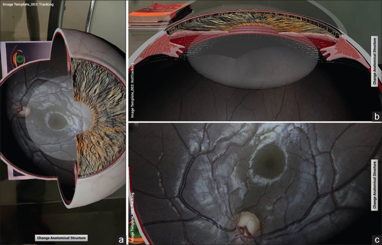
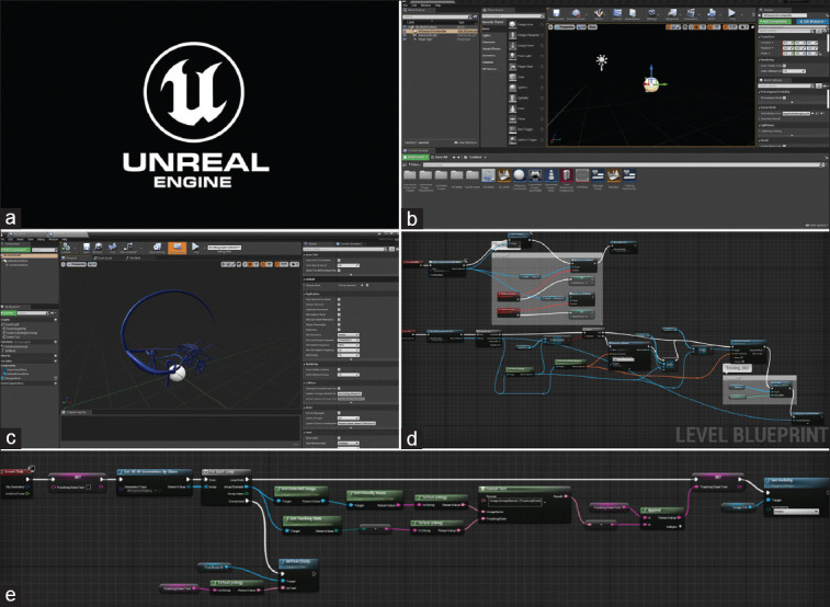
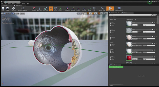
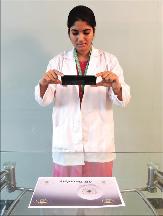
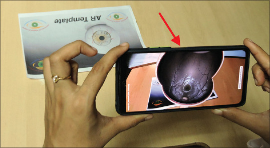
Augmented reality (AR) has come a long way from a science-fiction concept to a science-based reality. AR is a view of the real, physical world in which the elements are enhanced by computer-generated inputs. AR is available on mobile handsets, which constitutes an essential e-learning platform. Today, AR is a real technology and not a science-fiction concept. The use of an e-ophthalmology platform with AR will pave the pathway for new-age gameful pedagogy. In this manuscript, we present a newly innovated AR program named "Eye MG AR" to simplify ophthalmic concept learning and to serve as a new-age immersive 3D pedagogical tool for gameful learning.

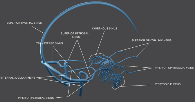
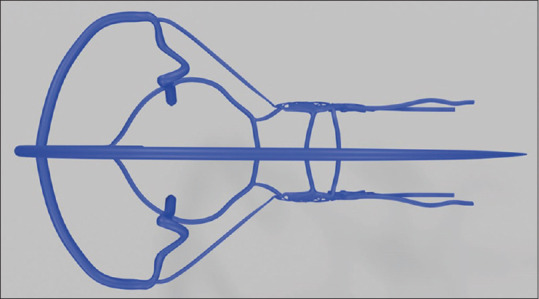
Keywords: 3D; 3D Eye Models; Augmented Reality; Cerebral Venous System; Pedagogy; e-Ophthalmology.


Conflict of interest statement

None

Figures

Figure 1
Figure 1
Image showing annotated three-dimensional (3D) model of the eyeball
Figure 2
Figure 2
Image showing the annotated 3D side view of the cerebral venous and dural sinuses model
Figure 3
Figure 3
Image showing the pedagogue viewing the eyeball model: (a) top view; (b) anterior segment; (c) posterior segment
Figure 4
Figure 4
(a) Logo of the AR software (Unreal Engine). (b) Main-level workspace in the Unreal Engine software. (c) Cerebral venous and dural sinuses model in the viewport created by us. (d) Level blueprint created by us in the software. (e) Widget blueprint created by us in the software
Figure 5
Figure 5
Image showing the eyeball model used inside Unreal Engine workspace
Figure 6
Figure 6
Image showing the cerebral venous and dural sinuses model used inside unreal engine workspace
Figure 7
Figure 7
Image showing the pedagogue beginning the augmented reality (AR) teaching session by focussing the mobile camera over the AR template
Figure 8
Figure 8
Image showing the AR template to be downloaded and printed as the base, for the model to appear
Figure 9
Figure 9
Image showing the borders within which the AR template should be fitted in the mobile screen
Figure 10
Figure 10
The pedagogue viewing the eyeball (red arrow) in the mobile screen over the AR template
Figure 11
Figure 11
The pedagogue viewing the cavernous sinus (red arrow) in the mobile screen over the AR template
Figure 12
Figure 12
Top view of the cerebral venous and dural sinuses when mobile is adjusted accordingly
Figure 13
Figure 13
Image showing the pedagogue. (a) Training the students using the Eye MG AR app. (b) Demonstrating the parts of the eyeball. (c) Focusing the optic disc
Figure 14
Figure 14
Image showing details of the Eye MG AR app on the Google Play Store, and the QR code for directly downloading the app by scanning it

References

    1. Gopalakrishnan S, Chouhan Suwalal S, Bhaskaran G, Raman R. Use of augmented reality technology for improving visual acuity of individuals with low vision. Indian J Ophthalmol. 2020;68:1136–42. - PMC - PubMed
    1. Iskander M, Ogunsola T, Ramachandran R, McGowan R, Al-Aswad LA. Virtual reality and augmented reality in ophthalmology:A contemporary prospective. Asia Pac J Ophthalmol (Phila) 2021;10:244–52. - PMC - PubMed
    1. Karakas AB, Govsa F, Ozer MA, Eraslan C. 3D brain imaging in vascular segmentation of cerebral venous sinuses. J Digit Imaging. 2019;32:314–21. - PMC - PubMed
    1. Cabrilo I, Bijlenga P, Schaller K. Augmented reality in the surgery of cerebral arteriovenous malformations:Technique assessment and considerations. Acta Neurochir (Wien) 2014;156:1769–74. - PubMed
    1. Eye MG AR - - Apps on Google Play [Internet] [[cited 2021 Nov 26]]. Available from: https://play.google.com/store/apps/details? id=com. EyeMG_AR&hl=en&gl=IN .