Review
. 2021 Dec 22;18(1):41.
doi: 10.1186/s12977-021-00583-z.

Rotten to the core: antivirals targeting the HIV-1 capsid core

Affiliations
Review

Rotten to the core: antivirals targeting the HIV-1 capsid core

William M McFadden et al. Retrovirology. .

Abstract

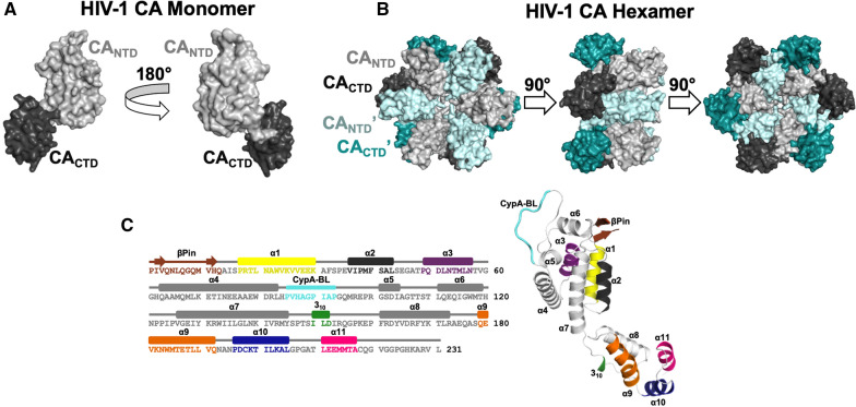
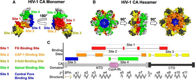
The capsid core of HIV-1 is a large macromolecular assembly that surrounds the viral genome and is an essential component of the infectious virus. In addition to its multiple roles throughout the viral life cycle, the capsid interacts with multiple host factors. Owing to its indispensable nature, the HIV-1 capsid has been the target of numerous antiretrovirals, though most capsid-targeting molecules have not had clinical success until recently. Lenacapavir, a long-acting drug that targets the HIV-1 capsid, is currently undergoing phase 2/3 clinical trials, making it the most successful capsid inhibitor to-date. In this review, we detail the role of the HIV-1 capsid protein in the virus life cycle, categorize antiviral compounds based on their targeting of five sites within the HIV-1 capsid, and discuss their molecular interactions and mechanisms of action. The diverse range of inhibition mechanisms provides insight into possible new strategies for designing novel HIV-1 drugs and furthers our understanding of HIV-1 biology.


Conflict of interest statement

The authors declare that they have no competing interests.

Figures

Fig. 1
Fig. 1
Structure of mature HIV-1 CA. A The HIV-1 CA monomer comprises CANTD (light gray) and CACTD (dark gray) domains (PDB ID: 4XFX). The view is rotated by 180°. B The same domains are shown in the context of the HIV-1 CA hexamer. Colors are as described in A, with neighboring CANTDʹ and CACTDʹ domains shown as light and dark cyan, respectively. The views are rotated by 90°. C Schematic outlining the sequence and secondary structural elements of HIV-1 CA (based on PDB ID: 4XFX) (Part C of this figure is based on Fig. 1 from Gres et al. [105]. Reprinted with permission from AAAS)
Fig. 2
Fig. 2
Binding sites of CA-targeting antivirals in mature HIV-1 CA monomers and hexamers. A The five CA-targeting antiviral binding sites are shown mapped onto the HIV-1 CA monomer (PDB ID: 4XFX). CANTDs are shown in light gray, CACTDs are shown in dark gray. Binding site 1 is shown in red, binding site 2 in orange, binding site 3 in yellow, binding site 4 in green, and binding site 5 in blue. The view is rotated by 180°. B The same domains and five CA-targeting antiviral binding sites are shown mapped onto the HIV-1 CA hexamer. Colors are as described in A. The views are rotated by 90°. C Schematic outlining the domains, binding sites, and secondary structure of HIV-1 CA. Compound binding sites are colored as described in A, and the Cyclophilin A binding loop (CypA-BL) is shown as pink circles. The CANTD and CACTD are shown as light and dark gray bars, respectively. The β-hairpin (βPin) and α-helices (α) shown as gray arrows and twists, respectively. Schematic generated by Protean 3D™ (V17.2.1) from DNASTAR, Inc
Fig. 3
Fig. 3
Binding interactions of PF74 at binding site 1 of HIV-1 CA. A Chemical structure of PF74. B PF74 binding at the CANTD–CACTD interfaces of adjacent subunits within a CA hexamer (PDB ID: 4XFZ). The larger box shows a close-up view of the PF74 binding site in relation to the threefold (triangle) and twofold (oval) interfaces. CANTDs and the corresponding CACTDs are colored by the same colors (light and dark, respectively). C CANTD and a CACTD of a neighboring subunit (CACTDʹ) participate in PF74 binding (gray and dark cyan ribbons; site 1 residues are shown in red cartoon and pink sticks). PF74 is shown as yellow sticks. D PF74 binding geometries in CA-PF74 (PDB ID: 4XFZ; PF74 in yellow), CAXL-PF74 (PDB ID: 4U0E; PF74 in beige), and CANTD-PF74 (PDB ID: 2XDE; PF74 in brown). E Threefold interface of CA-PF74 superposed onto CA (aligned based on residues 1–219). Helices α10 of CA-PF74 and CA are in yellow and dark gray. Water molecules are shown as red spheres in CA; no water molecules were present in CA-PF74 (Parts of this figure are based on Fig. 4 from Gres et al. [105]. Reprinted with permission from AAAS)
Fig. 4
Fig. 4
Host factor FG motifs share a common binding conformation with the PF74 benzyl group. Crystal structures of HIV-1 CA hexamers in complex with CPSF6 (PDB ID: 6AY9; blue cartoon and sticks), NUP153 (PDB ID: 6AYA; orange cartoon and sticks), and Sec24C (PDB ID: 6PU1; magenta cartoon and sticks) peptides were aligned to a crystal structure of HIV-1 CA hexamer in complex with PF74 (PDB ID: 4XFZ; yellow sticks). The HIV-1 CA protein is omitted for clarity. The benzyl group of PF74 binds in a similar manner to the benzyl group of the phenylalanine in the FG motif of each host factor peptide
Fig. 5
Fig. 5
Different inter-subunit interactions of various CA-targeting compounds at CA binding site 1. A Chemical structures of BI-2 and GS-6207 (PF74 is shown in Fig. 3A). B BI-2 only interacts with the N-terminal domain of HIV-1 CA. The crystal structure of HIV-1 CA hexamer in complex with BI-2 (PDB ID: 4U0F; purple sticks) was superposed to a crystal structure of HIV-1 CA hexamer in complex with PF74 (PDB ID: 4XFZ; yellow sticks). The CANTD of one CA monomer is shown in light gray cartoon with binding site 1 shown in red, the CACTD of a neighboring monomer (CACTDʹ) is shown in dark green. Residues interacting with PF74 and/or BI-2 are shown as sticks. C GS-6207 forms extended interactions with the CANTD and CACTD of a neighboring CA monomer (CANTDʹ show in light cyan cartoon and CACTDʹ shown in dark green). The crystal structure of HIV-1 CA hexamer in complex with GS-6207 (PDB ID: 6V2F; green sticks) was superposed to a crystal structure of HIV-1 CA hexamer in complex with PF74 (PDB ID: 4XFZ; yellow sticks). The CANTD of one CA monomer is shown in light gray cartoon with binding site 1 shown in red. The novel interaction with CANTDʹ is shown as a black dotted line. The R1 and R2 groups of PF74 (see Fig. 3A) superpose well to GS-6207, while the R3 group adopts a different conformation
Fig. 6
Fig. 6
HIV-1 antivirals targeting the CAP-1 binding site. A Two CA monomers from a CA hexamer (PDB ID: 4XFX) with CANTDs shown in light gray and light cyan, respective CACTDs in dark gray and dark cyan, and binding site 2 in orange. One subunit is outlined in red. B NMR structures of CANTD in complex with CAP-1 (PDB ID: 2JPR; CAP-1 in green sticks). Left: surface and cartoon views of CA site 2 interacting with pose 1 of CAP-1. Right: surface and cartoon views of CA site 2 interacting with pose 2 of CAP-1. C Crystal structure of CANTD in complex with BD-3 (PDB ID: 4E91; BD-3 in pink sticks). Left: surface and cartoon views of CA site 2 interacting with pose 1 of BD-3. Right: surface and cartoon views of CA site 2 interacting with pose 1 of BD-3
Fig. 7
Fig. 7
HIV-1 antivirals targeting the twofold binding site. A Two CA monomers from a CA hexamer (PDB ID: 4XFX) with CANTDs shown in light gray and light cyan, with the respective CACTDs in dark gray and dark cyan, and binding site 3 in yellow. B Crystal structure of CACTD in complex with CAI (PDB ID: 2BUO; CAI in cyan cartoon and sticks). Surface (left) and cartoon (right) views of CA site 3 interacting with CAI. C NMR structure of CACTD in complex with NYAD-13 (PDB ID: 2L6E; NYAD-13 in pink cartoon and sticks). Left: Surface (left) and cartoon (right) views of CA site 3 interacting with NYAD-13, which is stabilized through hydrocarbon stapling with (S)-2-(2ʹ-pentenyl) alanine (magenta cartoon and sticks)
Fig. 8
Fig. 8
HIV-1 antivirals targeting the apical binding site. A Chemical structure of benzimidazole compound 4 (from [195]). B Crystal structure of CANTD in complex with 4 (PDB ID: 4E91; compound 4 in orange sticks) in surface view. CANTD is shown in light gray, with site 4 colored in green. C Close-up view of compound 4 at site 4, colors as in (B). Residues having strong hydrophobic interactions with compound 4 are shown as light green sticks. Hydrogen bonds with the side chain of R132 are shown as black dotted lines
Fig. 9
Fig. 9
HIV-1 CA central pore binding site. A CA hexamer (PDB ID: 4XFX) is shown with CANTDs in light gray and light cyan, CACTDs in dark gray and dark cyan, and R18 side chains at the sixfold symmetry axis in alternating blue and light blue colors. B IP6 (brown sticks) is shown bound to CA hexamer (PDB ID: 6BHT). Right, IP6 above and below R18 ring. C Hexacarboxybenzene (HCB; magenta sticks) bound to CA hexamer (PDB ID: 5HGP). Right, HCB interacting with R18

References

    1. Global HIV & AIDS statistics—2020 fact sheet. https://www.unaids.org/en/resources/fact-sheet.
    1. Moir S, Chun T-W, Fauci AS. Pathogenic mechanisms of HIV disease. Annu Rev Pathol. 2011;6:223–248. - PubMed
    1. Weiss RA. How does HIV cause AIDS? Science. 1993;260:1273–1279. - PubMed
    1. Fanales-Belasio E, Raimondo M, Suligoi B, Buttò S. HIV virology and pathogenetic mechanisms of infection: a brief overview. Annali dell'Istituto superiore di sanita. 2010;46:5–14. - PubMed
    1. Pornillos O, Ganser-Pornillos BK. HIV-1 virion structure. In: Hope TJ, Stevenson M, Richman D, editors. Encyclopedia of AIDS. New York: Springer; 2014. pp. 1–6.

Publication types