. 2021 Dec 22;71(10):2011-2021.
doi: 10.1136/gutjnl-2020-323951. Online ahead of print.

Lactobacillus gallinarum modulates the gut microbiota and produces anti-cancer metabolites to protect against colorectal tumourigenesis

Affiliations

Lactobacillus gallinarum modulates the gut microbiota and produces anti-cancer metabolites to protect against colorectal tumourigenesis

Naoki Sugimura et al. Gut. .

Abstract

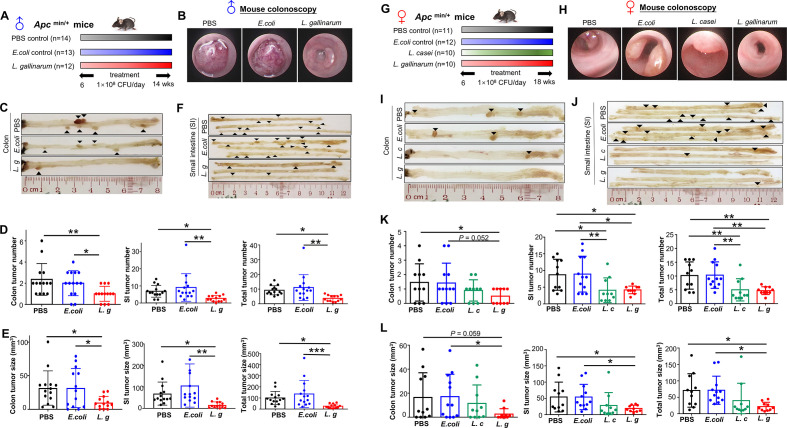
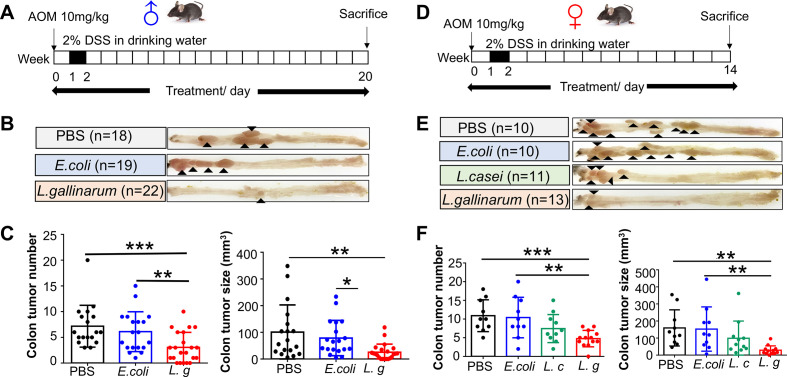
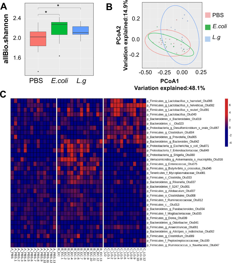
Objective: Using faecal shotgun metagenomic sequencing, we identified the depletion of Lactobacillus gallinarum in patients with colorectal cancer (CRC). We aimed to determine the potential antitumourigenic role of L. gallinarum in colorectal tumourigenesis.

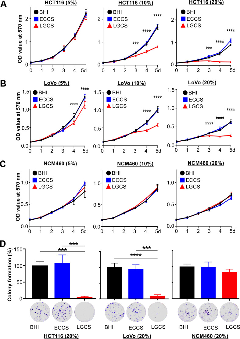
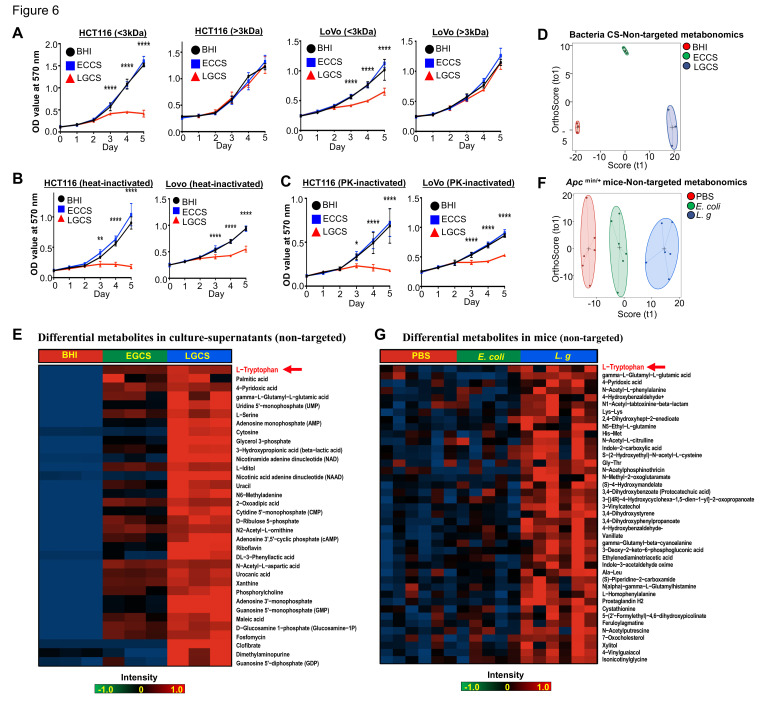
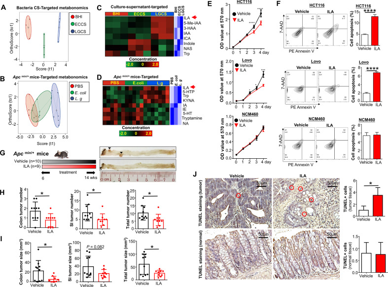
Design: The tumor-suppressive effect of L. gallinarum was assessed in murine models of CRC. CRC cell lines and organoids derived from patients with CRC were cultured with L. gallinarum or Escherichia coli MG1655 culture-supernatant to evaluate cell proliferation, apoptosis and cell cycle distribution. Gut microbiota was assessed by 16S ribosomal DNA sequencing. Antitumour molecule produced from L. gallinarum was identified by liquid chromatography mass spectrometry (LC-MS/MS) and targeted mass spectrometry.

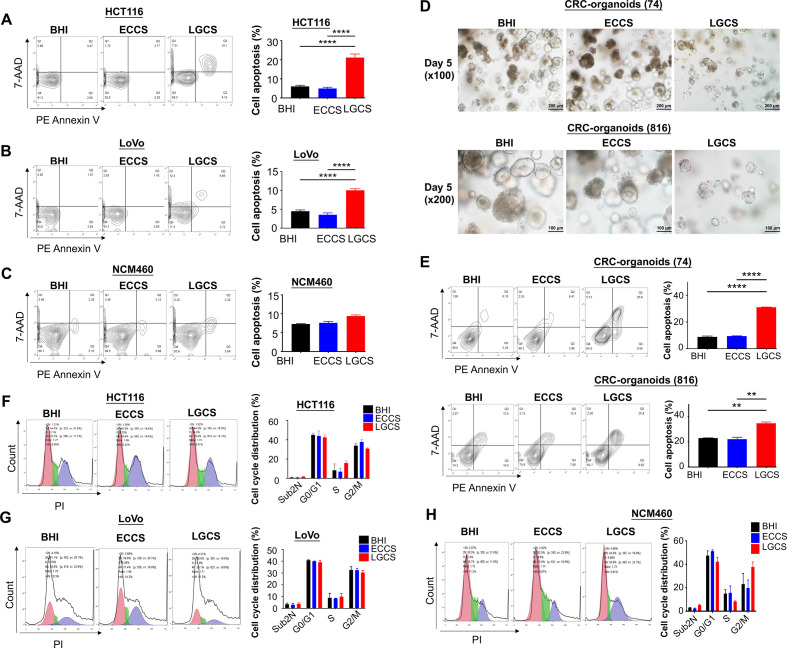
Results: L. gallinarum significantly reduced intestinal tumour number and size compared with E. coli MG1655 and phosphate-buffered saline in both male and female murine intestinal tumourigenesis models. Faecal microbial profiling revealed enrichment of probiotics and depletion of pathogenic bacteria in L. gallinarum-treated mice. Culturing CRC cells with L. gallinarum culture-supernatant (5%, 10% and 20%) concentration-dependently suppressed cell proliferation and colony formation. L. gallinarum culture-supernatant significantly promoted apoptosis in CRC cells and patient-derived CRC organoids, but not in normal colon epithelial cells. Only L. gallinarum culture-supernatant with fraction size <3 kDa suppressed proliferation in CRC cells. Using LC-MS/MS, enrichments of indole-3-lactic acid (ILA) was identified in both L. gallinarum culture-supernatant and the gut of L. gallinarum-treated mice. ILA displayed anti-CRC growth in vitro and inhibited intestinal tumourigenesis in vivo.

Conclusion: L. gallinarum protects against intestinal tumourigenesis by producing protective metabolites that can promote apoptosis of CRC cells.

Keywords: colorectal cancer; probiotics.


Conflict of interest statement

Competing interests: None declared.

Figures

Figure 1
Figure 1
L. gallinarum protects against intestinal tumourigenesis in Apc Min/+ mice. (A) Schematic diagram showing the experimental design, timeline of male Apc Min/+ mouse model. (B) Representative images of colon tumour from male Apc Min/+ mouse model. Colonoscopy confirmed that colon tumour size in L. gallinarum group was visually smaller than tumours in Escherichia coli MG1655 or PBS control groups. In both E. coli MG1655 and PBS control groups, colonoscope could not pass through the tumours. (C) Representative macroscopic images of colons from male Apc Min/+ mouse model. (D) Colon, small intestinal and total tumour number (colon+small intestinal) in male Apc Min/+ mice under different treatments. (E) Colon, small intestinal and total tumour size (colon+small intestinal) in male Apc Min/+ mice under different treatments. (F) Representative macroscopic images of small intestines from male Apc Min/+ mouse model. (G) Schematic diagram showing the experimental design, timeline of female Apc Min/+ mouse model. (H) Representative colonoscopic images of colon tumour from female Apc Min/+ mouse model. (I) Representative macroscopic images of colons from female Apc Min/+ mouse model. (J) Representative macroscopic images of small intestines from female Apc Min/+ mouse model. (K) Colon, small intestinal and total tumour number (colon+small intestinal) in female Apc Min/+ mice under different treatments. (L) Colon, small intestinal and total tumour size (colon+small intestinal) in female Apc Min/+ mice under different treatments. Each black triangle indicates one tumour location. P values are calculated by one-way analysis of variance. *P<0.05, **p<0.01, ***p<0.001. L. c, Lactobacillus casei; L. g, Lactobacillus gallinarum; PBS, phosphate-buffered saline; SI, small intestine.
Figure 2
Figure 2
L. gallinarum protects against intestinal tumourigenesis in AOM/DSS-induced CRC mice. (A) Schematic diagram showing the experimental design, timeline of male AOM/DSS mouse model. (B) Representative images of colon tumour from male AOM/DSS mouse model. (C) Colon tumour number and tumour size in male AOM/DSS mice under different treatments. (D) Schematic diagram showing the experimental design, timeline of female AOM/DSS mouse model. (E) Representative images of colon tumour from female AOM/DSS mouse model. (F) Colon tumour number and tumour size in female AOM/DSS mice under different treatments. Each black triangle indicates one tumour location. P values are calculated by one-way analysis of variance. *P<0.05, **p<0.01, ***p<0.001. AOM, azoxymethane; DSS, dextran sulfate sodium; L. c, Lactobacillus casei; L. g, Lactobacillus gallinarum; PBS, phosphate-buffered saline; SI, small intestine.
Figure 3
Figure 3
L. gallinarum modulates the gut microbiota of Apc Min/+ mice. 16S rDNA gene analysis of intestinal microbiome. (A) Alpha-diversity analysis of luminal microbiota OTU at various taxonomic ranks. (B) Principal coordinate analysis (PCoA) of β-diversity based on Bray-Curtis dissimilarity matrix of OTU-level compositional profiles. Ellipses represent 95% CIs. Solid diamond-shaped points in black denote species scores, which were calculated using the vegan R-CRAN package. (C) Heatmap of differentially abundant bacterial OTUs using one-way analysis of variance, at p<0.05. L. g, L. gallinarum; OTU, operational taxonomic unit.
Figure 4
Figure 4
L. gallinarum supernatant inhibits the viability of colon cancer cells. The proliferation of cells was measured by 3-(4,5-dimethylthiazoly-2-yl)-2,5-diphenyltetrazolium bromide assay. Different concentrations of culture supernatant were used for culturing CRC cell lines, HCT116 (5%, 10%, 20%) and LoVo (5%, 10%, 20%) and normal colonic epithelial cell line, NCM460 (5%, 10%, 20%). (A) The culture supernatant of L. gallinarum, especially in 10% and 20%, significantly suppressed the cell growth of HCT116 from day 4 to day 5. (B) The cell growth of LoVo was also significantly suppressed by the culture supernatant of L. gallinarum with different concentrations; both 5% and 10% from day 4 to day 5, and 20% from day 3 to day 5. (C) No change in cell growth could be observed in normal colonic epithelial cell line. (D) 20% LGCS suppressed colony formation of CRC cells. P values are calculated by two-way analysis of variance. ***P<0.001, ****p<0.0001. BHI, brain heart infusion; CRC, colorectal cancer; ECCS, Escherichia coli culture supernatant; LGCS, Lactobacillus gallinarum culture supernatant.
Figure 5
Figure 5
L. gallinarum supernatant promotes apoptosis instead of cell cycle arrest in CRC cells. (A) LGCS significantly promoted apoptosis including both early and late phases in two CRC cell lines HCT116, and (B) LoVo, but not in the normal colonic epithelial cell line (C) NCM460. (D) The size and number of CRC patient-derived organoids was visually reduced in medium containing 10% LGCS. (E) LGCS significantly promoted apoptosis including both early and late phases in CRC patient-derived organoids. (F) LGCS had no effect on cell cycle distribution in HCT116, (G) LoVo and (H) NCM460. P values are calculated by one-way analysis of variance. **P<0.01, ****p<0.0001. BHI, brain heart infusion; CRC, colorectal cancer; ECCS, Escherichia coli culture supernatant; LGCS, Lactobacillus gallinarum culture supernatant; PI, propidium iodide.
Figure 6
Figure 6
Antitumour molecules produced from L. gallinarum are non-protein with a molecular weight <3 kDa. (A) LMW-LGCS but not HMW-LGCS significantly suppressed cell growth of HCT116 and LoVo. (B) Decrease in proliferation of CRC cells was observed in heat-inactivated LGCS. (C) Decrease in proliferation of CRC cells was observed in PK-inactivated LGCS. (D) Score plots of PCA revealed clear separations of metabolites in culture supernatant of L. gallinarum, E. coli MG1655 and BHI groups. (E) Heatmap analysis revealed the abundance of different metabolites in LGCS, ECCS and BHI groups. (F) Score plots of PCA revealed clear separations among L. gallinarum, E. coli MG1655 and PBS-treated Apc Min/+ mice. (G) Heatmap analysis revealed the abundance of different metabolites in the gut of Apc Min/+ mice under different treatments. P values are calculated by two-way analysis of variance or Student’s t-test as appropriate. *P<0.05, **p<0.01, ****p<0.0001. BHI, brain heart infusion; CRC, colorectal cancer; ECCS, Escherichia coli culture supernatant; HMW, high molecular weight; LGCS, Lactobacillus gallinarum culture supernatant; LMW, low molecular weight; PBS, phosphate-buffered saline; PCA, principal component analysis; PK, proteinase K.
Figure 7
Figure 7
L. gallinarum produces and catabolises L-tryptophan to release ILA to protect against CRC. Targeted metabonomics on L-tryphtophan were performed on different culture supernatants and faecal samples from Apc Min/+ mice under different treatments. (A) Score plots of PCA revealed clear separations among culture supernatant of L. gallinarum, E. coli MG1655 and BHI groups. (B) Score plots of PCA revealed clear separations among faecal samples from L. gallinarum-treated, E. coli MG1655-treated and PBS Apc Min/+ mice. (C) Heatmap analysis revealed the abundance of different metabolites in LGCS, ECCS and BHI groups. (D) Heatmap analysis revealed the abundance of different metabolites in the gut of Apc Min/+ mice under different treatments. (E) The cell growth of CRC cells was significantly suppressed by ILA. (F) The cell apoptosis of CRC cells was significantly increased by ILA. (G) Schematic diagram showing the experimental design, timeline and representative macroscopic images of colons from of ILA-treated Apc Min/+ mouse model. (H) Colon, small intestinal and total tumour number (colon+small intestinal) in Apc Min/+ mice with or without ILA treatment. (I) Colon, small intestinal with or without ILA treatment. (J) TUNEL-positive staining cells in colon tissues of Apc Min/+ mice with or without ILA treatment. Each black triangle indicates one tumour location. P values are calculated by two-way analysis of variance or Student’s t-test as appropriate. *P<0.05, **p<0.01, ****p<0.0001. BHI, brain heart infusion; CRC, colorectal cancer; ECCS, Escherichia coli culture supernatant; ILA, indole-3-lactic acid; LGCS, Lactobacillus gallinarum culture supernatant; PBS, phosphate-buffered saline; PCA, principal component analysis.

References

    1. Siegel RL, Miller KD, Jemal A. Cancer statistics, 2019. CA Cancer J Clin 2019;69:7–34. 10.3322/caac.21551 - DOI - PubMed
    1. Rawla P, Sunkara T, Barsouk A. Epidemiology of colorectal cancer: incidence, mortality, survival, and risk factors. Prz Gastroenterol 2019;14:89–103. 10.5114/pg.2018.81072 - DOI - PMC - PubMed
    1. Li Q, Hu W, Liu W-X, et al. . Streptococcus thermophilus inhibits colorectal tumorigenesis through secreting β-galactosidase. Gastroenterology 2021;160:1179–93. 10.1053/j.gastro.2020.09.003 - DOI - PubMed
    1. Mao J, Qi S, Cui Y, et al. . Lactobacillus rhamnosus GG attenuates lipopolysaccharide-induced inflammation and barrier dysfunction by regulating MAPK/NF-κB signaling and modulating metabolome in the piglet intestine. J Nutr 2020;150:1313–23. 10.1093/jn/nxaa009 - DOI - PubMed
    1. Lin R, Sun Y, Mu P, et al. . Lactobacillus rhamnosus GG supplementation modulates the gut microbiota to promote butyrate production, protecting against deoxynivalenol exposure in nude mice. Biochem Pharmacol 2020;175:113868. 10.1016/j.bcp.2020.113868 - DOI - PubMed