. 2022 Aug;43(8):2081-2093.
doi: 10.1038/s41401-021-00833-y. Epub 2021 Dec 22.

NFAT inhibitor 11R-VIVIT ameliorates mouse renal fibrosis after ischemia-reperfusion-induced acute kidney injury

Affiliations

NFAT inhibitor 11R-VIVIT ameliorates mouse renal fibrosis after ischemia-reperfusion-induced acute kidney injury

Zhi-Yong Xie et al. Acta Pharmacol Sin. 2022 Aug.

Abstract

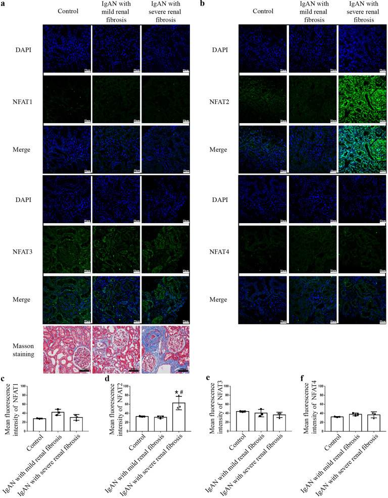
Acute kidney injury (AKI) with maladaptive tubular repair leads to renal fibrosis and progresses to chronic kidney disease (CKD). At present, there is no curative drug to interrupt AKI-to-CKD progression. The nuclear factor of the activated T cell (NFAT) family was initially identified as a transcription factor expressed in most immune cells and involved in the transcription of cytokine genes and other genes critical for the immune response. NFAT2 is also expressed in renal tubular epithelial cells (RTECs) and podocytes and plays an important regulatory role in the kidney. In this study, we investigated the renoprotective effect of 11R-VIVIT, a peptide inhibitor of NFAT, on renal fibrosis in the AKI-to-CKD transition and the underlying mechanisms. We first examined human renal biopsy tissues and found that the expression of NFAT2 was significantly increased in RTECs in patients with severe renal fibrosis. We then established a mouse model of AKI-to-CKD transition using bilateral ischemia-reperfusion injury (Bi-IRI). The mice were treated with 11R-VIVIT (5 mg/kg, i.p.) on Days 1, 3, 10, 17 and 24 after Bi-IRI. We showed that the expression of NFAT2 was markedly increased in RTECs in the AKI-to-CKD transition. 11R-VIVIT administration significantly inhibited the nuclear translocation of NFAT2 in RTECs, decreased the levels of serum creatinine and blood urea nitrogen, and attenuated renal tubulointerstitial fibrosis but had no toxic side effects on the heart and liver. In addition, we showed that 11R-VIVIT administration alleviated RTEC apoptosis after Bi-IRI. Consistently, preapplication of 11R-VIVIT (100 nM) and transfection with NFAT2-targeted siRNA markedly suppressed TGFβ-induced HK-2 cell apoptosis in vitro. In conclusion, 11R-VIVIT administration inhibits IRI-induced NFAT2 activation and prevents AKI-to-CKD progression. Inhibiting NFAT2 may be a promising new therapeutic strategy for preventing renal fibrosis after IR-AKI.

Keywords: 11R-VIVIT; HK-2 cells; NFAT2; acute kidney injury; apoptosis; chronic kidney disease; renal fibrosis; renal tubular epithelial cells.


Conflict of interest statement

The authors declare no competing interests.

Figures

Fig. 1
Fig. 1. NFAT2 is markedly increased in human renal tissue with severe fibrosis.
Immunostaining for NFAT1, NFAT2, NFAT3 and NFAT4 was performed on tissues from IgAN patients and adjacent normal tissues from renal cell carcinoma patients. Representative confocal images showed higher expression and more nuclear localization of NFAT2 in the renal tubules, interstitium and glomerulus in IgAN patients with severe renal fibrosis (n = 3) than in IgAN patients with mild renal fibrosis or in adjacent normal renal tissue from renal cell carcinoma patients (n = 3) (Scale bars = 50 μm). Masson staining was used to assess kidney fibrosis (scale bars = 100 μm). a Immunostaining for NFAT1 and NFAT3. b Immunostaining for NFAT2 and NFAT4. cf Quantitative analysis for the immunofluorescence of NFAT1, NFAT2, NFAT3 and NFAT4. *P < 0.05 vs. Control; #P < 0.05 vs. IgAN with mild renal fibrosis. NFAT1 nuclear factor of activated T cells 1, NFAT2 nuclear factor of activated T cells 2, NFAT3 nuclear factor of activated T cells 3, NFAT4 nuclear factor of activated T cells 4.
Fig. 2
Fig. 2. NFAT2 is increased in the renal tissue of AKI-to-CKD transition animal model.
a Scr and (b) serum BUN levels were measured in the sham-operated and IRI models. c Masson staining showed renal histopathology fibrosis. Scale bars = 100 μm. d Quantification of tubulointerstitial fibrosis. ef Protein expression of NFAT2 in the kidneys of sham-operated (2d, 14d, 28d) and IRI (2d, 14d, 28d) mice, as determined by Western blotting and densitometric analysis. *P < 0.05 vs. SHAM-2d, #P < 0.05 vs. SHAM-14d, &P < 0.05 vs. SHAM-28d. NFAT2 nuclear factor of activated T cells 2, AKI acute kidney injury, CKD chronic kidney disease, Scr serum creatinine, BUN blood urea nitrogen, IRI ischemia-reperfusion injury, NFAT1 nuclear factor of activated T cells 1, NFAT3 nuclear factor of activated T cells 3, NFAT4 nuclear factor of activated T cells 4.
Fig. 3
Fig. 3. NFAT2 is activated in the RTECs of AKI-to-CKD transition animal model.
a Immunostaining for NFAT1, NFAT2, NFAT3, NFAT4 (green) and DAPI (blue) was performed in frozen renal sections from sham-operated (2d, 14d, 28d) and IRI (2d, 14d, 28d) mice. Scale bars = 50 μm. be Quantitative analysis for the immunofluorescence of NFAT1, NFAT2, NFAT3 and NFAT4. *P < 0.05 vs. SHAM-2d, #P < 0.05 vs. SHAM-14d, &P < 0.05 vs. SHAM-28d. NFAT2 nuclear factor of activated T cells 2, RTECs renal tubular epithelial cells, AKI acute kidney injury, CKD chronic kidney disease, NFAT1 nuclear factor of activated T cells 1, NFAT3 nuclear factor of activated T cells 3, NFAT4 nuclear factor of activated T cells 4, IRI ischemia-reperfusion injury.
Fig. 4
Fig. 4. 11R-VIVIT inhibits the nuclear localization and dephosphorylation of NFAT2 in RTECs in an AKI-to-CKD progression model.
a Double immunofluorescence staining of NFAT2 (green) and DAPI (blue) and merged images of kidneys from the sham-operated, IRI and IRI + 11R-VIVIT treatment groups. Scale bars = 20 μm. b Quantification of the percentage of renal tubular epithelial cells with NFAT2 expression in the nucleus. *P < 0.05 vs. IRI-2d, #P < 0.05 vs. IRI-14d, &P < 0.05 vs. IRI-28d. c Protein expression of p-NFAT2 (Ser172) in the sham-operated, IRI and IRI + 11R-VIVIT treatment groups on the 2nd, 14th and 28th day. d The quantitative results of p-NFAT2 (Ser172) were normalized to GAPDH. NFAT2 nuclear factor of activated T cells 2, RTECs renal tubular epithelial cells, AKI acute kidney injury, CKD chronic kidney disease, IRI ischemia-reperfusion injury, p-NFAT2 phosphorylated nuclear factor of activated T cells 2.
Fig. 5
Fig. 5. 11R-VIVIT attenuates tubulointerstitial fibrosis in an animal model of AKI-to-CKD progression.
a Scr and (b) serum BUN levels were determined in the sham-operated, IRI and IRI + 11R-VIVIT treatment groups on the 2nd, 14th and 28th day. cd α-SMA and fibronectin mRNA expression was measured in each group by RT-qPCR on the 2nd, 14th and 28th day. e Masson staining showed renal histopathological fibrosis in each group on the 2nd, 14th and 28th day. Scale bars = 100 μm. f Quantification of tubulointerstitial fibrosis. g Protein expression of α-SMA and fibronectin in each group on the 2nd, 14th and 28th day. hi The quantitative results of α-SMA and fibronectin were normalized to GAPDH. *P < 0.05 vs. IRI-2d, #P < 0.05 vs. IRI-14d, &P < 0.05 vs. IRI-28d. AKI acute kidney injury, CKD chronic kidney disease, Scr serum creatinine, BUN blood urea nitrogen, IRI ischemia-reperfusion injury, RT-qPCR reverse transcription-quantitative polymerase chain reaction.
Fig. 6
Fig. 6. 11R-VIVIT attenuates tubular epithelial cell apoptosis in an animal model of AKI-to-CKD progression.
a TUNEL staining of frozen renal tissue sections from the sham-operated, IRI and IRI + 11R-VIVIT treatment groups on the 2nd, 14th and 28th day. Scale bars = 50 μm. b Quantification of TUNEL‑positive tubular epithelial cells in the sham-operated, IRI and IRI + 11R-VIVIT treatment groups on the 2nd, 14th and 28th day. c Western blot analysis of the expression of caspase-3 and C‑caspase-3 in each group on the 2nd, 14th and 28th day. de The quantitative results of C-caspase-3 and caspase-3 were normalized to β-actin. f Western blot analysis of the expression of Bax in each group on the 2nd, 14th and 28th day. g The quantitative results of Bax were normalized to β-actin. *P < 0.05 vs. IRI-2d, #P < 0.05 vs. IRI-14d, &P < 0.05 vs. IRI-28d. AKI acute kidney injury, CKD chronic kidney disease, RTECs renal tubular epithelial cells, IRI ischemia-reperfusion injury, C‑caspase‑3 cleaved-caspase‑3.
Fig. 7
Fig. 7. NFAT2-targeted siRNA attenuates RTEC apoptosis in vitro following TGFβ stimulation.
HK-2 cells in the TGFβ group were stimulated with 5 ng/mL TGFβ for 72 h. The scramble+TGFβ and siRNA-NFAT2 + TGFβ groups were transfected with 5 nM scramble or siRNA-NFAT2 respectively, prior to TGFβ stimulation. ac NFAT2-targeted siRNA significantly reduced the mRNA and protein levels of NFAT2. Three sequences of the NFAT2-targeted siRNA were used in the present study, and sequence 001 induced significant reductions in the protein and mRNA levels of NFAT2. d The protein expression of α-SMA and fibronectin in HK-2 cells. ef The quantitative results of α-SMA and fibronectin were normalized to GAPDH. g Apoptosis was examined by flow cytometry. h Quantification of RTEC apoptosis by flow cytometry. i Western blot analysis of the expression of caspase-3 and C‑caspase-3 in HK-2 cells. j The quantitative results of caspase-3 and C‑caspase-3 were normalized to β-actin. k Western blot analysis of the expression of Bax in HK-2 cells. l The quantitative results of Bax were normalized to β-actin. m TUNEL staining of HK-2 cells in the CON, TGFβ, Scramble+TGFβ and siRNA-NFAT2 + TGFβ group. n Quantification of TUNEL‑positive RTECs. Scale bars = 20 μm. &P < 0.05 vs. CON and scramble, *P < 0.05 vs. TGFβ, #P < 0.05 vs. scramble+ TGFβ. NFAT2 nuclear factor of activated T cells 2, RTECs renal tubular epithelial cells, TGFβ transforming growth factor beta, CON control, C‑caspase‑3 cleaved caspase‑3.
Fig. 8
Fig. 8. 11R-VIVIT attenuates TGFβ‑induced apoptosis in HK-2 cells.
a TGFβ treatment increased total NFAT2 expression in HK-2 cells. b TGFβ treatment decreased p-NFAT2 (Ser172) expression and 11R-VIVIT could increase the protein expression of p-NFAT2 (Ser172). c Quantification of NFAT2 expression normalized to GAPDH. d Quantification of p-NFAT2 (Ser172) expression normalized to GAPDH. ef TGFβ treatment increased nuclear NFAT2 expression in HK-2 cells, and 11R-VIVIT inhibited the nuclear localization of NFAT2. g Quantification of NFAT2 expression in the cytoplasmic fraction HK-2 cells; The results were normalized to GAPDH. h Quantification of NFAT2 expression in the nuclear fraction of HK-2 cells; The results were normalized to Histone H3. i The protein expression of α-SMA and fibronectin in HK-2 cells. jk The quantitative results of α-SMA and fibronectin were normalized to GAPDH. l Apoptosis was examined by flow cytometry. m Quantification of RTEC apoptosis by flow cytometry. n Western blot analysis of the expression of caspase-3 and C‑caspase-3 in HK-2 cells. o The quantitative results of caspase-3 and C‑caspase-3 were normalized to β-actin. p Western blot analysis of the expression of Bax in HK-2 cells. q The quantitative results of Bax were normalized to β-actin. r TUNEL staining of HK-2 cells in the CON, TGFβ and 11R-VIVIT + TGFβ group. s Quantification of TUNEL‑positive tubular epithelial cells. Scale bars = 20 μm. *P < 0.05 vs. TGFβ. TGFβ transforming growth factor beta, NFAT2 nuclear factor of activated T cells 2, p-NFAT2 phosphorylated nuclear factor of activated T cells 2, RTECs renal tubular epithelial cells, C‑caspase‑3 cleaved caspase‑3.

References

    1. Heung M, Steffick DE, Zivin K, Gillespie BW, Banerjee T, Hsu CY, et al. Acute kidney injury recovery pattern and subsequent risk of CKD: an analysis of veterans health administration data. Am J Kidney Dis. 2017;77:742–52. - PMC - PubMed
    1. See EJ, Jayasinghe K, Glassford N, Bailey M, Johnson DW, Polkinghorne KR, et al. Long-term risk of adverse outcomes after acute kidney injury: a systematic review and meta-analysis of cohort studies using consensus definitions of exposure. Kidney Int. 2019;95:160–72. doi: 10.1016/j.kint.2018.08.036. - DOI - PubMed
    1. Boor P, Ostendorf T, Floege J. Renal fibrosis: novel insights into mechanisms and therapeutic targets. Nat Rev Nephrol. 2010;7:743–57. - PubMed
    1. Chen DQ, Feng YL, Chen L, Liu JR, Wang M, Vaziri ND, et al. Poricoic acid A enhances melatonin inhibition of AKI-to-CKD transition by regulating Gas6/AxlNFκB/Nrf2 axis. Free Radic Biol Med. 2019;134:484–97. doi: 10.1016/j.freeradbiomed.2019.01.046. - DOI - PubMed
    1. Chen DQ, Cao G, Zhao H, Chen L, Yang T, Wang M, et al. Combined melatonin and poricoic acid A inhibits renal fibrosis through modulating the interaction of Smad3 and β-catenin pathway in AKI-to-CKD continuum. Ther Adv Chronic Dis. 2019;10:2040622319869116. - PMC - PubMed