. 2021 Dec 1;332(Pt 2):112925.
doi: 10.1016/j.sna.2021.112925. Epub 2021 Jun 17.

Acoustics at the nanoscale (nanoacoustics): A comprehensive literature review.: Part II: Nanoacoustics for biomedical imaging and therapy

Affiliations

Acoustics at the nanoscale (nanoacoustics): A comprehensive literature review.: Part II: Nanoacoustics for biomedical imaging and therapy

Chang Peng et al. Sens Actuators A Phys. .

Abstract

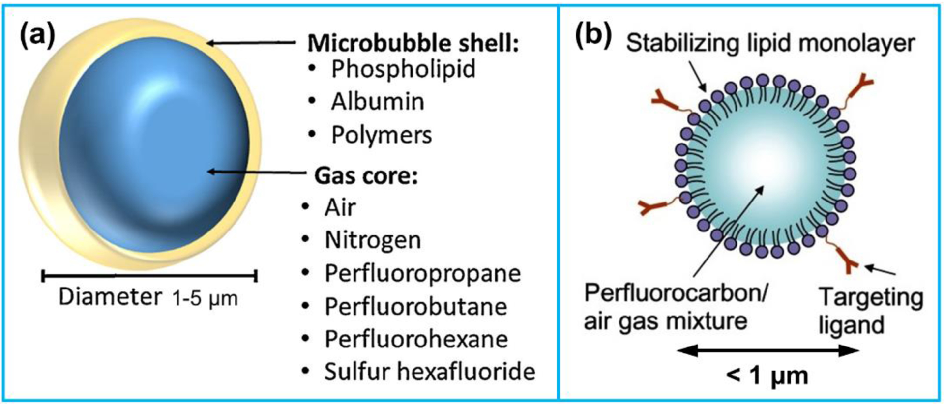
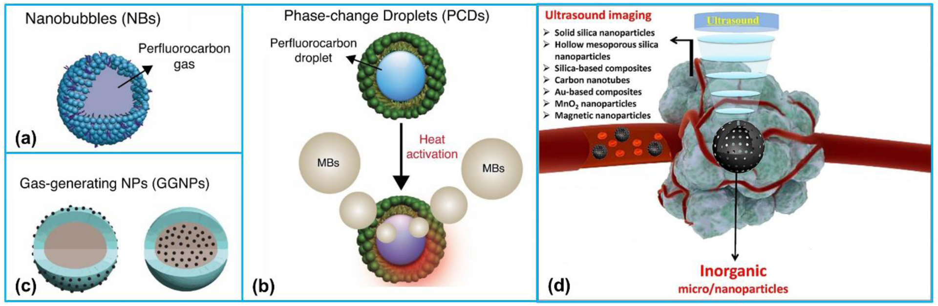
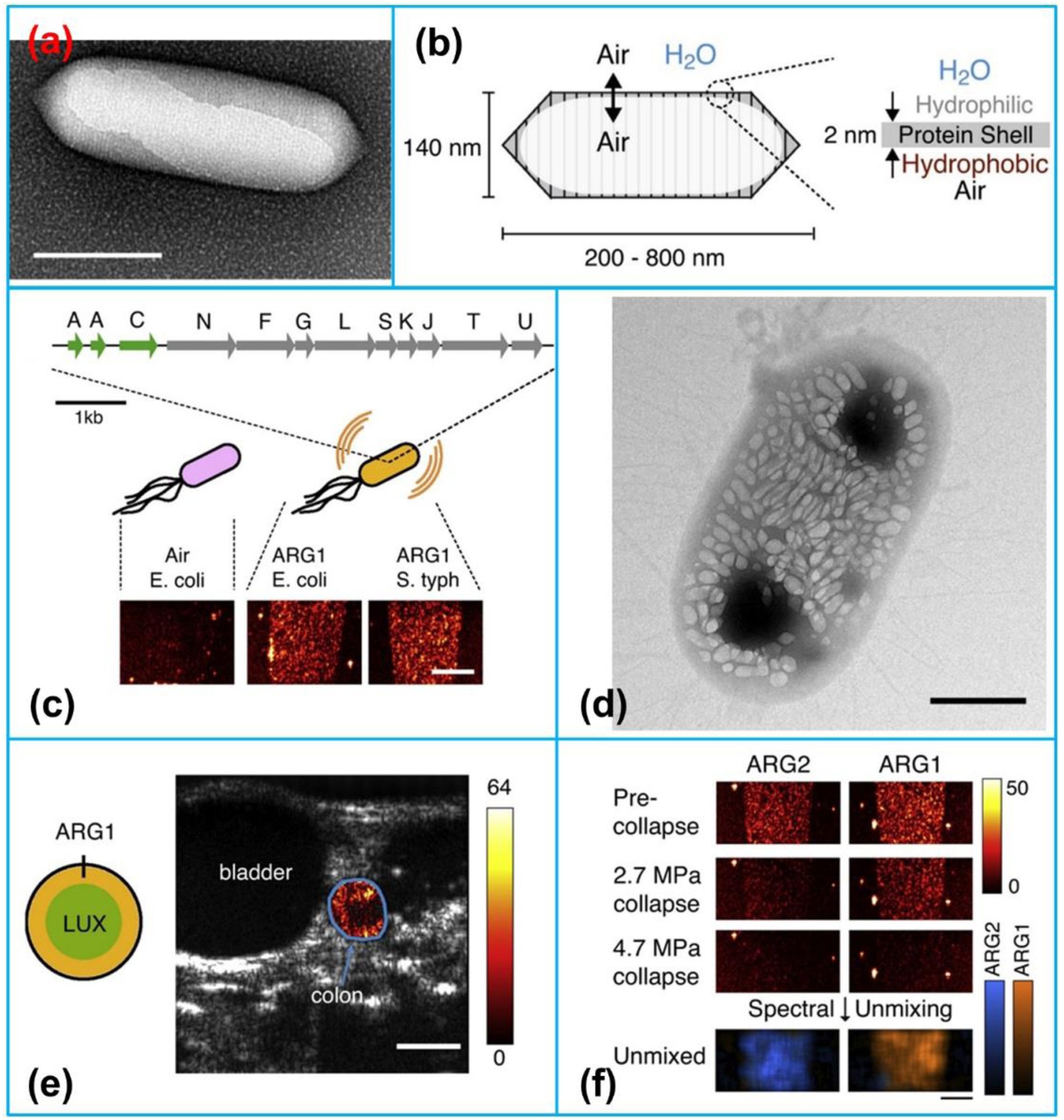
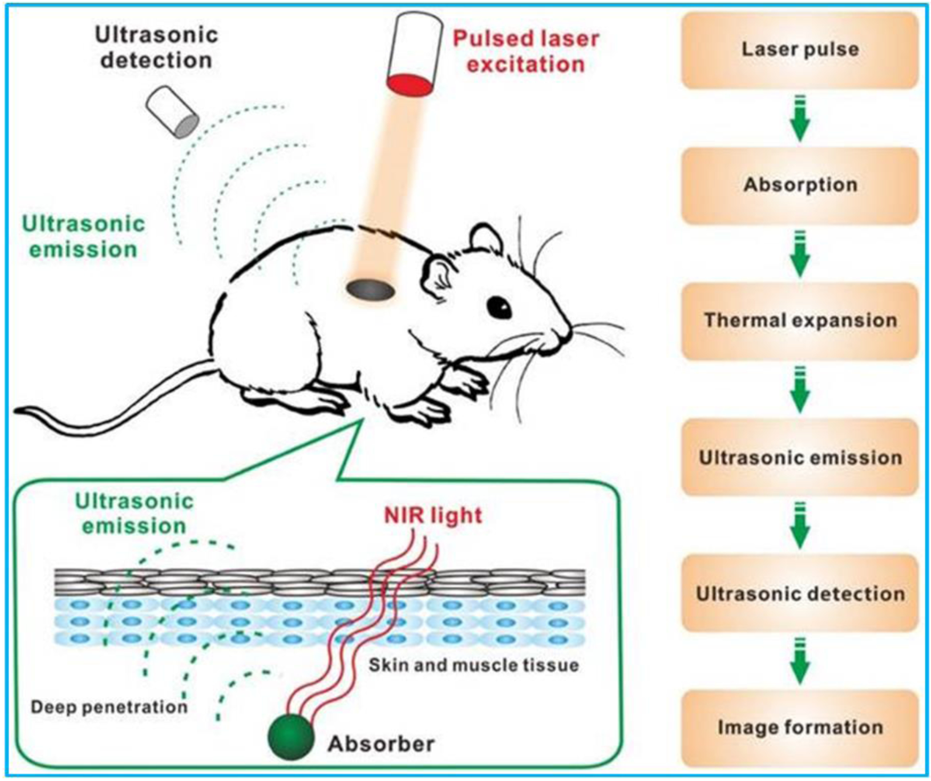
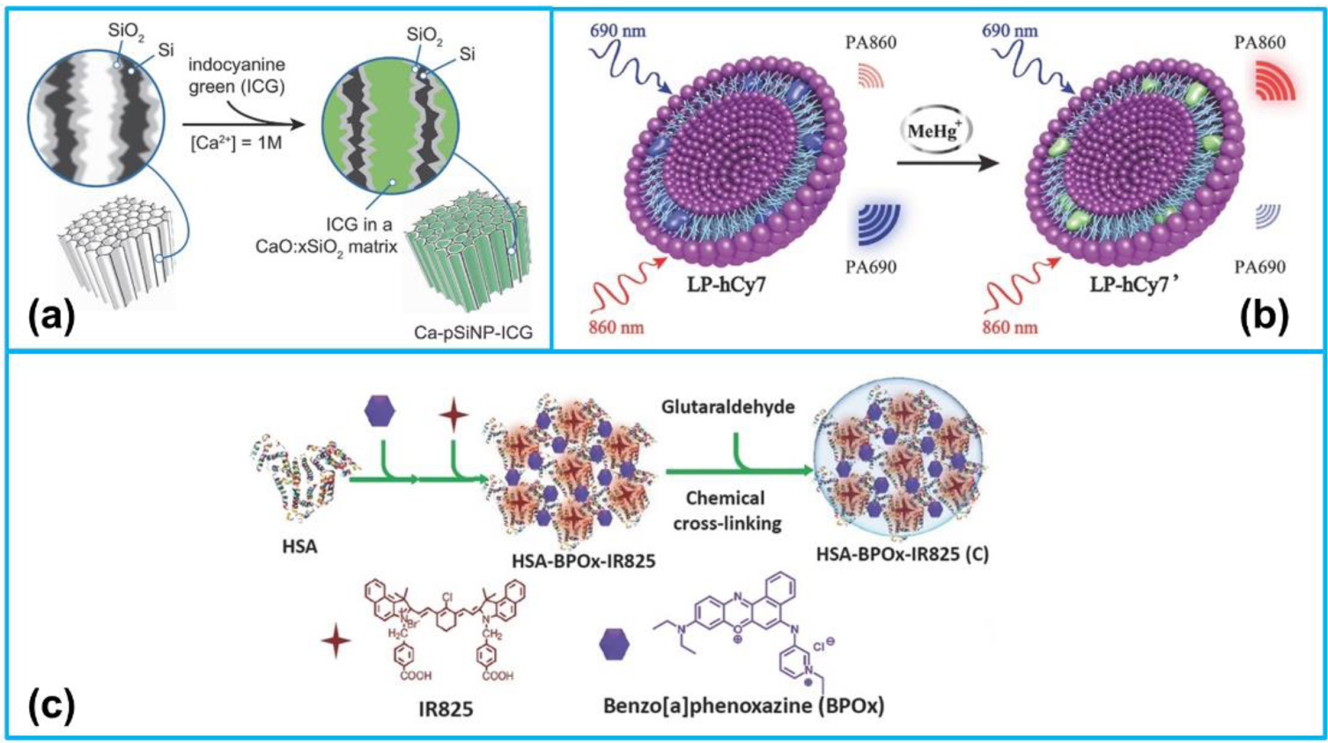
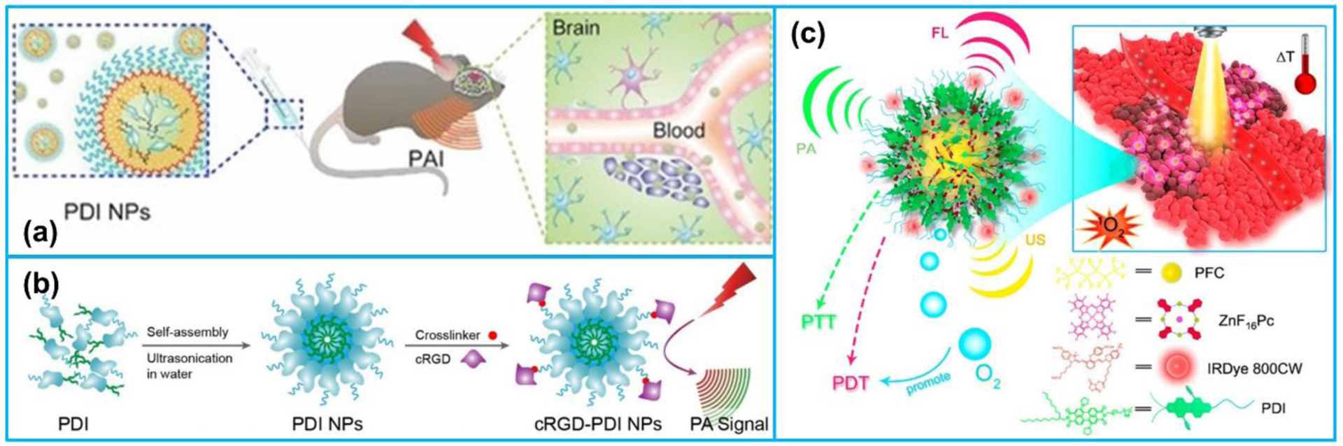
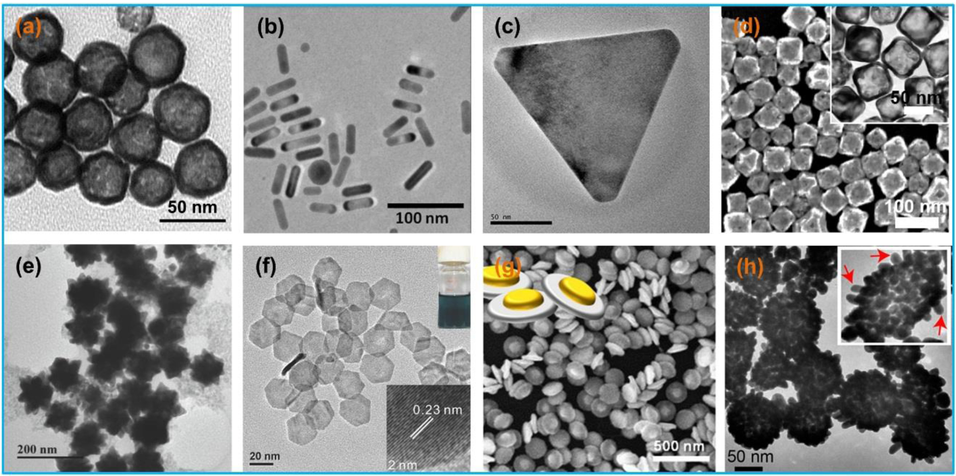
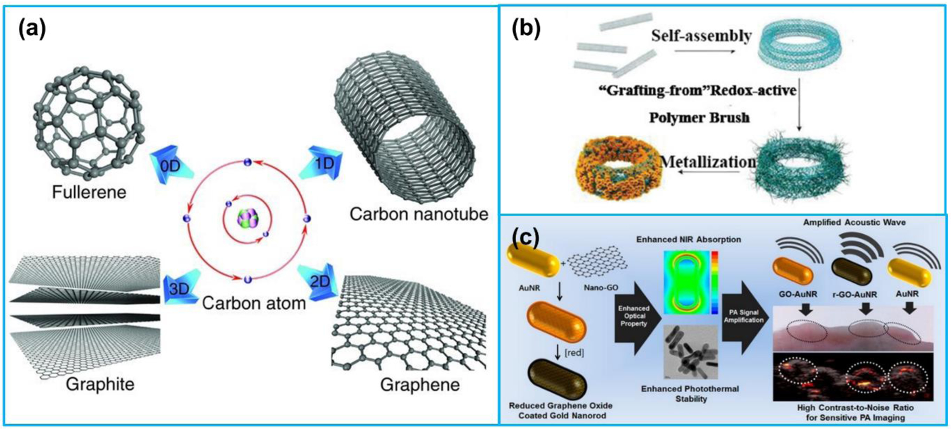
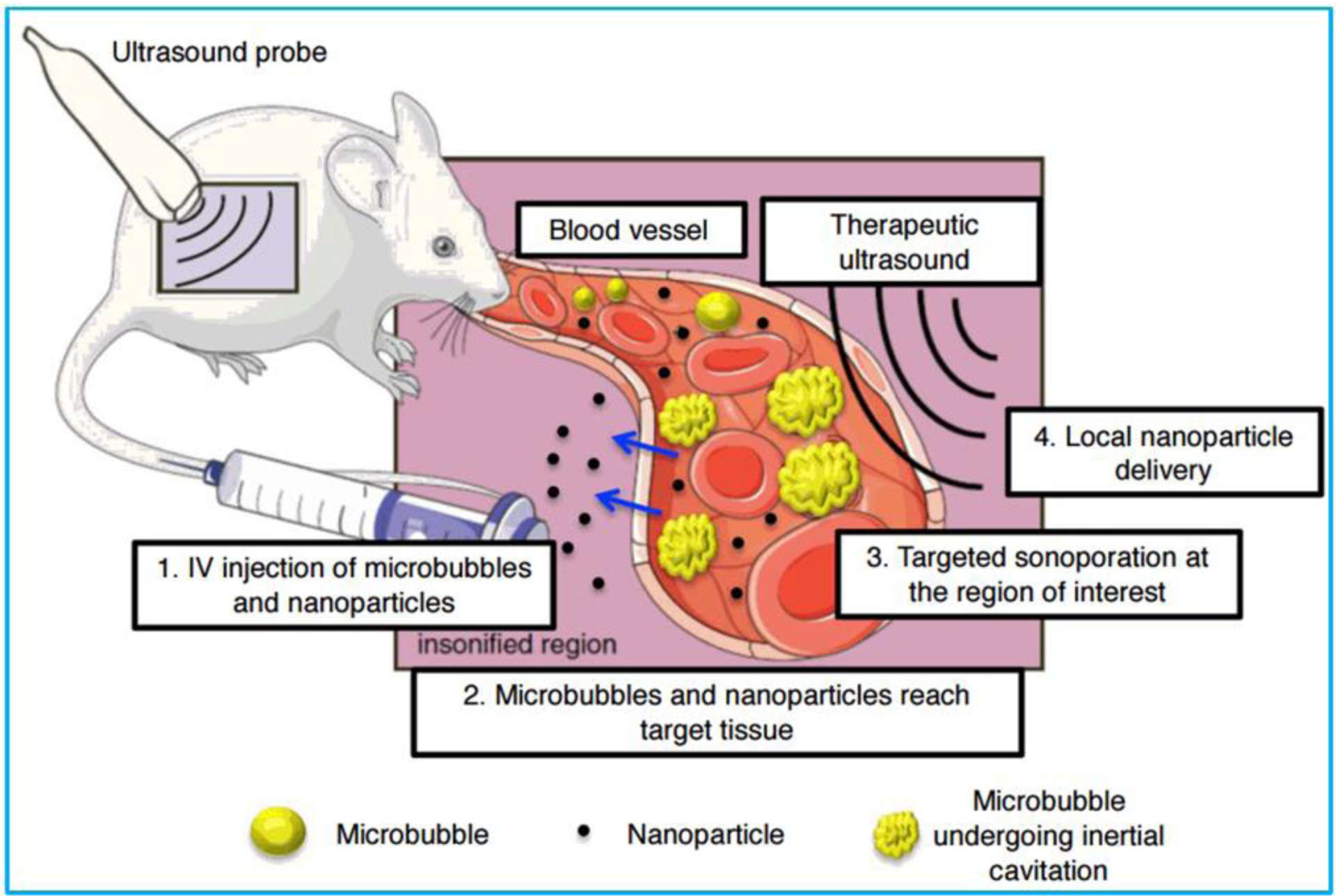
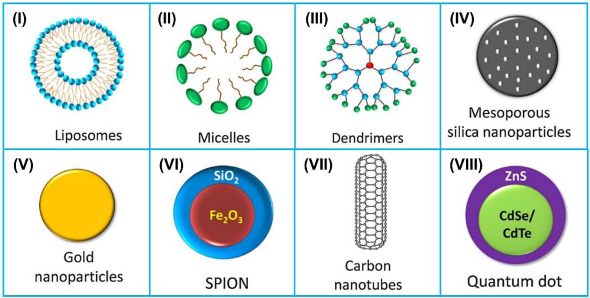
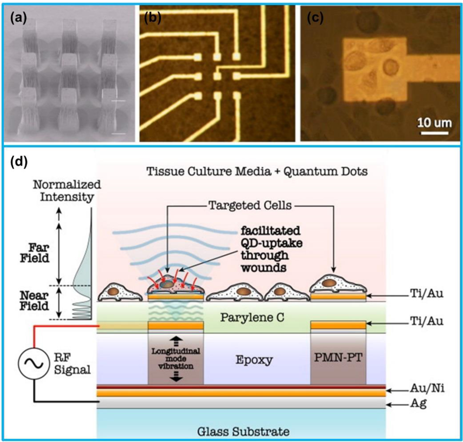
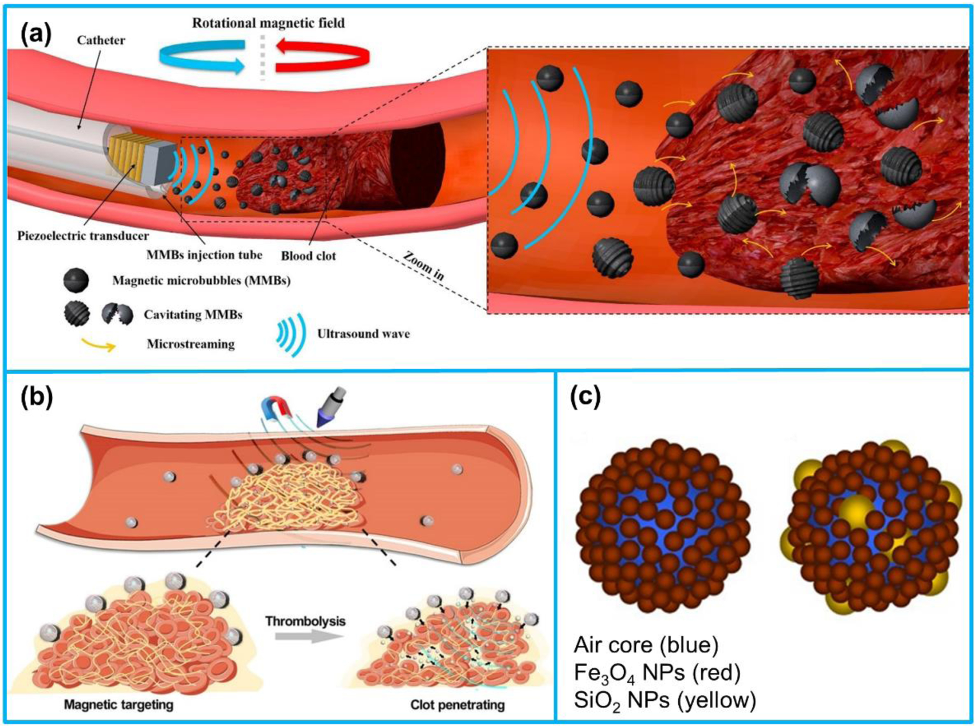
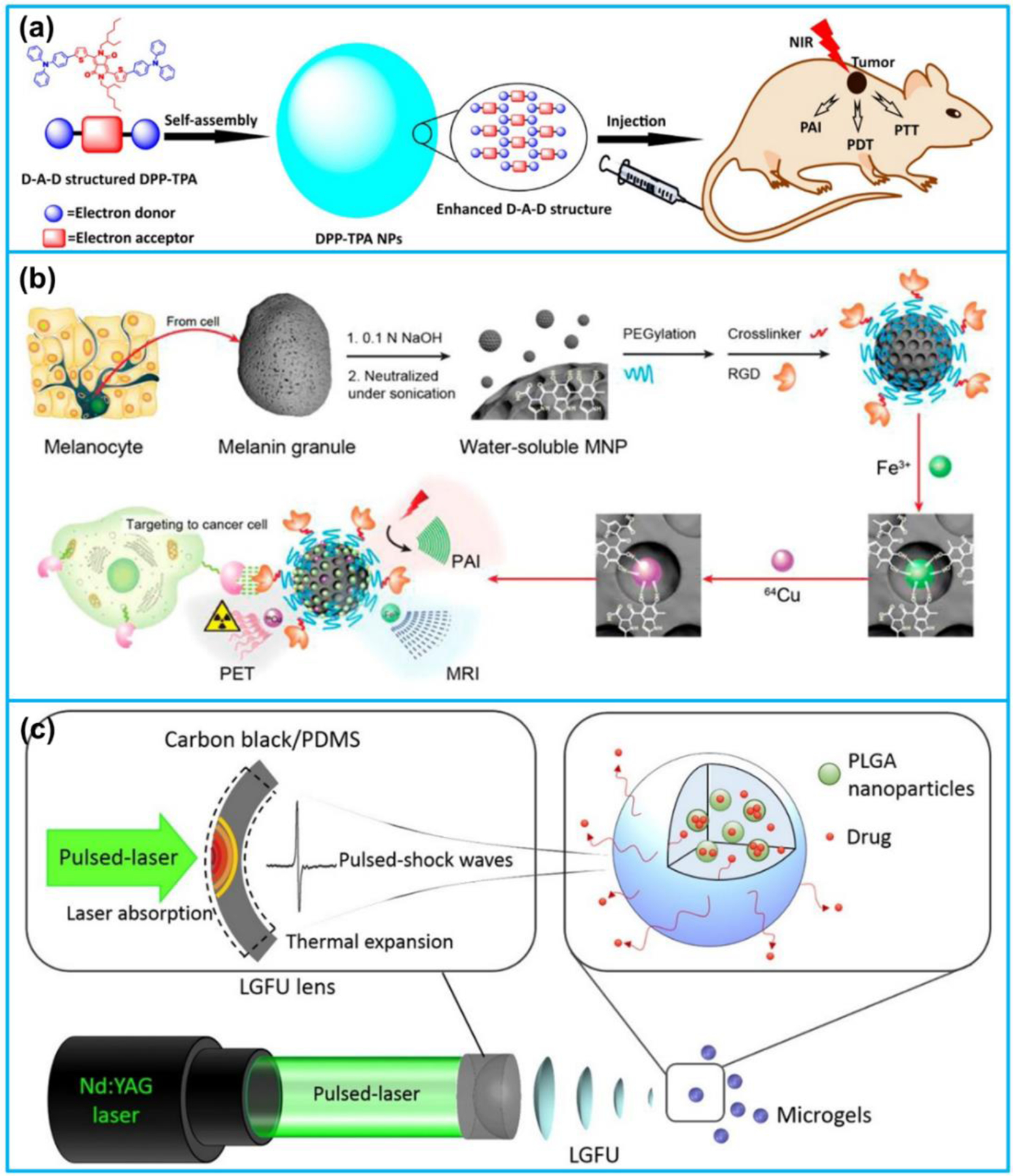
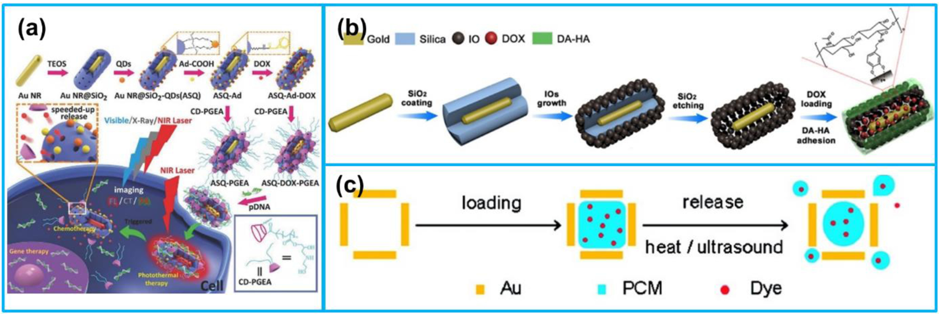
In the past decade, acoustics at the nanoscale (i.e., nanoacoustics) has evolved rapidly with continuous and substantial expansion of capabilities and refinement of techniques. Motivated by research innovations in the last decade, for the first time, recent advancements of acoustics-associated nanomaterials/nanostructures and nanodevices for different applications are outlined in this comprehensive review, which is written in two parts. As part II of this two-part review, this paper concentrates on nanoacoustics in biomedical imaging and therapy applications, including molecular ultrasound imaging, photoacoustic imaging, ultrasound-mediated drug delivery and therapy, and photoacoustic drug delivery and therapy. Firstly, the recent developments of nanosized ultrasound and photoacoustic contrast agents as well as their various imaging applications are examined. Secondly, different types of nanomaterials/nanostructures as nanocarriers for ultrasound and photoacoustic therapies are discussed. Finally, a discussion of challenges and future research directions are provided for nanoacoustics in medical imaging and therapy.

Keywords: acoustics; biomedical applications; imaging; laser ultrasound; nanoacoustics; nanomaterials; nanotechnology; photoacoustics; sonothrombolysis; therapy.


Conflict of interest statement

Declaration of competing interest Xiaoning Jiang has a financial interest in SonoVascular, Inc., who licensed an intravascular sonothrombolysis technology from North Carolina State University.

Figures

Figure 1.
Figure 1.
(a) Structure of a microbubble. Reproduced with permission [16]. Copyright 2015, Elsevier. (b) Composition of a nanosized ultrasound contrast agent. Reproduced with permission [19]. Copyright 2013, Dove Medical Press Ltd.
Figure 2.
Figure 2.
Schematic representation of organic and inorganic nanosized UCAs for ultrasound imaging. Organic UCAs: (a) nanobubbles, (b) phase-change droplets, and (c) gas-generating nanoparticles. (a)-(c) Reproduced with permission [29]. Copyright 2018, Elsevier. (d) The summary of inorganic nanoparticles. Reproduced with permission [30]. Copyright 2017, Elsevier.
Figure 3.
Figure 3.
(a) Transmission electron microscopy (TEM) image of a gas vesicle (scale bars, 150 nm). (b) Composition of a gas vesicle. (c) Engineered gene, ARG1, comprising genes from A. flos-aquae (green) and B. megaterium (gray) to make the gas vesicles detectable by ultrasound in heterologous host. (d) TEM image of an E. coli Nissle 1917 cell expressing ARG1 (scale bar, 50 nm). (e) Ultrasound image of a live mouse with ARG1-expressing E. coli arranged in the colon (scale bar, 2.5 mm). (f) Ultrasound images of ARG1 and ARG2 before and after the application of two different collapse pressures. Reproduced with permission [83]. Copyright 2018, Elsevier.
Figure 4.
Figure 4.
Schematic of working principle of PA imaging. Reproduced with permission [85]. Copyright 2016, Ivyspring International Publisher.
Figure 5.
Figure 5.
(a) Procedure used to simultaneously load and seal CG into the pores of porous silicon nanoparticles (pSiNPs). Reproduced with permission [102]. Copyright 2018, Wiley-VCH. (b) Mechanism of ratiometric PA imaging of MeHg+. Reproduced with permission [114]. Copyright 2017, Wiley-VCH. (c) The preparation process of C-HSA-BPOx-IR825. Reproduced with permission [111]. Copyright 2015, Wiley-VCH.
Figure 6.
Figure 6.
(a) PA imaging process of brain tumor in vivo by PDI NPs. Reproduced with permission [98]. Copyright 2015, Wiley-VCH. (b) The preparation of cRGD-PDI NPs. Reproduced with permission [105]. Copyright 2017, American Chemical Society. (c) IRDye800CW-labeled photosensitizer ZnF16Pc-loaded PDI photoacoustic nanodroplet (PS-PDI-PAnD). Reproduced with permission [116]. Copyright 2018, American Chemical Society.
Figure 7.
Figure 7.
Microscopy photographs of different types of gold nanoparticles: (a) nanospheres, (b) nanorods, (c) nanoprisms, (d) nanocages, (e) nanostars, (f) nanoplates, (g) nanodisks, and (h) nanoshells. (a) Reproduced with permission [148]. Copyright 2009, Elsevier. (b) Reproduced with permission [149]. Copyright 2012, American Chemical Society. (c) Reproduced with permission [126]. Copyright 2015, American Chemical Society. (d) Reproduced with permission [131]. Copyright 2017, Elsevier. (e) Reproduced with permission [150]. Copyright 2018, Wiley-VCH. (f) Reproduced with permission [135]. Copyright 2014, Wiley-VCH. (g) Reproduced with permission [139]. Copyright 2017, American Chemical Society. (h) Reproduced with permission [142]. Copyright 2017, American Chemical Society.
Figure 8.
Figure 8.
(a) Structures of 0D fullerene, 1D carbon nanotube, 2D graphene and 3D graphite formed by the carbon atoms. (b) Preparation of CNT ring Au nanoparticles (CNTR @ AuNPs). (c) Preparation and application of reduced graphene oxide Au nanorods (rGO-AuNRs). (a) Reproduced with permission [167]. Copyright 2011, Wiley-VCH. (b) Reproduced with permission [164]. Copyright 2016, American Chemical Society. (c) Reproduced with permission [165]. Copyright 2015, American Chemical Society.
Figure 9.
Figure 9.
Schematic illustration of the mechanism of ultrasound-mediated nanoparticle delivery. Reproduced with permission [177]. Copyright 2017, Korean Society of Ultrasound in Medicine.
Figure 10.
Figure 10.
Schematic illustration of the 8 most reported nanocarriers: (I) liposomes, (II) micelles, (III) dendrimers, (IV) meso-porous silica nanoparticles (MSNs), (V) gold nanoparticles (AuNPs), (VI) super paramagnetic iron oxide nanoparticles (SPIONs), (VII) carbon nanotubes (CNTs), and (VIII) quantum dots (QDs). Reproduced with permission [193]. Copyright 2019, Elsevier.
Figure 11.
Figure 11.
(a) SEM mage of a 3 × 3 micro-transducer array (each area 25 μm × 25 μm). (b) Electrode above each micro-transducer. (c) Human melanoma cells above a micro-transducer. (d) Schematic illustration of ultrasound-mediated quantum dots (QDs) delivery at cellular level using the micro-transducer array. (a)-(d) Reproduced with permission [187]. Copyright 2011, Elsevier.
Figure 12.
Figure 12.
(a) Illustration of magnetic microbubbles (MMBs)-mediated sonothrombolysis. (b) Schematic view of SiO2-tPA shelled MMBs for targeted tPA delivery and controlled release. (c) Composition of MMBs. (a) Reproduced with permission [245]. Copyright 2019, Elsevier. (b) Reproduced with permission [186]. Copyright 2020, American Association for the Advancement of Science. (c) Reproduced with permission [243]. Copyright 2016, Springer Nature.
Figure 13.
Figure 13.
(a) D-A-D structured DPP-TPA NPs as theranostic agents for PA imaging-guided PDT/PTT. (b) Multimodality molecular imaging of MNPs. (c) Laser-generated-focused ultrasound-mediated drug delivery. (a) Reproduced with permission [261]. Copyright 2017, American Chemical Society. (b) Reproduced with permission [263]. Copyright 2014, American Chemical Society. (c) Reproduced with permission [248]. Copyright 2015, Elsevier.
Figure 14.
Figure 14.
(a) Preparation processes of multimodal imaging-guided therapeutic platforms. (b) Preparation of GNR @ IOs-DOX nanocapsules. (c) Loading the hollow interior of an AuNC with a dye doped PCM and then releasing t from the AuNC. (a) Reproduced with permission [270]. Copyright 2016, Wiley-VCH. (b) Reproduced with permission [271]. Copyright 2016, American Chemical Society. (c) Reproduced with permission [130]. Copyright 2011, American Chemical Society.

References

    1. Cuenca AG, Jiang H, Hochwald SN, Delano M, Cance WG, Grobmyer SR, Emerging implications of nanotechnology on cancer diagnostics and therapeutics, Cancer. 107 (2006) 459–466. - PubMed
    1. Farokhzad OC, Langer R, Nanomedicine: developing smarter therapeutic and diagnostic modalities, Adv. Drug Deliv. Rev 58 (2006) 1456–1459. - PubMed
    1. Nie S, Xing Y, Kim GJ, Simons JW, Nanotechnology applications in cancer, Annu. Rev. Biomed. Eng 9 (2007) 257–288. - PubMed
    1. Wang X, Yang L, Chen Z, Shin DM, Application of nanotechnology in cancer therapy and imaging, CA. Cancer J. Clin 58 (2008) 97–110. - PubMed
    1. Barreto JA, O’Malley W, Kubeil M, Graham B, Stephan H, Spiccia L, Nanomaterials: applications in cancer imaging and therapy, Adv. Mater 23 (2011) H18–H40. - PubMed

LinkOut - more resources