

Histopathological Analysis of Decellularized Porcine Small Intestinal Submucosa after Treatment of Skin Ulcer
- PMID: 34938643
- PMCID: PMC8687724
- DOI: 10.1097/GOX.0000000000003967
Histopathological Analysis of Decellularized Porcine Small Intestinal Submucosa after Treatment of Skin Ulcer
Abstract
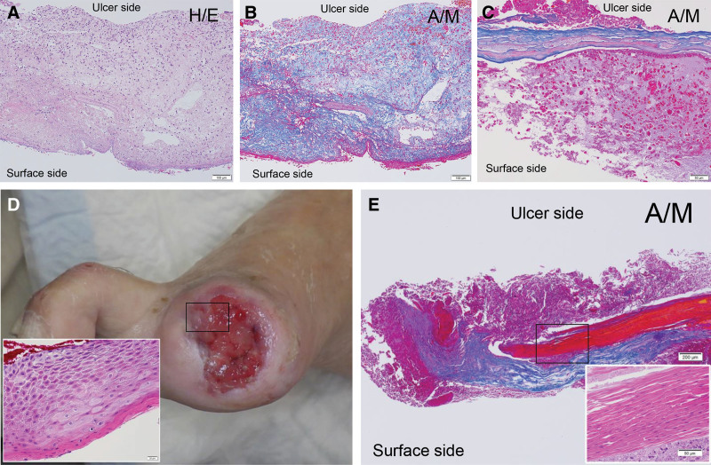
Background: Decellularized porcine small intestinal submucosa (SIS), commercialized as an extracellular matrix rich in cell-inducing substrates and factors, has been clinically applied to treat intractable skin ulcers and has shown therapeutic effects. The SIS reportedly induces cell infiltration and integrates with the ulcer bed after 3-7 days of application. The attached SIS degenerates over time, and the remaining mass appears as slough, below which is granulation tissue that is essential for healing. This study aimed to determine whether the slough should be removed in clinical settings.
Methods: Five patients with intractable skin ulcers were included in this case series. Seven days after applying a two-layer fenestrated-type SIS to the ulcer, the removed slough was histopathologically examined.
Results: The collagen fibers of the SIS somewhat degenerated, and inflammatory cell infiltration was observed from the ulcer side to the surface side of the SIS. Neovascularization was similarly observed on the ulcer side. The degree of inflammatory cell infiltration decreased from the ulcer side to the surface side, whereas pus (ie, aggregates of neutrophils) was observed on the surface and ulcer edges. Additionally, the removed slough contained regenerative epithelium on the ulcer side of the remaining collagen fibers.
Conclusions: After treating intractable skin ulcers using SIS, we recommend removal of the upper surface and ulcer edge of the degenerated SIS or slough to prevent infection and preservation of the lower side of the degenerated SIS to maintain the granulation tissue and regenerative epithelium.
Copyright © 2021 The Authors. Published by Wolters Kluwer Health, Inc. on behalf of The American Society of Plastic Surgeons.
Conflict of interest statement
Disclosure: The authors have no financial interest to declare in relation to the content of this article.
Figures


References
-
- Salgado RM, Bravo L, García M, et al. . Histomorphometric analysis of early epithelialization and dermal changes in mid-partial-thickness burn wounds in humans treated with porcine small intestinal submucosa and silver-containing hydrofiber. J Burn Care Res. 2014;35:e330–e337. - PubMed
-
- Badylak SF. Small intestinal submucosa (SIS): a biomaterial conducive to smart tissue remodeling. Bell E, ed. In: Tissue Engineering. Boston, MA: Birkhäuser; 1993:179–189.
-
- Hodde J, Janis A, Ernst D, et al. . Effects of sterilization on an extracellular matrix scaffold: part I. Composition and matrix architecture. J Mater Sci Mater Med. 2007;18:537–543. - PubMed
LinkOut - more resources
Full Text Sources