Review
. 2022 Mar 30;32(3):182-200.
doi: 10.1093/glycob/cwab105.

How microbial glycosyl hydrolase activity in the gut mucosa initiates microbial cross-feeding

Affiliations
Review

How microbial glycosyl hydrolase activity in the gut mucosa initiates microbial cross-feeding

Maryse D Berkhout et al. Glycobiology. .

Abstract

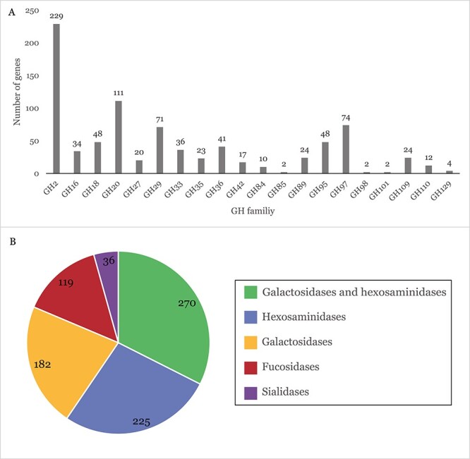
The intestinal epithelium is protected from direct contact with gut microbes by a mucus layer. This mucus layer consists of secreted mucin glycoproteins. The outer mucus layer in the large intestine forms a niche that attracts specific gut microbiota members of which several gut commensals can degrade mucin. Mucin glycan degradation is a complex process that requires a broad range of glycan degrading enzymes, as mucin glycans are intricate and diverse molecules. Consequently, it is hypothesized that microbial mucin breakdown requires concerted action of various enzymes in a network of multiple resident microbes in the gut mucosa. This review investigates the evolutionary relationships of microbial carbohydrate-active enzymes that are potentially involved in mucin glycan degradation and focuses on the role that microbial enzymes play in the degradation of gut mucin glycans in microbial cross-feeding and syntrophic interactions.

Keywords: CAZymes; glycosidases; gut microbiota; mucin; syntrophic interactions.


Figures

Fig. 1
Fig. 1
The predominant mucin glycan core structures found in the human gut and a hypothetical mucin glycan. (A) The bottle brush-like structure of a mucin glycoprotein: A secreted MUC2 mucin consists of a protein core (red) and branched, complex glycan extensions (blue). (B) The host intestinal epithelial cells secrete mucin glycans that form a mucus layer to avoid direct contact with bacteria. The mucus consists of an inner layer, which is virtually impenetrable to bacteria, and an outer layer, which forms a niche for specific microbiota. (C) The predominant core structures in the gut. Core 1 consists of galactose that is β1–3-linked to GalNAc. Core 2 contains an additional GlcNAc that is β1–6-linked to GalNAc. Core 3 consists of GlcNAc β1–3 linked to GalNAc. Core 3 can subsequently be extended to core 4 through the β1–6 linkage of another GlcNAc to the GalNAc. (D) A hypothetical mucin glycan that consists of core 2, is extended by several GlcNAc and galactose subunits, and is terminated by sialic acid (Neu5Ac), sulfate (SO3) and fucose. Mucin glycans are always O-linked to a serine or threonine residue on the protein backbone.
Fig. 2
Fig. 2
Functional distribution of GHs implicated in mucin glycan degradation in a consortium of primary mucin glycan degraders (the PMD consortium, Table II). (A) Number of genes encoding GH family members that are implicated in mucin glycan degradation in the PMD consortium of mucin glycan degraders that are included in the CAZy database. (B) Number of genes within the PMD consortium encoding GH family members grouped by their function. Two families (GH2 and GH36) comprise both galactosidases and hexosaminidases and are therefore shown separately.
Fig. 3
Fig. 3
A hypothetical mucin glycan and the enzyme classes that are required to hydrolyze the bonds between the subunits.
Fig. 4
Fig. 4
Cooperative mucin glycan degradation of a mucin glycan degrader and a butyrate-producing partner organism results in the production of SCFAs. Extracellular CAZymes of the mucin glycan degrader release shorter glycans from mucin. These glycans are imported into the cell, where further degradation takes place. This results in the production of SCFAs propionate and acetate. Subsequently, acetate can be taken up by a partner organism, which produces butyrate from acetate. Butyrate is secreted and becomes available to the host.
Fig. 5
Fig. 5
Cross-feeding of Bifidobacterium spp. with butyrate producer A. hallii: A three-species mucin coculture of B. bifidum, B. breve or B. infantis and A. hallii produces SCFAs propionate and butyrate. Mucin glycan degradation by B. bifidum releases fucose, acetate and lactate. B. infantis or B. breve can use fucose to produce acetate, lactate and 1,2-propanediol. A. hallii uses 1,2-propanediol to produce propionate and uses acetate and lactate to produce butyrate (Bunesova et al. 2018).

References

    1. Van den Abbeele P, Belzer C, Goossens M, Kleerebezem M, De Vos WM, Thas O, De Weirdt R, Kerckhof FM, Van De Wiele T. 2013. Butyrate-producing clostridium cluster XIVa species specifically colonize mucins in an in vitro gut model. ISME J. 7(5):949–961. - PMC - PubMed
    1. Almagro-Moreno S, Boyd EF. 2009. Insights into the evolution of sialic acid catabolism among bacteria. BMC Evol Biol. 9(1):1–16. - PMC - PubMed
    1. Ambrogi V, Bottacini F, O’Sullivan J, O’Connell Motherway M, Linqiu C, Schoemaker B, Schoterman M, van Sinderen D. 2019. Characterization of GH2 and GH42 β-galactosidases derived from bifidobacterial infant isolates. AMB Express. 9(1). - PMC - PubMed
    1. Arike L, Hansson GC. 2016. The densely O-glycosylated MUC2 mucin protein protects the intestine and provides food for the commensal bacteria. J Mol Biol. 428(16):3221–3229. - PMC - PubMed
    1. Belzer C, Chia LW, Aalvink S, Chamlagain B, Piironen V, Knol J, de Vos WM. 2017. Microbial metabolic networks at the mucus layer lead to diet-independent butyrate and vitamin B12 production by intestinal symbionts. MBio. 8(5):1–14. - PMC - PubMed

Publication types

MeSH terms