

The Reinforced Ma-Griffith Method Combined with Minimally Invasive Small-Incision Suture for Acute Achilles Tendon Rupture
- PMID: 34939329
- PMCID: PMC8867432
- DOI: 10.1111/os.13140
The Reinforced Ma-Griffith Method Combined with Minimally Invasive Small-Incision Suture for Acute Achilles Tendon Rupture
Abstract
Objective: To evaluate the treatment effects of the reinforced Ma-Griffith method combined with a minimally invasive small incision(M-G/MISI) in the treatment of acute Achilles tendon rupture.
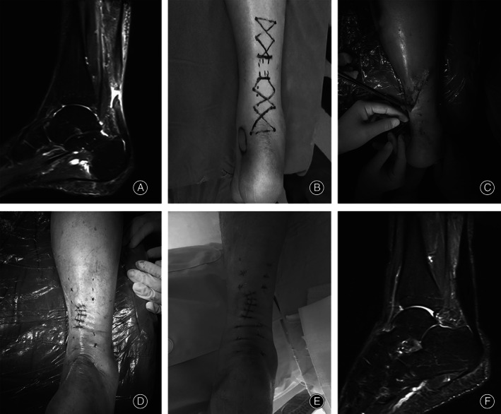
Methods: From January 2012 to January 2020, a retrospective study was carried out on thirty-one patients with acute Achilles tendon ruptures that were treated using the M-G/MISI. Patient with acute Achilles tendon rupture was operated on in the prone position. The M-G/MISI begin with making a small incision to debride the stumps of ruptured tendon. Then M-G/MISI was used to suture the distal and proximal Achilles tendons with the help of a epidural puncture needle and polydioxanone synthetic absorbable suture (PDS) Ⅱ line. Finally the stumps of ruptured tendon was reattached. After the surgery, the affected limb was fixed with either a plaster slab below the knee brace or a functional brace. Removal of plaster external fixation and partial weight-bearing with crutches five weeks after the operation; Complete weight-bearing nine weeks after the operation; jogging permitted 12 weeks after the operation; Patients were allowed to resume normal activities six months after the operation.
Results: All 31 patients in this study were male. Nineteen of these patients had Achilles tendon rupture on the right lower extremity, while 12 had ruptures on the left lower extremity. The patients had a mean age of 33.35 ± 7.13 years (range, 18-52 years). The mean operation time was 79.58 ± 22.67 minutes (range, 40-167 minutes). The mean time from injury to operation was 4.19 ± 2.01 days (range, 1-8 days), and the mean hospital stay was 9.87 ± 3.88 days (range, 5-22 days). The new technique had a small incision with a mean length of 3.94 ± 1.82 cm (range, 2-6 cm). The mean intraoperative blood loss was 16.77 ± 13.76 mL (range, 10-50 mL) and the mean follow-up time was 21.35 ± 10.18 months (range, 6-50 months). No wound infection, fistula, skin necrosis, sural nerve damage, deep venous thrombosis or tendon re-rupture was found. One year after the surgery, all patients reported 97.00 (range, 93-100 points) AOFAS ankle-hindfoot score points and the mean ATRS was 97.39 (range, 91-100) points.
Conclusion: The reinforced Ma-Griffith method, combined with a minimally invasive small incision suture, is a simple, effective, minimally invasive technique and low-cost surgical method for the treatment of acute Achilles tendon rupture.
Keywords: Achilles tendon; Minimally invasive surgery; Reinforced Ma-Griffith method; Rupture; Small incision.
© 2021 The Authors. Orthopaedic Surgery published by Chinese Orthopaedic Association and John Wiley & Sons Australia, Ltd.
Figures


References
-
- Ames PR, Longo UG, Denaro V, Maffulli N. Achilles tendon problems: not just an orthopaedic issue. Disabil Rehabil, 2008, 30: 1646–1650. - PubMed
-
- Levi N. The incidence of Achilles tendon rupture in Copenhagen. Injury, 1997, 28: 311–313. - PubMed
-
- Beddy P, Dunne R, de Blacam C. Achilles wiiitis. Am J Roentgenol, 2009, 192: W79. - PubMed
-
- Park YH, Lim JW, Choi GW, Kim HJ. Quantitative magnetic resonance imaging analysis of the common site of acute Achilles tendon rupture: 5 to 8 cm above the distal end of the calcaneal insertion. Am J Sports Med, 2019, 47: 2374–2379. - PubMed
MeSH terms
Grants and funding
LinkOut - more resources
Full Text Sources
Miscellaneous
