Case Reports
. 2021 Dec 12;28(6):5295-5303.
doi: 10.3390/curroncol28060442.

Carcinosarcoma, a Rare Malignant Neoplasm of the Pancreas

Affiliations
Case Reports

Carcinosarcoma, a Rare Malignant Neoplasm of the Pancreas

Jaffar Khan et al. Curr Oncol. .

Abstract

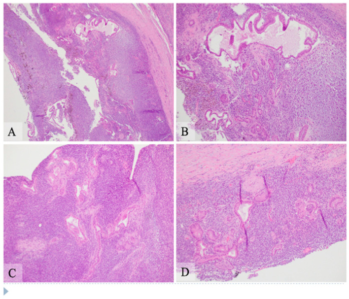
Carcinosarcoma of the pancreas is a rare entity with poor prognosis. Here, we report a case of pancreatic carcinosarcoma in a 68-year-old male patient who underwent a pancreatoduodenectomy for a unilocular cystic mass in the head of the pancreas. Histologically, the lesion showed a biphasic tumor with a carcinoma component and a spindle cell sarcomatous component, which were intimately intermingled. Most of the carcinoma components are well-differentiated ductal adenocarcinoma with small areas of moderately to poorly differentiated ductal adenocarcinoma. The sarcomatous component is a high-grade highly cellular spindle cell tumor with frequent mitosis and apoptosis. Immunohistochemical studies demonstrated that the carcinomatous component was positive for epithelial markers and cyclin D1, and the sarcomatous component was negative for these markers while positive for vimentin, p16, and DOG1 with patchy positivity for S100. Other markers, including SOX10, CD117, Melan A, HMB45, actin, desmin, myogenin, beta-catenin, TLE1, and p53, were negative in both components. Molecular studies demonstrated that the tumor was microsatellite stable. Whole exome next generation sequencing analysis was performed and no pathogenic alterations in the genes were identified.

Keywords: biphasic; carcinosarcoma; malignant neoplasm; pancreas; pancreatic ductal carcinoma.


Conflict of interest statement

The authors declare no conflict of interest.

Figures

Figure 1
Figure 1
Hematoxylin and eosin stain shows pancreatic carcinosarcoma with the well differentiated epithelial component and the spindle cell component. The left lower corner of panels (A,B), the left upper corner of panel (C) and the bottom portion of panel (D) are the cystic areas. (A) Low power view (original magnification 40×). Majority of the cyst wall is composed of sheets of spindle cells with scattered epithelial component with small or large duct structures. There are foci of hemosiderin deposition, mostly in the spindle cell areas, suggestive of previous hemorrhage. (B) Intermediate power view (original magnification 100×) of an upper area in panel A. The ducts are lined by a single layer of columnar cells with basally located round nuclei with minimal atypia and moderate amount of eosinophilic cytoplasm without mucinous contents. (C,D) Additional intermediate power view (original magnification 100×) of areas with well differentiated ductal adenocarcinoma closely intermixed with the spindle cell componentt. The epithelium of the ducts shows minimal atypia and moderate amount of eosinophilic cytoplasm. The spindle cells intimately surround the epithelial component with hyperchromatic and elongated nuclei and scant amount of cytoplasm.
Figure 2
Figure 2
Hematoxylin and eosin stain shows pancreatic carcinosarcoma with moderately differentiated ductal adenocarcinoma and the spindle cell component, intermediate power view (AD, original magnification 200×). (A) The epithelial component shows complex structure with convoluted glandular structures which has single layer of cells with moderate amount of eosinophilic cytoplasm and mildly enlarged nuclei. (B) The epithelial component shows multilayers of cells with cribriform-type interconnection. The nuclei of cells are moderately enlarged, hyperchromatic and irregular. (C) A duct (upper middle area) with simple to multilayered epithelial cells and adjacent scattered smaller and irregular ducts with hyperchromatic nuclei. The duct in the right lower corner has focal necrosis with inflammatory cells in the lumen. (D) Some areas of the tumor show small and angulated ducts embedded in a desmoplastic stromal background, simulating the typical morphology of a conventional pancreatic ductal carcinoma.
Figure 3
Figure 3
Hematoxylin and eosin stain shows pancreatic carcinosarcoma with poorly differentiated ductal adenocarcinoma and spindle cell component, intermediate and high power view ((A,B) original magnification 200×, (C,D) original magnification 400×). (A) Well to moderately differentiated ductal structures (left side) with transition to poorly formed ducts and to small sheets of epithelioid cells with no apparent structure. (B) Scattered small and poorly formed ductal structures are intermixed with infiltrative small sheets of single cells. (C) Small to poorly formed ducts (central area) are surrounded by small nests to single cells with vesicular nuclei, prominent nucleoli and small amount of eosinophilic to clear cytoplasm. (D) An atypical duct with surrounding spindle cells component. The ductal epithelium has hyperchromatic nuclei, some of which show conspicuous nucleoli. The spindle cell component is highly cellular with compact elongated cells with hyperchromatic nuclei and scant cytoplasm. Mitosis and apoptosis are present in the spindle cell area.
Figure 4
Figure 4
Immunohistochemical analysis of pancreatic carcinosarcoma (original magnification 200×. (A) Pancytokeratin AE1/AE3 highlights the epithelial component, but the spindle cell component is negative. (B) CK19 is positive in the ductal epithelial cells and is negative in the spindle cell component. (C) Vimentin is strongly and diffusely positive in the spindle cell component but is negative in the epithelial component. (D) S100 shows patchy positivity in the spindle cell component but not in the epithelial component. (E) SOX10 is negative in both the spindle cell and epithelial components. (F) DOG1 shows strong and diffuse positivity in the spindle cell component but not in the epithelial component. (G) p16 is positive in the spindle cell component and negative in the epithelial components. (H) Cyclin D1 shows patchy positivity in the epithelial component but is negative in the spindle cell component. (I) Ki-67 demonstrates high level of nuclear staining (about 20%) in the spindle cell component with low level staining (about 2%) in the epithelial component.

References

    1. Ruess D.A., Kayser C., Neubauer J., Fichtner-Feigl S., Hopt U.T., Wittel U.A. Carcinosarcoma of the Pancreas: Case Report with Comprehensive Literature Review. Pancreas. 2017;46:1225–1233. doi: 10.1097/MPA.0000000000000904. - DOI - PubMed
    1. Bosman F. World Health Organization Classification of Tumours of the Digestive System. 5th ed. IARC Press; Lyon, France: 2019. Ductal adenocarcinoma variants and mixed neoplasms of the pancreas; pp. 328–329.
    1. Millis J.M., Chang B., Zinner M.J., Barsky S.H. Malignant mixed tumor (carcinosarcoma) of the pancreas: A case report supporting organ-induced differentiation of malignancy. Surgery. 1994;115:132–137. - PubMed
    1. Alhatem A., Quinn P.L., Xia W., Chokshi R.J. Pancreatic Carcinosarcoma Clinical Outcome Analysis of the National Cancer Institute Database. J. Surg. Res. 2021;259:62–70. doi: 10.1016/j.jss.2020.11.033. - DOI - PubMed
    1. Cantrell L.A., Blank S.V., Duska L.R. Uterine carcinosarcoma: A review of the literature. Gynecol. Oncol. 2015;137:581–588. doi: 10.1016/j.ygyno.2015.03.041. - DOI - PubMed

Publication types