

Optimal Breast Density Characterization Using a Three-Dimensional Automated Breast Densitometry System
- PMID: 34940087
- PMCID: PMC8700257
- DOI: 10.3390/curroncol28060448
Optimal Breast Density Characterization Using a Three-Dimensional Automated Breast Densitometry System
Abstract
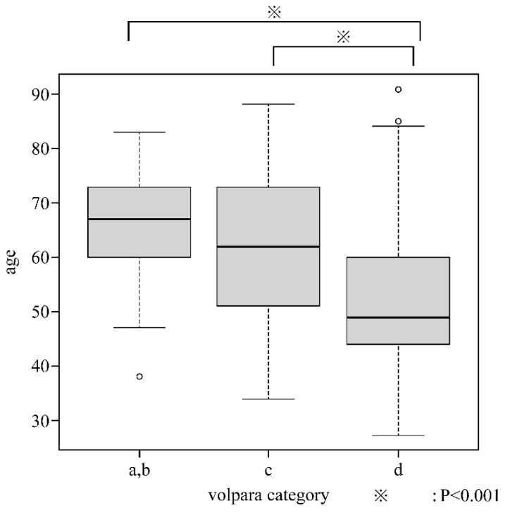
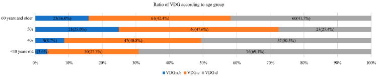
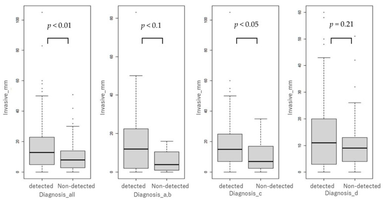
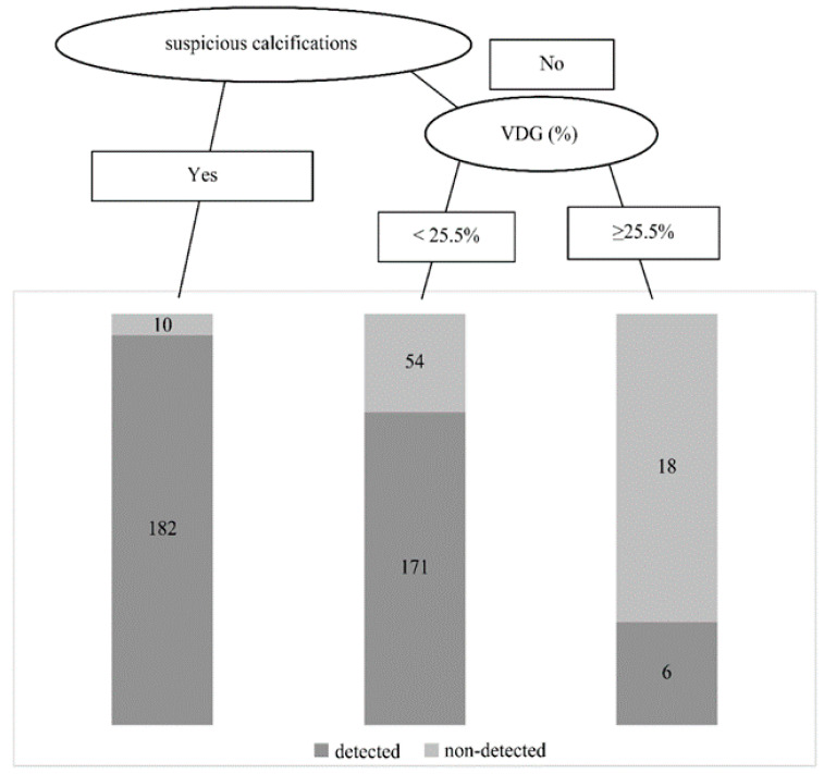
Dense breasts are a risk factor for breast cancer. Assessment of breast density is important and radiologist-dependent. We objectively measured mammographic density using the three-dimensional automatic mammographic density measurement device Volpara™ and examined the criteria for combined use of ultrasonography (US). Of 1227 patients who underwent primary breast cancer surgery between January 2019 and April 2021 at our hospital, 441 were included. A case series study was conducted based on patient age, diagnostic accuracy, effects of mammography (MMG) combined with US, size of invasion, and calcifications. The mean density of both breasts according to the Volpara Density Grade (VDG) was 0-3.4% in 2 patients, 3.5-7.4% in 55 patients, 7.5-15.4% in 173 patients, and ≥15.5% in 211 patients. Breast density tended to be higher in younger patients. Diagnostic accuracy of MMG tended to decrease with increasing breast density. US detection rates were not associated with VDG on MMG and were favorable at all densities. The risk of a non-detected result was high in patients without malignant suspicious calcifications. Supplementary use of US for patients without suspicious calcifications on MMG and high breast density, particularly ≥25.5%, could improve the breast cancer detection rate.
Keywords: Volpara; breast density; breast neoplasms; mammography; ultrasonography.
Conflict of interest statement
The authors declare no conflict of interest.
Figures
References
MeSH terms
LinkOut - more resources
Full Text Sources
Medical
Miscellaneous
