Comparative Study
. 2021 Dec 10;11(12):510.
doi: 10.3390/bios11120510.

Lateral Flow Immunoassay of SARS-CoV-2 Antigen with SERS-Based Registration: Development and Comparison with Traditional Immunoassays

Affiliations
Comparative Study

Lateral Flow Immunoassay of SARS-CoV-2 Antigen with SERS-Based Registration: Development and Comparison with Traditional Immunoassays

Kseniya V Serebrennikova et al. Biosensors (Basel). .

Abstract

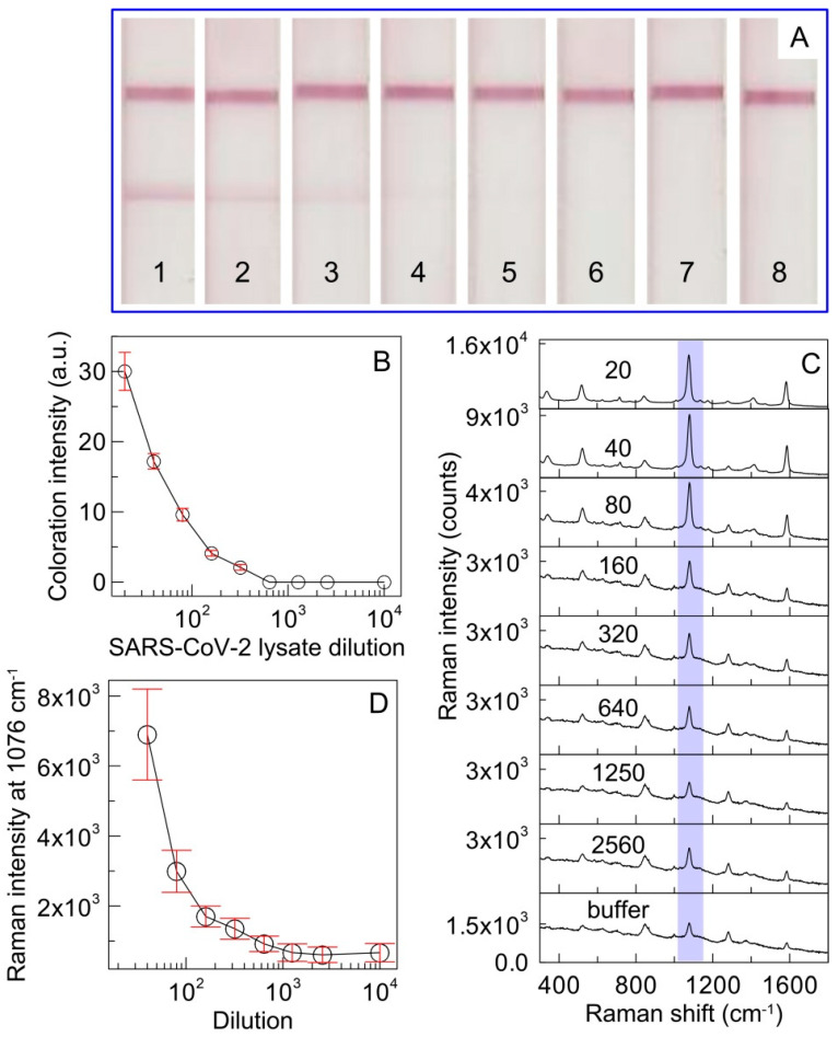
The current COVID-19 pandemic has increased the demand for pathogen detection methods that combine low detection limits with rapid results. Despite the significant progress in methods and devices for nucleic acid amplification, immunochemical methods are still preferred for mass testing without specialized laboratories and highly qualified personnel. The most widely used immunoassays are microplate enzyme-linked immunosorbent assay (ELISA) with photometric detection and lateral flow immunoassay (LFIA) with visual results assessment. However, the disadvantage of ELISA is its considerable duration, and that of LFIA is its low sensitivity. In this study, the modified LFIA of a specific antigen of the causative agent of COVID-19, spike receptor-binding domain, was developed and characterized. This modified LFIA includes the use of gold nanoparticles with immobilized antibodies and 4-mercaptobenzoic acid as surface-enhanced Raman scattering (SERS) nanotag and registration of the nanotag binding by SERS spectrometry. To enhance the sensitivity of LFIA-SERS analysis, we determined the optimal compositions of SERS nanotags and membranes used in LFIA. For benchmark comparison, ELISA and conventional colorimetric LFIA were used with the same immune reagents. The proposed method combines a low detection limit of 0.1 ng/mL (at 0.4 ng/mL for ELISA and 1 ng/mL for qualitative LFIA) with a short assay time equal to 20 min (at 3.5 h for ELISA and 15 min for LFIA). The results obtained demonstrate the promise of using the SERS effects in membrane immuno-analytical systems.

Keywords: Raman spectra; SARS-CoV-2; immunochromatography; surface antigen; test strips.


Conflict of interest statement

The authors declare no conflict of interest.

Figures

Figure 1
Figure 1
Schematic principle of SERS-based LFIA for RBD protein detection using SERS nanotag.
Figure 2
Figure 2
TEM image of bare AuNPs (A) and MBA-modified AuNP (B). (C) Absorbance spectra of bare AuNP (1) and AuMBA at 1 µM (2), 3 µM (3), 5 µM (4), and 10 µM (5) concentrations. The inset shows an enlarged portion of spectra.
Figure 3
Figure 3
(A) Dependence of the Raman intensity on the concentration of MBA in the AuMBA probe and SERS nanotag. (B) Optimization of analytical membrane. (C) Images of test strips after conventional procedure of LFIA using SERS nanotags with 1 µM (1), 3 µM (2), and 5 µM (3) of MBA. (D) Optimization of antibody concentration to prepare SERS nanotags; the positive control contains 10 or 30 ng/mL of RBD. PBS containing 1% v/v Tween 20 is used as a negative control.
Figure 4
Figure 4
(A) Photographic images of LFIA strips after application of RBD at concentrations 0 (1), 0.01 (2). 0.03 (3), 0.1 (4), 0.3 (5), 1 (6), 3 (7), 10 (8), 30 (9), and 100 ng/mL (10). The top and bottom lines correspond to the control and test zones, respectively. (B) Calibration curve obtained after conventional LFIA procedure using SERS nanotags. The error bars indicate the STD for three measurements; (C) SERS spectra measured in the test line for RBD concentrations from 0.01 to 100 ng/mL. (D) Calibration curve of SERS-LFIA for RBD. The bars show the STD of the Raman signal at 1076 cm−1, measured from three independent SERS-based LFIA runs.
Figure 5
Figure 5
(A) Photographic images of LFIA strips after application of the lysate at sequential two-fold dilutions of 20 (1), 40 (2), 80 (3), 160 (4), 320 (5), 640 (6), 1280 (7), and 2560 (8); (B) Calibration curve obtained after conventional LFIA procedure using SERS nanotags as immunoprobe for spike RBD detection in SARS-CoV-2 viral lysate. The error bars indicate the STD for three measurements; (C) SERS spectra measured in the TZ for different lysate dilutions. The numbers mean the dilution. (D) Dependence of the SERS intensity on the lysate dilution. The bars show the STD of the Raman signal at 1076 cm−1, measured at five points in the middle of the test line.
Figure 6
Figure 6
Calibration curves for spike RBD protein using ELISA (A) and standard AuNP-based LFIA (B). The error bars indicate the STD for three measurements.

References

    1. Gorbalenya A.E., Baker S.C., Baric R.S., de Groot R.J., Drosten C., Gulyaeva A.A., Haagmans B.L., Lauber C., Leontovich A.M., Neuman B.W., et al. The species severe acute respiratory syndrome-related coronavirus: Classifying 2019-nCoV and naming it SARS-CoV-2. Nat. Microbiol. 2020;5:536–544. doi: 10.1038/s41564-020-0695-z. - DOI - PMC - PubMed
    1. Synowiec A., Szczepański A., Barreto-Duran E., Lie L.K., Pyrc K. Severe acute respiratory syndrome coronavirus 2 (SARS-CoV-2): A systemic infection. Clin. Microbiol. Rev. 2021;34:e00133-20. doi: 10.1128/CMR.00133-20. - DOI - PMC - PubMed
    1. Mariano G., Farthing R.J., Lale-Farjat S.L.M., Bergeron J.R.C. Structural characterization of SARS-CoV-2: Where we are, and where we need to be. Front. Mol. Biosci. 2020;7:605236. doi: 10.3389/fmolb.2020.605236. - DOI - PMC - PubMed
    1. Chen W., Feng P., Liu K., Wu M., Lin H. Computational identification of small interfering RNA targets in SARS-CoV-2. Virol. Sin. 2020;35:359–361. doi: 10.1007/s12250-020-00221-6. - DOI - PMC - PubMed
    1. Wassie G.T., Azene A.G., Bantie G.M., Dessie G., Aragaw A.M. Incubation period of severe acute respiratory syndrome novel coronavirus 2 that causes coronavirus disease 2019: A systematic review and meta-analysis. Curr. Ther. Res. Clin. Exp. 2020;93:100607. doi: 10.1016/j.curtheres.2020.100607. - DOI - PMC - PubMed

Publication types