. 2021 Dec 3;19(12):693.
doi: 10.3390/md19120693.

Eisenia bicyclis Extract Repairs UVB-Induced Skin Photoaging In Vitro and In Vivo: Photoprotective Effects

Affiliations

Eisenia bicyclis Extract Repairs UVB-Induced Skin Photoaging In Vitro and In Vivo: Photoprotective Effects

Se-In Choi et al. Mar Drugs. .

Abstract

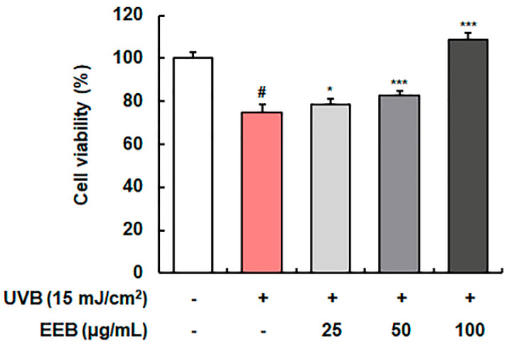
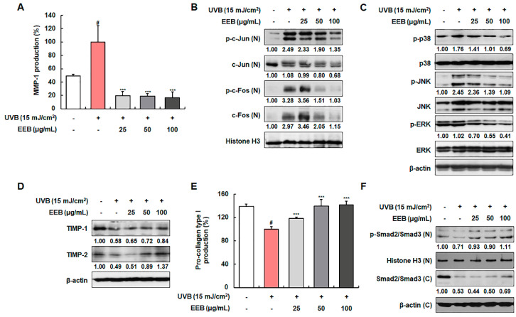
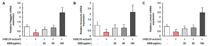
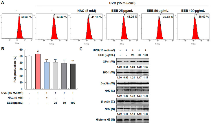
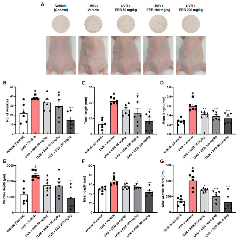
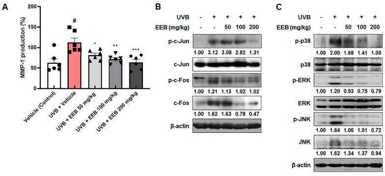
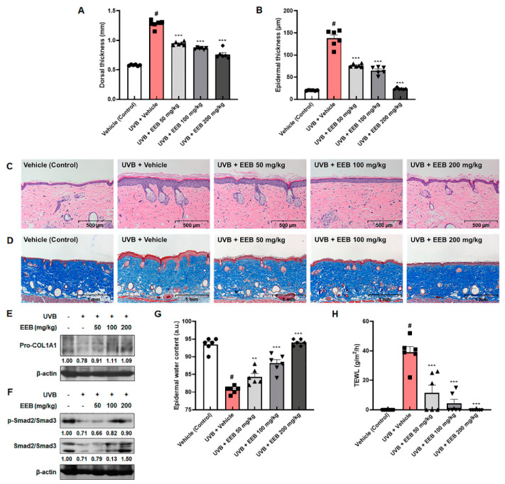
Chronic exposure to ultraviolet B (UVB) is a major cause of skin aging. The aim of the present study was to determine the photoprotective effect of a 30% ethanol extract of Eisenia bicyclis (Kjellman) Setchell (EEB) against UVB-induced skin aging. By treating human dermal fibroblasts (Hs68) with EEB after UVB irradiation, we found that EEB had a cytoprotective effect. EEB treatment significantly decreased UVB-induced matrix metalloproteinase-1 (MMP-1) production by suppressing the activation of mitogen-activated protein kinase (MAPK)/activator protein 1 (AP-1) signaling and enhancing the protein expression of tissue inhibitors of metalloproteinases (TIMPs). EEB was also found to recover the UVB-induced degradation of pro-collagen by upregulating Smad signaling. Moreover, EEB increased the mRNA expression of filaggrin, involucrin, and loricrin in UVB-irradiated human epidermal keratinocytes (HaCaT). EEB decreased UVB-induced reactive oxygen species (ROS) generation by upregulating glutathione peroxidase 1 (GPx1) and heme oxygenase-1 (HO-1) expression via nuclear factor erythroid-2-related factor 2 (Nrf2) activation in Hs68 cells. In a UVB-induced HR-1 hairless mouse model, the oral administration of EEB mitigated photoaging lesions including wrinkle formation, skin thickness, and skin dryness by downregulating MMP-1 production and upregulating the expression of pro-collagen type I alpha 1 chain (pro-COL1A1). Collectively, our findings revealed that EEB prevents UVB-induced skin damage by regulating MMP-1 and pro-collagen type I production through MAPK/AP-1 and Smad pathways.

Keywords: AP-1; Eisenia bicyclis; MAPK; MMP-1; Smad; UVB; collagen; photoaging.


Conflict of interest statement

The authors declare no conflict of interest.

Figures

Figure 1
Figure 1
UPLC chromatogram of EEB detected at 230 nm.
Figure 2
Figure 2
Effect of EEB on UVB-damaged cell protection in Hs68 cells. UVB-irradiated cells were treated with various concentrations of EEB (25, 50, and 100 µg/mL). Cell viability was analyzed using the 3-(4,5-dimethylthiazol-2-yl)-2,5-diphenyl tetrazolium bromide (MTT) assay. Values are represented as mean ± standard deviation (SD). # p < 0.05 vs. the non-UVB-irradiated control group; * p < 0.05 and *** p < 0.001 vs. the UVB-irradiated group.
Figure 3
Figure 3
Effects of EEB on the production of MMP-1 and pro-collagen type I, the activation of MAPK/AP-1 and Smad signaling pathways, and TIMPs expression in UVB-irradiated Hs68 fibroblasts. Cells were irradiated with UVB and treated with various concentrations of EEB (25, 50, and 100 µg/mL). (A, E) The MMP-1 and pro-collagen type I levels in the cell culture media were determined using ELISA kits. Western blot analysis was conducted to determine the protein expression in whole-cell lysates and cytosolic and nuclear fractions. Protein expression of (B) p-c-Jun, total c-Jun, p-c-Fos, total c-Fos, (C) p-p38, total p38, p-JNK, total JNK, p-ERK, total ERK, (D) TIMP-1, TIMP-2, (F) p-Smad2/Smad3, and total Smad2/Smad3. Protein levels of AP-1 pathway and p-Smad2/Smad3 normalized to Histone H3, phosphorylated protein levels of MAPK pathway normalized to total form, and protein levels of TIMPs and total Smad2/Smad3 normalized to β-actin were determined using Bio-Rad Quantity One software (Basic; Bio-Rad Laboratories Inc., Hercules, CA, USA). Values are represented as mean ± SD. # p < 0.05 vs. the non-UVB-irradiated control group; *** p < 0.001 vs. the UVB-irradiated group.
Figure 4
Figure 4
Effects of EEB on skin moisturization in UVB-induced HaCaT keratinocytes. Cells were irradiated with UVB and then treated with various concentrations of EEB (25, 50, and 100 µg/mL). Total cellular RNA was extracted from EEB-treated HaCaT cells. The mRNA levels of (A) filaggrin, (B) involucrin, and (C) loricrin were determined by quantitative real-time RT-PCR (qRT-PCR) and adjusted to GAPDH. Values are represented as mean ± SD. # p < 0.05 vs. the non-UVB-irradiated control group; *** p < 0.001 vs. the UVB-irradiated group.
Figure 5
Figure 5
Effects of EEB on ROS production and the antioxidant enzymes expression through Nrf2 signaling in UVB-induced Hs68 cells. Cells were irradiated with UVB and then treated with various concentrations of EEB (25, 50, and 100 µg/mL). (A, B) Productions of intracellular ROS were stained with H2DCFDA and analyzed by flow cytometry. (C) Whole-cell lysates and cytosolic and nuclear fractions were analyzed by Western blotting to determine the levels. Protein expression of GPx1, HO-1, and Nrf2. Protein levels of GPx1, HO-1, and cytosolic fraction Nrf2 normalized to β-actin and nucleus fraction Nrf2 normalized to Histone H3 were determined using Bio-Rad Quantity One software (Basic; Bio-Rad Laboratories Inc., Hercules, CA, USA). Values are represented as mean ± SD. # p < 0.05 vs. the non-UVB-irradiated control group; *** p < 0.001 vs. the UVB-irradiated group.
Figure 6
Figure 6
Effects of EEB on wrinkle formation in the dorsal skin of UVB-irradiated HR-1 hairless mice. HR-1 hairless mice were orally administrated with various doses of EEB (50, 100, and 200 mg/kg, p.o.) every 6 days and were irradiated with UVB three times per week for 8 weeks (60 mJ/cm2 to 120 mJ/cm2). (A) Images of the dorsal skin of mice and skin replicas. The replicas were analyzed by Visioline® VL-650. (B) Number of wrinkles, (C) total length (mm), (D) mean length (mm), (E) wrinkle depth (µm), (F) mean depth (µm), and (G) max wrinkle depth (µm) were analyzed. Each symbol in the figures represents an individual mouse of its own group. Values are represented as mean ± standard error of the mean (SEM; n = 6). # p < 0.05 vs. the vehicle-treated control group; * p < 0.05, ** p < 0.01, and *** p < 0.001 vs. the UVB only-treated group.
Figure 7
Figure 7
Effects of EEB on MMP-1 production and the MAPK/AP-1 signaling pathway in the dorsal skin of UVB-irradiated HR-1 hairless mice. HR-1 hairless mice were orally administrated various doses of EEB (50, 100, and 200 mg/kg, p.o.) every 6 days and irradiated with UVB three times per week for 8 weeks (60 mJ/cm2 to 120 mJ/cm2). (A) MMP-1 production levels in dorsal skin tissue-lysates were determined with ELISA kits. Each symbol in the figures represents an individual mouse of its own group. Dorsal skin tissue-lysates were analyzed by Western blotting to determine the protein levels. Protein expression of (B) p-c-Jun, total c-Jun, p-c-Fos, total c-Fos, (C) p-p38, total p38, p-ERK, total ERK, p-JNK, and total JNK. Protein levels of p-c-Jun, p-p38, and p-ERK normalized to total form and p-c-Fos, c-Fos, p-JNK, and JNK normalized to β-actin were determined using Bio-Rad Quantity One software (Basic; Bio-Rad Laboratories Inc., Hercules, CA, USA). Values are represented as mean ± SEM (n = 6). # p < 0.05 vs. the vehicle-treated control group; * p < 0.05, ** p < 0.01, and *** p < 0.001 vs. the UVB only-treated group.
Figure 8
Figure 8
Effects of EEB on skin thickening, collagen degradation, and skin hydration in the dorsal skin of UVB-irradiated HR-1 hairless mice. HR-1 hairless mice were orally administrated with various doses of EEB (50, 100, and 200 mg/kg, p.o.) every 6 days and irradiated with UVB three times per week for 8 weeks (60 mJ/cm2 to 120 mJ/cm2). (A) Dorsal skin thickness was measured using a caliper. (B) Histogram of epidermal thickness measured using the (C) H&E-stained images of dorsal skin tissues. Scale bar = 500 µm. (D) Masson’s trichrome-stained images of dorsal skin tissues. Scale bar = 1 mm. Dorsal skin tissue-lysates were analyzed by Western blotting to determine the protein levels. Protein expression of (E) Pro-COL1A1, (F) p-Smad2/Smad3, and total Smad2/Smad3. (G) The epidermal water content and (H) TEWL were measured using the GPSkin Barrier® in the dorsal skin of UVB-irradiated HR-1 hairless mice. Each symbol in the figures represents an individual mouse of its own group. Protein levels of pro-COL1A1 and Smad pathway normalized to β-actin were determined using Bio-Rad Quantity One software (Basic; Bio-Rad Laboratories Inc., Hercules, CA, USA). Values are represented as mean ± SEM (n = 6). # p < 0.05 vs. the vehicle-treated control group; ** p < 0.01 and *** p < 0.001 vs. the UVB only-treated group.

References

    1. Sanches Silveira J.E., Myaki Pedroso D.M. UV light and skin aging. Rev. Environ. Health. 2014;29:243–254. doi: 10.1515/reveh-2014-0058. - DOI - PubMed
    1. Kageyama H., Waditee-Sirisattha R. Antioxidative, Anti-Inflammatory, and Anti-Aging Properties of Mycosporine-Like Amino Acids: Molecular and Cellular Mechanisms in the Protection of Skin-Aging. Mar. Drugs. 2019;17:222. doi: 10.3390/md17040222. - DOI - PMC - PubMed
    1. Deng M., Xu Y., Yu Z., Wang X., Cai Y., Zheng H., Li W., Zhang W. Protective Effect of Fat Extract on UVB-Induced Photoaging In Vitro and In Vivo. Oxid. Med. Cell. Longev. 2019;2019:6146942. doi: 10.1155/2019/6146942. - DOI - PMC - PubMed
    1. Zhang J., Wang X., Vikash V., Ye Q., Wu D., Liu Y., Dong W. ROS and ROS-Mediated Cellular Signaling. Oxid. Med. Cell. Longev. 2016;2016:4350965. doi: 10.1155/2016/4350965. - DOI - PMC - PubMed
    1. Hwang E., Gao W., Xiao Y.K., Ngo H.T.T., Yi T.H. Helianthus annuus L. flower prevents UVB-induced photodamage in human dermal fibroblasts by regulating the MAPK/AP-1, NFAT, and Nrf2 signaling pathways. J. Cell. Biochem. 2019;120:601–612. doi: 10.1002/jcb.27417. - DOI - PubMed