. 2021 Dec 15;148(24):dev199866.
doi: 10.1242/dev.199866. Epub 2021 Dec 23.

A stop or go switch: glycogen synthase kinase 3β phosphorylation of the kinesin 1 motor domain at Ser314 halts motility without detaching from microtubules

Affiliations

A stop or go switch: glycogen synthase kinase 3β phosphorylation of the kinesin 1 motor domain at Ser314 halts motility without detaching from microtubules

Rupkatha Banerjee et al. Development. .

Abstract

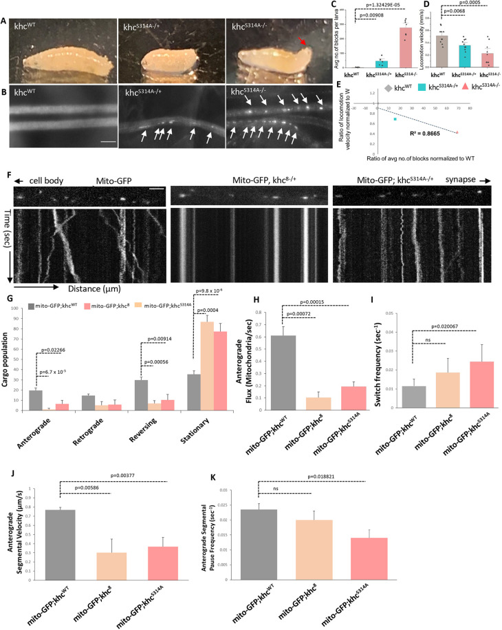
It is more than 25 years since the discovery that kinesin 1 is phosphorylated by several protein kinases. However, fundamental questions still remain as to how specific protein kinase(s) contribute to particular motor functions under physiological conditions. Because, within an whole organism, kinase cascades display considerable crosstalk and play multiple roles in cell homeostasis, deciphering which kinase(s) is/are involved in a particular process has been challenging. Previously, we found that GSK3β plays a role in motor function. Here, we report that a particular site on kinesin 1 motor domain (KHC), S314, is phosphorylated by GSK3β in vivo. The GSK3β-phosphomimetic-KHCS314D stalled kinesin 1 motility without dissociating from microtubules, indicating that constitutive GSK3β phosphorylation of the motor domain acts as a STOP. In contrast, uncoordinated mitochondrial motility was observed in CRISPR/Cas9-GSK3β non-phosphorylatable-KHCS314A Drosophila larval axons, owing to decreased kinesin 1 attachment to microtubules and/or membranes, and reduced ATPase activity. Together, we propose that GSK3β phosphorylation fine-tunes kinesin 1 movement in vivo via differential phosphorylation, unraveling the complex in vivo regulatory mechanisms that exist during axonal motility of cargos attached to multiple kinesin 1 and dynein motors.

Keywords: Drosophila; Axonal transport; CRISPR/Cas9; GSK3β; Kinesin 1; Phosphorylation; Processivity.


Conflict of interest statement

Competing interests The authors declare no competing or financial interests.

Figures

Fig. 1.
Fig. 1.
GSK3β phosphorylates KHC at Ser314. (A-C) Schematic of Drosophila KHC. Motor domain, green; putative conserved GSK3β phosphorylation sites, S209, 242 and 314, yellow. Secondary structures showing the location of these sites: blue, α-helices; pink, unstructured loops; magenta, β-sheets. (D) Schematic of GST-KHC fusion proteins. (E) KHCWT (KHC339/KHC410), KHCS209A, KHCS242A and KHCS242/S209A/S242A showed a γ-32P positive band, similar to khcWT, which were eliminated with CT99021. No bands were seen in KHCS314A, KHCS242A/S314A, KHCS242A/S314A and KHC S24209A/S242A/S314A.
Fig. 2.
Fig. 2.
KHCS314A does not influence microtubule binding but perturbs kinesin 1 motility and ATPase activity. KHCS314D stops motility (A) Schematic of the in vitro microtubule (MT)-binding assay. (B) MAPF is in the pellet (P), while BSA is in the soluble (S) fraction. (C) khcWT is found in the P. (D) Without MTs, KHCWT (KHC410) is mostly in the S. With MTs, KHCWT, KHCS209A, KHCS242A, KHCS314A and KHCS314D are in the P. (E) Representative images from movies (120 s). Trajectories show MT gliding. KHCS314A disrupts motility, while KHCS314D does not move. Scale bar: 5 µm. (F) MT gliding velocity for KHCS314A (P=0.0283, Student's t-test, P= 0.0465, Welch's t-test) and KHCS314D (P=0.0024, Student's t-test; P= 0.0090, Welch's t-test) is significantly decreased compared with KHCWT. (G) The average run lengths were significantly decreased in KHCS314A (P=0.0017, Student's t-test; P=0.000, Welch's t-test) and in KHCS314D (P=2.5×10⁻⁶, Student's t-test; P=2.8×10⁻⁵, Welch's t-test). (H) The total distance traveled by MTs was also significantly decreased in KHCS314A (P=2.3×10⁻⁵, Student's t-test; P=0.0005, Welch's t-test) and in KHCS314D (P=1.9×10⁻⁶, Student's t-test; P=0.0001-Welch's t-test). n=6 (two or three assays). Data are mean±s.e.m.. Only Student's t-test P-values are shown on graphs. (I) KHCS314A (P=0.0003) and KHCS314D (P=7×10⁻⁵) show significant decreases in % ATPase activity. (J) Table summarizing observations for GST-KHC mutants. Previous observations from Stewart et al. (1993) are also included for comparison. Upward arrowhead, increasing effects; downward arrowhead, decreasing effect; one upward and one downward, wild type. (K) Proposed model for how KHCS314A and KHCS314D disrupt kinesin 1 function in vitro without affecting MT binding. KHCS314A causes uncoordinated motility by decreasing ATP hydrolysis. KHCS314D stops motility by loss/reduction of ATP hydrolysis.
Fig. 3.
Fig. 3.
Drosophila CRISPR/Cas9 khcS314A larvae show posterior paralysis, axonal blockages and perturbed mitochondrial motility. (A) khcS314A−/− larvae tail-flip and show posterior paralysis (arrow). (B) khcS314A−/− and khcS314A−/+ larval segmental nerves show CSP-positive axonal blockages (arrows). Scale bar: 10 µm. (C) A significant amount of axonal blocks are seen in khcS314A−/+ (P=0.00908) and khcS314A−/− (P=1.32429E−05). n=8 larvae/genotype. Data are mean±s.e.m. (D) khcS314A−/+ (P=0.0068) and khcS314A−/− (P=0.0005) show significantly impaired crawling behaviors. n=15 larvae/genotype. (E) Ratio of the average number of axonal blocks (x-axis) plotted against the ratio of average larval velocity (y-axis) for khcS314A−/+ and khcS314A−/− show a strong correlation (R2=0.8665). n=8 larvae/genotype. (F) Wild type show robust bi-directional mitochondrial (Mito-GFP) motility. Mito-GFP;khc8−/+ and mito-GFP;khcS314A−/+ show disrupted mitochondrial motility. Note the stalled tracks in the kymographs. y-axis, time (s); x-axis, distance (µm). Scale bar: 5 µm. (G) A significant decrease is seen in the percent of anterogradely moving mitochondria in mito-GFP;khcS314A−/+ (P=0.02266) and mito-GFP;khc8−/+ larvae (P=6.7×10⁻⁵). (H) Mito-GFP;khcS314A−/+ (P=0.00015) and mito-GFP;khc8−/+ larvae (P=0.00072) show significant decreases in the average anterograde mitochondrial flux. (I) Mito-GFP;khcS314A−/+ (P=0.020067) show a significant increase in switch frequency. (J) Mito-GFP;khcS314A−/+ (P=0.003777) and mito-GFP;khc8−/+ (P=0.00586) show significant decreases in anterograde segmental velocities. (K) Mito-GFP;khcS314A−/+ (P=0.018821) show significant decreases in the anterograde segmental pause frequency. n=5 larvae, >120 mitochondria. Data are mean±s.e.m. Two-sample two-sided Student's t-test was used.
Fig. 4.
Fig. 4.
Endogenous fly khcS314A shows decreased MT and membrane binding with reduced ATPase activity. (A) Endogenous khcS314A isolated from heterozygous khcS314A−/+ is in P (pellet) and S (soluble) fractions. High molecular weight (HMW) khcS314A is in the P. (B) Schematic diagram of the membrane floatation assay. Membrane/vesicle fraction (35/8) is the layer between 35% and 8% sucrose. Decreased kinesin 1, dynein and active GSK3β are bound to membranes in khcS314A. The ratios of the intensity of proteins in PNS (post-nuclear supernatant) and membranes (the 35/8 fraction) show significant decreases in active GSK3β (P=0.0068), KHC (P=0.0090), KLC (P=0.0124) and DIC (P=0.0001) levels in khcS314A−/+ membranes. y-axis, intensity of proteins normalized to Rab5 in arbitrary units (AU). Data are mean±s.e.m. Two-sample two-sided Student's t-test with *P<0.05 and **P<0.005. (C) Active GSK3β co-immunoprecipitates with khcWT, but not with khcS314A (box). PD, pull down; PD (no 1°Ab), no antibody control; FT, flow through. PNS and 1% input are also shown. (D) khcS314A−/+ shows significant decreases in %ATPase activity (P=0.0024). (E) Table summarizing observations from khcS314A, sggM11, sggACTIVE and khc17. (F) Proposed model for how khcS314A affects kinesin 1 function in vivo in an organism.

References

    1. Amanchy, R., Periaswamy, B., Mathivanan, S., Reddy, R., Tattikota, S. G. and Pandey, A. (2007). A curated compendium of phosphorylation motifs. Nat. Biotechnol. 25, 285-286. 10.1038/nbt0307-285 - DOI - PubMed
    1. Banerjee, R., Rudloff, Z., Naylor, C., Yu, M. C. and Gunawardena, S. (2018). The presenilin loop region is essential for glycogen synthase kinase 3 beta (GSK3beta) mediated functions on motor proteins during axonal transport. Hum. Mol. Genet. 27, 2986-3001. 10.1093/hmg/ddy190 - DOI - PMC - PubMed
    1. Bloom, G. S., Wagner, M. C., Pfister, K. K. and Brady, S. T. (1988). Native structure and physical properties of bovine brain kinesin and identification of the ATP-binding subunit polypeptide. Biochemistry 27, 3409-3416. 10.1021/bi00409a043 - DOI - PubMed
    1. Brendza, K. M., Rose, D. J., Gilbert, S. P. and Saxton, W. M. (1999). Lethal kinesin mutations reveal amino acids important for ATPase activation and structural coupling. J. Biol. Chem. 274, 31506-31514. 10.1074/jbc.274.44.31506 - DOI - PMC - PubMed
    1. Cahu, J., Olichon, A., Hentrich, C., Schek, H., Drinjakovic, J., Zhang, C., Doherty-Kirby, A., Lajoie, G. and Surrey, T. (2008). Phosphorylation by Cdk1 increases the binding of Eg5 to microtubules in vitro and in Xenopus egg extract spindles. PLoS ONE 3, e3936. 10.1371/journal.pone.0003936 - DOI - PMC - PubMed

Publication types