

Differentiation of Cerebral Neoplasms with Vessel Size Imaging (VSI)
- PMID: 34940899
- PMCID: PMC8894153
- DOI: 10.1007/s00062-021-01129-8
Differentiation of Cerebral Neoplasms with Vessel Size Imaging (VSI)
Abstract
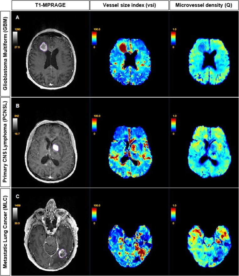
Purpose: Cerebral neoplasms of various histological origins may show comparable appearances on conventional Magnetic Resonance Imaging (MRI). Vessel size imaging (VSI) is an MRI technique that enables noninvasive assessment of microvasculature by providing quantitative estimates of microvessel size and density. In this study, we evaluated the potential of VSI to differentiate between brain tumor types based on their microvascular morphology.
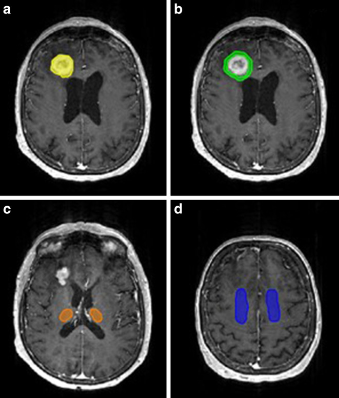
Methods: Using a clinical 3T MRI scanner, VSI was performed on 25 patients with cerebral neoplasms, 10 with glioblastoma multiforme (GBM), 8 with primary CNS lymphoma (PCNSL) and 7 with cerebral lung cancer metastasis (MLC). Following the postprocessing of VSI maps, mean vessel diameter (vessel size index, vsi) and microvessel density (Q) were compared across tumors, peritumoral areas, and healthy tissues.
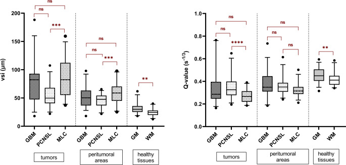
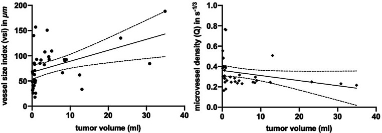
Results: The MLC tumors have larger and less dense microvasculature compared to PCNSLs in terms of vsi and Q (p = 0.0004 and p < 0.0001, respectively). GBM tumors have higher yet non-significantly different vsi values than PCNSLs (p = 0.065) and non-significant differences in Q. No statistically significant differences in vsi or Q were present between GBMs and MLCs. GBM tumor volume was positively correlated with vsi (r = 0.502, p = 0.0017) and negatively correlated with Q (r = -0.531, p = 0.0007).
Conclusion: Conventional MRI parameters are helpful in differentiating between PCNSLs, GBMs, and MLCs. Additionally incorporating VSI parameters into the diagnostic protocol could help in further differentiating between PCNSLs and metastases and potentially between PCNSLs and GBMs. Future studies in larger patient cohorts are required to establish diagnostic cut-off values for VSI.
Keywords: Brain imaging; Brain tumors; Differential diagnosis; Magnetic resonance imaging; Microvasculature.
© 2021. The Author(s).
Conflict of interest statement
Financial interests: J.B. Fiebach reports consulting and advisory board fees from Abbvie, AC Immune, Artemida, BioClinica, Biogen, BMS, Brainomix, Cerevast, Daiichi-Sankyo, EISAI, F. Hoffmann-La Roche AG, Eli Lilly, Guerbet, Ionis Pharmaceuticals, IQVIA, Janssen, Julius Clinical, jung diagnostics, Lysogene, Merck, Nicolab, Premier Research, and Tau Rx, outside the submitted work. E. Kellner is a shareholder of and receives fees from VEObrain GmbH, Freiburg, Germany. Non-financial interests: none. A. Foda, A. Gunawardana, X. Gao, M. Janz, A. Kufner, A.A. Khalil, R. Geran, R. Mekle, and I. Galinovic declare that they have no competing interests.
Figures
References
MeSH terms
LinkOut - more resources
Full Text Sources
Medical
