. 2022 Apr;9(2):481-495.
doi: 10.1007/s40744-021-00413-w. Epub 2021 Dec 23.

Ultrasound Features of Adhesive Capsulitis

Affiliations

Ultrasound Features of Adhesive Capsulitis

Salvatore Massimo Stella et al. Rheumatol Ther. 2022 Apr.

Abstract

Introduction: Adhesive capsulitis (AC), which is characterised by shoulder pain and a limited range of motion (ROM), is usually diagnosed on the basis of clinical suspicion, with imaging only being used to exclude other causes of similar symptoms. The aim of this study was to identify and describe the typical ultrasound (US) features of AC in a group of patients with shoulder pain and stiffness.

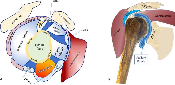
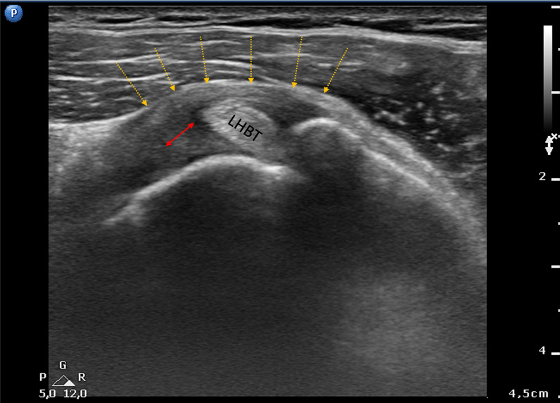
Methods: This was a cross-sectional study of 1486 patients with AC in which two experienced US specialists examined the axillary pouch (AP), the coracohumeral ligament (CHL), the superior glenohumeral ligament (SGHL), and the long head of the biceps tendon (LHBT), and dynamically visualised the infraspinatus tendon during passive external rotation (PER) during a US evaluation of shoulder ROM.

Results: AC was confirmed in 106 patients (7.1%). Thickening of the AP of more than 4 mm was observed in 93.4% of the patients, whereas 6.6% showed AP thickening of less than 4 mm but more than 60% of the thickening in the contralateral shoulder. Effusion within the LHBT sheath was detected in 71% of the patients, and thickening of the CHL or SGHL in 88%. The dynamic study of the infraspinatus tendon showed reduced sliding with folding towards the joint capsule in 73% of cases, thus changing the tendon's profile from flat to concave during PER. The reduced tendon sliding was associated with a bouncing movement that returned the tendon to its baseline resting position in 41.5% of cases.

Conclusions: We believe a sufficiently experienced US specialist can confirm a clinical diagnosis of AC by carrying out a comparative study of APs, evaluating the thickness of the CHL and SGHL, and detecting reduced sliding of the infraspinatus tendon.

Keywords: Adhesive capsulitis; Axillary pouch; Coracohumeral ligament; Frozen shoulder; Ultrasound.


Figures

Fig. 1
Fig. 1
Normal axillary pouch in oblique axial (a) and coronal scans (b). HH humeral head, P axillary pouch, IGHL inferior glenohumeral ligament, SN surgical neck
Fig. 2
Fig. 2
a Schematic drawing of the sagittal section of the axillary pouch (AP) and glenohumeral ligaments. b Coronal view of the axillary pouch in slight abduction and neutral rotation. A-C acromioclavicular, IGHL inferior glenohumeral ligament, SAB subacromial bursa, SGHL superior glenohumeral ligament
Fig. 3
Fig. 3
a Oblique axial section: red arrows show the thickness of the axillary pouch in a patient with AC (AP thickening between the calipers). b Coronal section: red arrows show the thickness of the AP in another patient with AC. The dashed blue arrows indicate a small amount of joint effusion entrapped in the AP. HH humeral head, SN surgical neck
Fig. 4
Fig. 4
Axillary pouch thickening was less than 4 mm (yellow arrows) in a small percentage of patients, but there was a significant, greater than 60% difference from the thickness of the contralateral  axillary pouch
Fig. 5
Fig. 5
Axial section of the normally concave rotator interval showing conspicuous coracohumeral ligament thickening (red double arrow) with a rounded and convex profile (yellow dotted arrows). LHBT long head of the biceps tendon
Fig. 6
Fig. 6
Axial section of the rotator interval of a right shoulder in a patient with AC. Coracohumeral ligament (CHL) thickness with a “pseudo-double” tendon appearance due to the smaller false tendon, which is the CHL lateral to the LHBT. CHL coracohumeral ligament, GT greater tuberosity, LHBT long head of the biceps tendon, LT lesser tubercle
Fig. 7
Fig. 7
Dynamic study of the infraspinatus tendon (white arrows) sliding backwards during passive external rotation governed by the US specialist (T1–T2). Note the characteristic change in the tendon from a flat to a concave profile (white arrowhead). The sliding of the tendon folds it towards the joint capsule because they are closely contiguous. HH humeral head
Fig. 8
Fig. 8
The folding may be minimal or more pronounced and was found only on the affected side and never on the contralateral side

References

    1. Duplay ES. De la peri-arthrite scapulo humerale et des raideurs de l’epaule qui en son la consequence. Arch Gen Med. 1872;20:31–42.
    1. Codman E. The shoulder: rupture of the supraspinatus tendon and other lesions in or about the subcromial bursa. Boston: Todd; 1934.
    1. Neviaser JS. Adhesive capsulitis of the shoulder: a study of the pathological findings in periarthritis of the shoulder. J Am Acad Orthop Surg. 1945;27:211–222.
    1. Fields BK, Skalski MR, Patel DB, et al. Adhesive capsulitis: review of imaging findings, pathophysiology, clinical presentation, and treatment options. Skeletal Radiol. 2019;48:1171–1184. doi: 10.1007/s00256-018-3139-6. - DOI - PubMed
    1. Binder A, Bulgen D, Hazleman B, Roberts S. Frozen shoulder: a long-term prospective study. Ann Rheum Dis. 1984;43:361–364. doi: 10.1136/ard.43.3.361. - DOI - PMC - PubMed