. 2022 Apr;31(4):797-810.
doi: 10.1002/pro.4267. Epub 2021 Dec 31.

A novel and unique ATP hydrolysis to AMP by a human Hsp70 Binding immunoglobin protein (BiP)

Affiliations

A novel and unique ATP hydrolysis to AMP by a human Hsp70 Binding immunoglobin protein (BiP)

Hongtao Li et al. Protein Sci. 2022 Apr.

Abstract

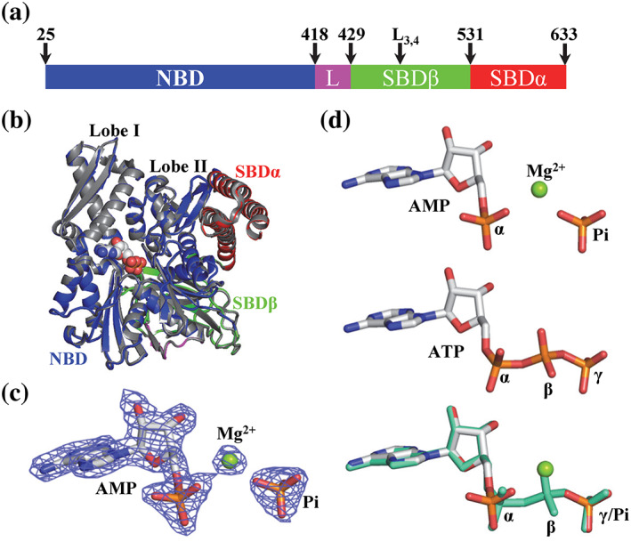
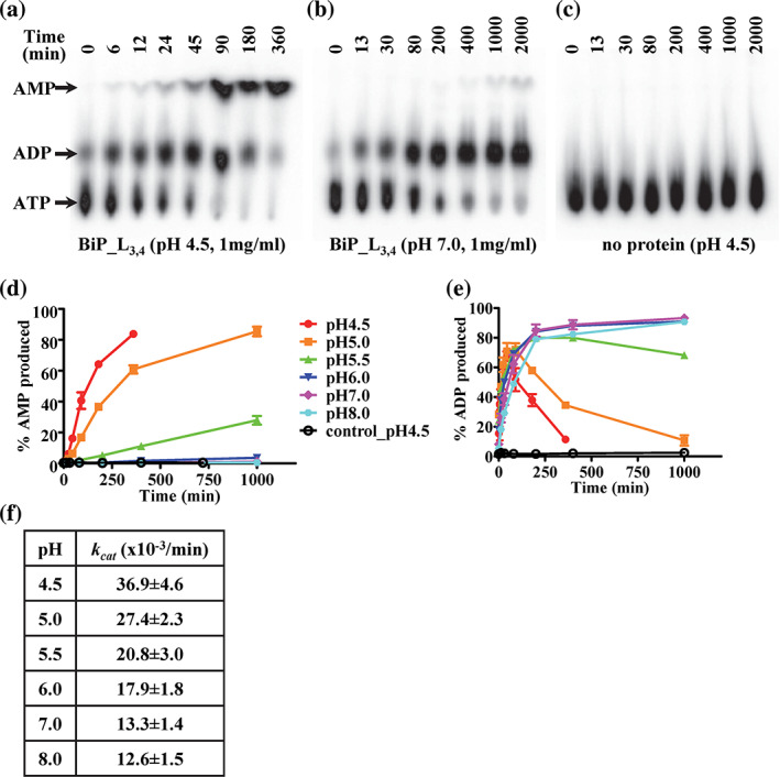
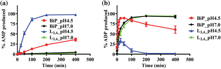
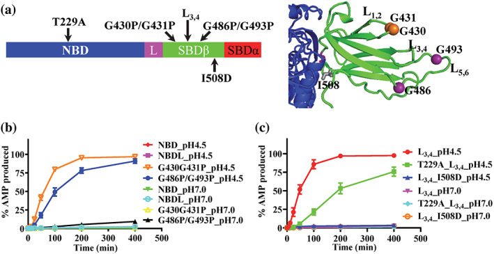
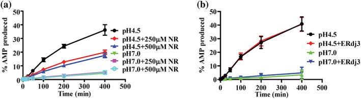
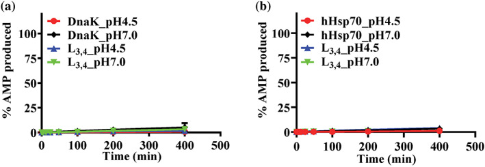
Hsp70s are ubiquitous and highly conserved molecular chaperones. They play crucial roles in maintaining cellular protein homeostasis. It is well established that Hsp70s use the energy of ATP hydrolysis to ADP to power the chaperone activity regardless of the cellular locations and isoforms. Binding immunoglobin protein (BiP), the major member of Hsp70s in the endoplasmic reticulum, is essential for protein folding and quality control. Unexpectedly, our structural analysis of BiP demonstrated a novel ATP hydrolysis to AMP during crystallization under the acidic conditions. Our biochemical studies confirmed this newly discovered ATP to AMP hydrolysis in solutions. Unlike the canonical ATP to ADP hydrolysis observed for Hsp70s, this ATP hydrolysis to AMP depends on the substrate-binding domain of BiP and is inhibited by the binding of a peptide substrate. Intriguingly, this ATP to AMP hydrolysis is unique to BiP, not shared by two representative Hsp70 proteins from the cytosol. Taken together, this novel and unique ATP to AMP hydrolysis may provide a potentially new direction for understanding the activity and cellular function of BiP.

Keywords: ADP; AMP; ATP; ATPase; BiP; Hsp70; heat shock proteins (HSPs); molecular chaperones; protein folding.


Conflict of interest statement

The authors declare no potential conflicts of interest.

Figures

FIGURE 1
FIGURE 1
AMP is bound in the nucleotide‐binding pocket of a new Binding immunoglobin protein (BiP) crystal structure, BiP‐AMP. (a) Domain organization of BiP. The residue numbers at the domain boundaries and the L3,4 mutation are labeled on the top. L: the inter‐domain linker. (b) The overall structure of the new BiP‐AMP structure is almost identical to the previously published BiP‐ATP2 structure (PDB: 6ASY). The domain coloring of the BiP‐AMP structure is the same as in (a). The BiP‐ATP2 structure is in gray. The AMP and Pi in the BiP‐AMP structure were shown as spheres. The lobes I and II of the NBD are labeled. (c) The electron density for the bound AMP and Pi in the BiP‐AMP structure is well‐resolved. A 2Fo‐Fc map contoured at 2.0 sigma is shown as blue mesh. The AMP and Pi are shown as sticks and the bound Mg ion as a green ball. (d) Comparison of the AMP‐Pi in the BiP‐AMP structure with the ATP in the BiP‐ATP2 structure. Top: AMP‐Pi; Middle: ATP; bottom: superposition of AMP‐Pi (same color as the top panel) with ATP (green). The AMP‐Pi and ATP are shown as sticks and the bound Mg ion for AMP is highlighted as a green ball. The three phosphates in ATP are labeled as α, β, and γ. The α phosphate and free Pi are labeled as α and Pi, respectively
FIGURE 2
FIGURE 2
The Binding immunoglobin protein (BiP)‐L3,4 protein is able to hydrolyze ATP to AMP in solutions. (a–c) ATP is efficiently hydrolyzed to AMP by the BiP_L3,4 protein at pH 4.5 (a), but not pH 7.0 (b). Thin‐layer chromatography (TLC) plates were used to separate ATP, ADP, and AMP. The position of each nucleotide is indicated at the left. The time points for each reaction were labeled on the top of the TLC plates. (c) The no protein control for pH 4.5. (d,e) The ATP hydrolysis to AMP and ADP by the BiP_L3,4 protein (1 mg/ml) at a series of pH conditions. (d) AMP hydrolysis; (e) ADP hydrolysis. The pH values are labeled on the right in (d). control_pH 4.5: buffer only was used as a control for pH 4.5. The percentages of hydrolysis were plotted as a function of reaction time (mean ± SEM from three independent experiments with more than two different protein purifications). (f) The catalytic constants (k cat ) of the overall ATP hydrolysis by the BiP_L3,4 protein at 1 mg/ml. The k cat values were calculated by fitting data from (d) and (e) with a first‐order rate equation by nonlinear regression analysis. The summation of ADP and AMP produced was used for calculating the amount of overall ATP hydrolysis
FIGURE 3
FIGURE 3
The wild‐type (WT) Binding immunoglobin protein (BiP) protein hydrolyzes ATP to AMP at a lower rate than that of the BiP_L3,4 protein. (a,b) The ATP hydrolysis to AMP and ADP, respectively. BiP: the WT BiP protein. L3,4: the BiP_L3,4 protein. pH 4.5 and 7.0 were tested. The data were presented as the percentages of AMP produced (mean ± SEM from more than three independent experiments using at least two different protein purifications)
FIGURE 4
FIGURE 4
The nucleotide‐binding domain–substrate‐binding domain (NBD–SBD) contacts are required for the ATP to AMP hydrolysis by Binding immunoglobin protein (BiP). (a) The locations of the BiP mutations. Left, the BiP mutations were labeled in the domain organization of BiP. The domain coloring and labeling were the same as Figure 1a. Right, ribbon diagram of the BiP‐AMP structure. Only the SBDβ (green) and the part of the NBD (blue) forming contacts with SBDβ were shown. (b) The ATP hydrolysis to AMP was not observed without SBD but enhanced by the G430P/G431P and G486P/G493P mutations in the peptide‐binding site. (c) The T229A and I508D mutations compromised the ATP to AMP hydrolysis by BiP. For both (b) and (c), the AMP production was plotted over time at pH 4.5 and pH 7.0. Mean ± SEM from three independent experiments with more than two different protein purifications were used
FIGURE 5
FIGURE 5
The influences of peptide substrate and ERdj3 on the ATP to AMP hydrolysis of Binding immunoglobin protein (BiP). (a) The NR peptide inhibits the ATP to AMP hydrolysis by BiP. The ATP hydrolysis to AMP by the wild‐type (WT) BiP protein was analyzed at pH 4.5 and pH 7.0 in the absence or presence of two different concentrations of the NR peptide (250 and 500 μM). Each data point was mean ± SEM from three independent experiments using more than two different protein purifications. (b) The Hsp40 cochaperone ERdj3 showed little influence on the ATP to AMP hydrolysis of BiP. The hydrolysis to AMP by the WT BiP protein was assayed in the presence of ERdj3 at pH 4.5 and 7.0. The data were presented as mean ± SD from three independent experiments using at least two different protein purifications)
FIGURE 6
FIGURE 6
The ATP to AMP hydrolysis is not conserved in either DnaK or hHsp70. (a,b) Little hydrolysis to AMP was observed for DnaK (a) and hHsp70 (b). Both the wild‐type (WT) and corresponding L3,4 mutant proteins were tested. In (a), DnaK: the WT DnaK protein; L3,4: the DnaK protein carrying an analogous L3,4 mutation. In (b), hHsp70: the WT hHsp70 protein; L3,4: the hHsp70 protein carrying an analogous L3,4 mutation. The data were presented as the percentage of AMP produced (mean ± SEM from more than three independent experiments using at least two different protein purifications)

References

    1. Mayer MP, Bukau B. Hsp70 chaperones: Cellular functions and molecular mechanism. Cell Mol Life Sci. 2005;62:670–684. - PMC - PubMed
    1. Brodsky JL, Chiosis G. Hsp70 molecular chaperones: Emerging roles in human disease and identification of small molecule modulators. Curr Top Med Chem. 2006;6:1215–1225. - PubMed
    1. Young JC. Mechanisms of the Hsp70 chaperone system. Biochem Cell Biol. 2010;88:291–300. - PMC - PubMed
    1. Rosenzweig R, Nillegoda NB, Mayer MP, Bukau B. The Hsp70 chaperone network. Nat Rev Mol Cell Biol. 2019;20:665–680. - PubMed
    1. Liu Q, Liang C, Zhou L. Structural and functional analysis of the Hsp70/Hsp40 chaperone system. Protein Sci. 2020;29:378–390. - PMC - PubMed

Publication types