. 2021 Dec 24;374(6575):eabl5450.
doi: 10.1126/science.abl5450. Epub 2021 Dec 24.

Fibrin is a critical regulator of neutrophil effector function at the oral mucosal barrier

Affiliations

Fibrin is a critical regulator of neutrophil effector function at the oral mucosal barrier

Lakmali M Silva et al. Science. .

Abstract

Tissue-specific cues are critical for homeostasis at mucosal barriers. Here, we report that the clotting factor fibrin is a critical regulator of neutrophil function at the oral mucosal barrier. We demonstrate that commensal microbiota trigger extravascular fibrin deposition in the oral mucosa. Fibrin engages neutrophils through the αMβ2 integrin receptor and activates effector functions, including the production of reactive oxygen species and neutrophil extracellular trap formation. These immune-protective neutrophil functions become tissue damaging in the context of impaired plasmin-mediated fibrinolysis in mice and humans. Concordantly, genetic polymorphisms in PLG, encoding plasminogen, are associated with common forms of periodontal disease. Thus, fibrin is a critical regulator of neutrophil effector function, and fibrin-neutrophil engagement may be a pathogenic instigator for a prevalent mucosal disease.


Figures

Fig. 1.
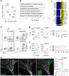
Fig. 1.. Defective fibrinolysis triggers oral mucosal immunopathology.
(A) Plg-deficient mice (Plg−/−) display periodontal bone loss at the age of 12 and 24 weeks compared with Plg+/+ and Plg+/− littermates. (B) μCT visualization of periodontal bone loss in 24-weeks-old Plg+/+ and Plg−/− maxillae (black arrows depict the distance between alveolar bone crest and cementoenamel junction). (C) Mice with catalytically inactive plasmin (PlgS743A/S743A) develop significantly increased spontaneous periodontal bone loss. (D) Fraser–Lendrum staining of Plg+/+ and Plg−/− oral mucosal tissue sections (Green: collagen; magenta: keratin, fibrin; and orange: erythrocytes; black arrow depicts fibrin deposition). Scale bar: 100 μm and inset scale bar: 50 μm. (E) Immunofluorescence (IF) staining for fibrin(ogen) (gray) in Plg+/−;Fga+/−, Plg−/−;Fga+/−, Plg+/−;Fga−/− and Plg−/−;Fga−/− oral tissue sections. Scale bar: 50 μm. (F) Bone loss measurements in Plg+/−;Fga+/−, Plg−/−;Fga+/−, Plg+/−;Fga−/− and Plg−/−;Fga−/− mice maxillae. (G) Difference (Δ) in bone loss between Plg−/− and Plg+/+ mice under SPF and GF conditions. (H) Fraser–Lendrum staining of Plg+/+ and Plg−/− GF mice oral mucosal tissue sections. Scale bar: 100 μm and inset scale bar: 50; and (I) Measurements of mucosal edema and attachment loss in Plg+/+ and Plg−/− under SPF and GF conditions.
Fig. 2.
Fig. 2.. Lesions of mucosal immunopathology in Plg deficiency are dominated by neutrophils.
(A) Principal coordinates analysis (PCoA) plot showing the distribution of differentially expressed genes in oral mucosa of 12-week-old Plg+/+ and Plg−/− mice. (B) Gene ontology (GO) biological processes upregulated in Plg−/− mice in ascending order of P-value. (C) Heat maps showing significantly differentially expressed genes involved in granulopoiesis/neutrophil recruitment, myeloid cell activation and inflammation/extracellular matrix disassembly in Plg−/− compared with Plg+/+ gingival tissues. (D) Flow cytometry analysis of 12- and 24-week-old mouse oral mucosal tissues. Contour plots show changes in neutrophil and monocyte/macrophage populations in percentages. Absolute count and percentages of (E) neutrophils and (F) monocytes/macrophages in 12- and 24-week-old Plg+/+ and Plg−/− mouse gingiva. (G) Fibrin(ogen) (green) and Ly6G (neutrophils; magenta) IF staining of 24-week-old Plg−/− mouse oral mucosal sections showing extravascular localization of fibrin deposits and neutrophils. Yellow arrowheads indicate blood vessels. Scale bar: 50 μm and quantification of number of neutrophils per 105 μm2 of stained tissue sections.
Fig. 3.
Fig. 3.. Myeloid integrin αMβ2-binding motif on fibrin(ogen) mediates plasminogen deficiency-induced oral immunopathology.
(A) Bone loss measurement in Plg+/−;Fgg+/390-396A, Plg−/−;Fgg+/390-396A, Plg+/−;Fgg390-396A/390-396A, Plg−/−;Fgg390-396A/390-396A mice maxillae. (B) μCT visualization of periodontal bone loss of maxilla (black arrows depict the distance between alveolar bone crest and cemento–enamel junction). (C) IF staining of fibrin(ogen) (green) and Ly6G (neutrophils; magenta) in Plg+/+;Fga+/+, Plg−/−;Fga+/+, Plg−/−;Fgg390-396A/390-396A and Plg−/−;Fga−/− mouse gingival sections. Scale bar: 50 μm. Graphs below show mean fluorescence intensity (MFI) of fibrin(ogen)/DAPI staining, and number of neutrophils per 105 μm2 of stained tissue sections. (D) Flow cytometry analysis of 24-week-old gingiva. Contour plots show neutrophil and monocyte/macrophage populations in percentages. (E) Counts of neutrophils in 24-week-old Plg+/−;Fgg+/390-396A, Plg−/−;Fgg+/390-396A, Plg−/−;Fgg390-396A/390-396A and Plg+/−;Fgg390-396A/390-396A mouse gingival sections. (F) Bone loss measurement in wild-type 10- and 24-week-old maxillae. (G) Bone loss measurement in Fgg+/+, Fgg+/390-396A and Fgg390-396A/390-396A mice maxillae at 24 weeks of age.
Fig. 4.
Fig. 4.. Fibrin(ogen)-neutrophil interaction through αMβ2-binding triggers key neutrophil effector functions.
(A) Human neutrophil binding to Fgg390-396A/390-396A fibrin compared to wild-type fibrin. Scale bar: 200 μm. (B) Binding of wild-type and αM integrin-deficient (αM−/−) mouse neutrophils to wild-type fibrin. Scale bar: 200 μm. (C to H) Neutrophils were plated on wild-type or Fgg390-396A fibrin. (C) Representative graph of neutrophil reactive oxygen species (ROS) production over time (15 nM MPO). (D) ROS production at 5-hour time point (n=15). (E) Neutrophil extracellular traps (NETs; magenta) at 4- and 5-hour time points for neutrophils plated on wild-type and Fgg390-396A fibrin. Scale bars: 50 μm. (F) Cit-H3 and MPO staining (gray) of NETs at 5-hour time point. Scale bar: 50 μm. (G) NETosis progression over time (1 to 5 hours) (see Movie 1). (H) Quantification of NETosing cells at 5-hour time point (n=7).
Fig. 5.
Fig. 5.. NETosis mediates periodontal immunopathology in Plg-deficient mice.
(A) Immunofluorescence staining of Cit-H3 and MPO (yellow) in Plg+/+, Plg−/− and Plg−/−;Fgg390-396A/390-396A mouse oral mucosal tissue sections. Scale bars: 50 μm. Quantification of percentage of Cit-H3 and MPO stained area, respectively. (B and C) Plg−/−-control (Ctrl) and Plg−/−-DNase I-treated mice. (B) Bone loss and (C) staining for Cit-H3 and MPO (yellow) in gingiva. Scale bars: 50 μm (D) Plg+/−;Ela+/− and Plg−/−;Ela+/−, Plg−/−;Ela−/− and Plg+/−;Ela−/− mice bone loss measurements. (E and F) siRNA treatment. (E) Plasma fibrinogen levels. (F) Bone-loss measurements of Plg−/−-siLuc- and Plg−/−-siFbg-treated mice.
Fig. 6.
Fig. 6.. Fibrin–neutrophil axis as an initiator in common forms of periodontitis.
(A) Fraser–Lendrum staining for fibrin(ogen) and immunohistochemistry (IHC) staining of MPO in oral mucosal tissues from patients with severe periodontitis and healthy controls. Scale bars: 100 μm. Regional association plots of PLG (area ±110-kb flanking PLG) with (C) severe disease, and (D) high Aa subgingival colonization in the dental atherosclerosis risk in communities study (D-ARIC) population.

Comment in

References

    1. Tefs K. et al., Molecular and clinical spectrum of type I plasminogen deficiency: A series of 50 patients. Blood 108, 3021–3026 (2006). - PubMed
    1. Schuster V, Hugle B, Tefs K, Plasminogen deficiency. J Thromb Haemost 5, 2315–2322 (2007). - PubMed
    1. Frimodt-Moller J, Conjunctivitis ligneosa combined with a dental affection. Report of a case. Acta Ophthalmol (Copenh) 51, 34–38 (1973). - PubMed
    1. Kurtulus Waschulewski I. et al., Immunohistochemical analysis of the gingiva with periodontitis of type I plasminogen deficiency compared to gingiva with gingivitis and periodontitis and healthy gingiva. Arch Oral Biol 72, 75–86 (2016). - PubMed
    1. Ertas U, Saruhan N, Gunhan O, Ligneous periodontitis in a child with plasminogen deficiency. Niger J Clin Pract 20, 1656–1658 (2017). - PubMed

Publication types

MeSH terms