Immunophenotyping of an Unusual Mixed-Type Extraskeletal Osteosarcoma in a Dog
- PMID: 34941834
- PMCID: PMC8707392
- DOI: 10.3390/vetsci8120307
Immunophenotyping of an Unusual Mixed-Type Extraskeletal Osteosarcoma in a Dog
Abstract
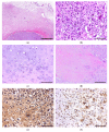
A 6-year-old female Maltese dog presented with a cervical mass without pain. The tumor was surrounded by a thick fibrous tissue and consisted of an osteoid matrix with osteoblasts and two distinct areas: a mesenchymal cell-rich lesion with numerous multinucleated giant cells and a chondroid matrix-rich lesion. The tumor cells exhibited heterogeneous protein expression, including a positive expression of vimentin, cytokeratin, RANKL, CRLR, SOX9, and collagen 2, and was diagnosed as extraskeletal osteosarcoma. Despite its malignancy, the dog showed no sign of recurrence or metastasis three months after the resection. Further analysis of the tumor cells revealed a high expression of proliferation- and metastasis-related biomarkers in the absence of angiogenesis-related biomarkers, suggesting that the lack of angiogenesis and the elevated tumor-associated fibrosis resulted in a hypoxic tumor microenvironment and prevented metastasis.
Keywords: dog; extraskeletal; hypoxia; immunohistochemistry; osteosarcoma; prognosis.
Conflict of interest statement
The authors declare no conflict of interest.
Figures


References
-
- WHO Classification of Tumours Editorial Board, editor. Soft Tissue and Bone Tumours. 5th ed. International Agency for Research on Cancer; Lyon, France: 2020.
-
- Salas S., de Pinieux G., Gomez-Brouchet A., Larrousserie F., Leroy X., Aubert S., Decouvelaere A.V., Giorgi R., Fernandez C., Bouvier C. Ezrin immunohistochemical expression in cartilaginous tumours: A useful tool for differential diagnosis between chondroblastic osteosarcoma and chondrosarcoma. Virchows Arch. 2009;454:81–87. doi: 10.1007/s00428-008-0692-8. - DOI - PubMed
-
- Gomez-Brouchet A., Mourcin F., Gourraud P.A., Bouvier C., De Pinieux G., Le Guelec S., Brousset P., Delisle M.B., Schiff C. Galectin-1 is a powerful marker to distinguish chondroblastic osteosarcoma and conventional chondrosarcoma. Hum. Pathol. 2010;41:1220–1230. doi: 10.1016/j.humpath.2009.10.028. - DOI - PubMed
Publication types
Grants and funding
LinkOut - more resources
Full Text Sources
Research Materials

