. 2022 Jan 6;82(1):90-105.e13.
doi: 10.1016/j.molcel.2021.11.031. Epub 2021 Dec 22.

APC7 mediates ubiquitin signaling in constitutive heterochromatin in the developing mammalian brain

Affiliations

APC7 mediates ubiquitin signaling in constitutive heterochromatin in the developing mammalian brain

Cole J Ferguson et al. Mol Cell. .

Abstract

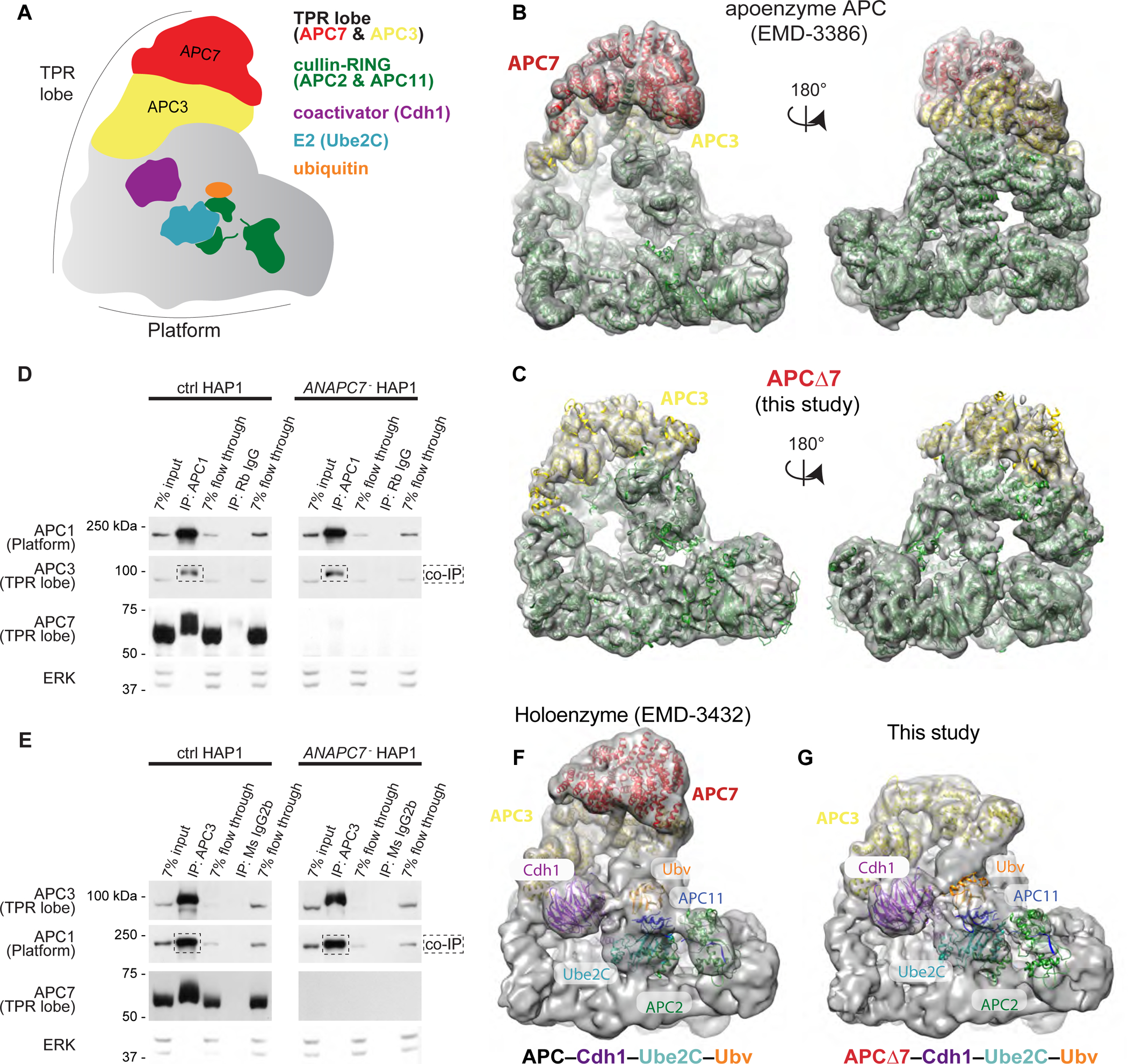
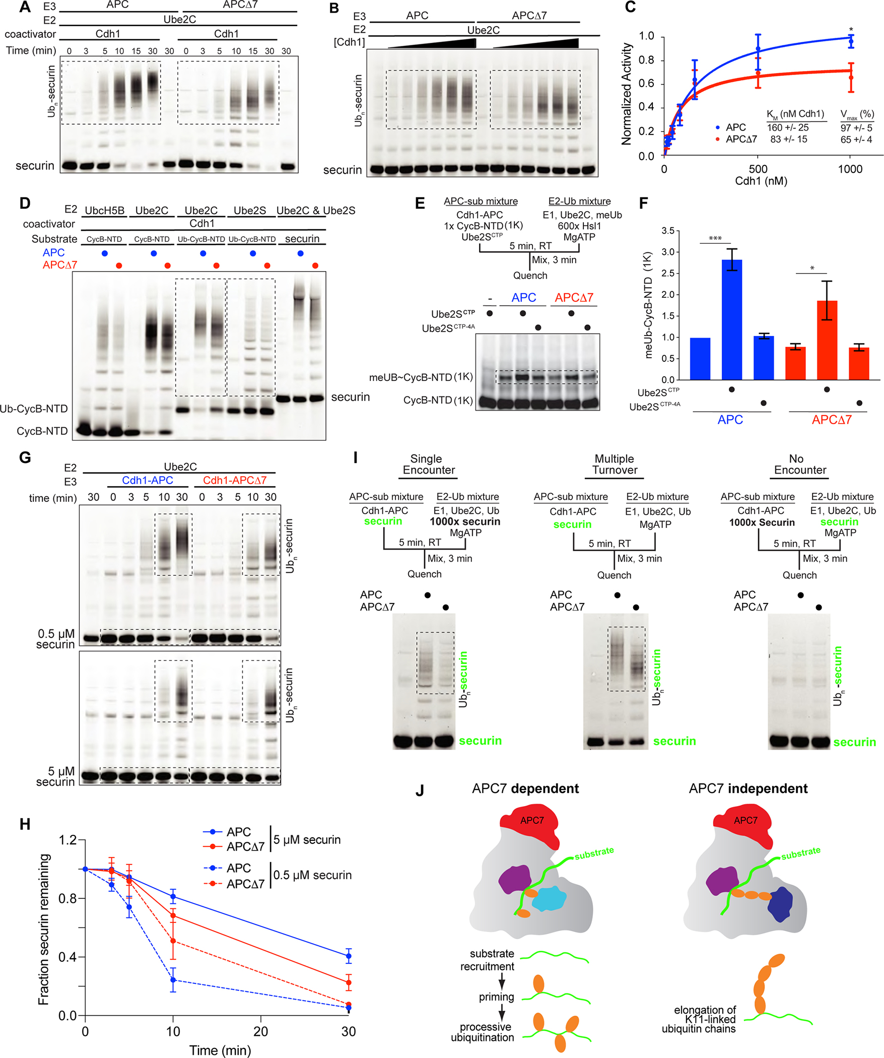
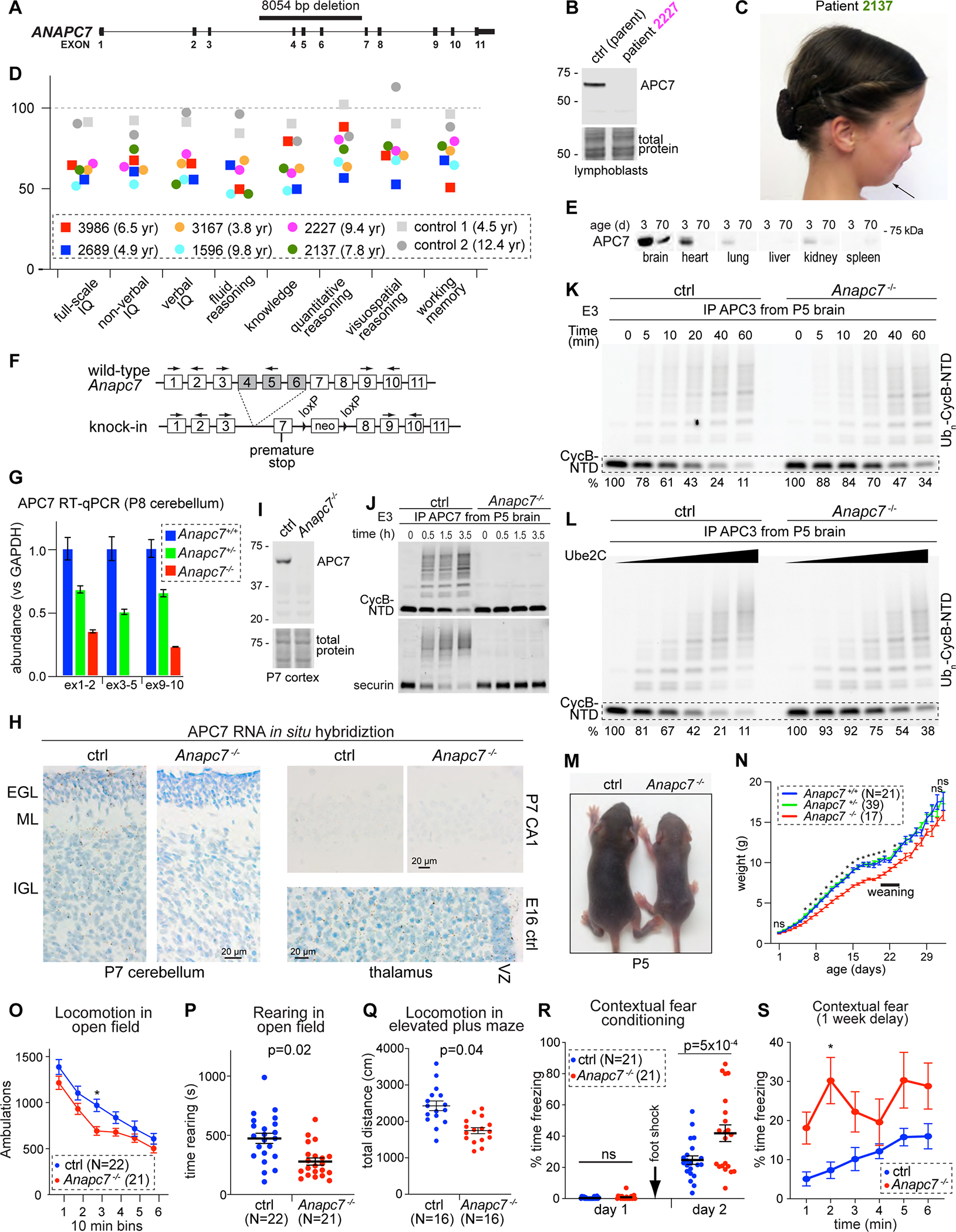
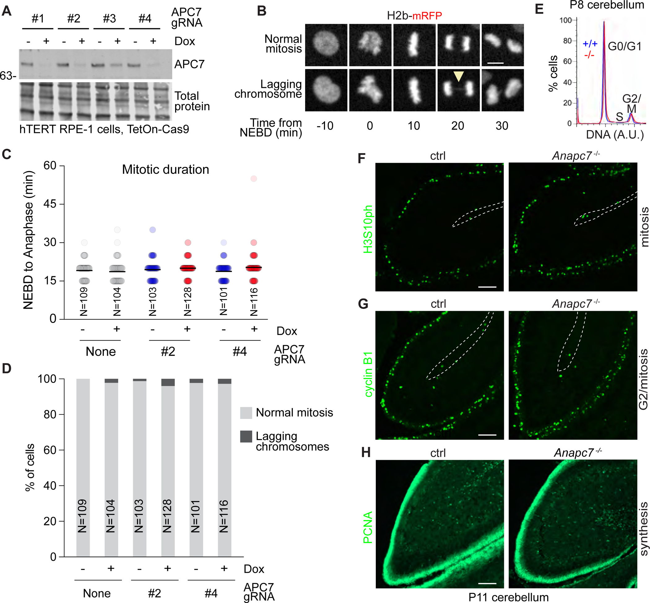
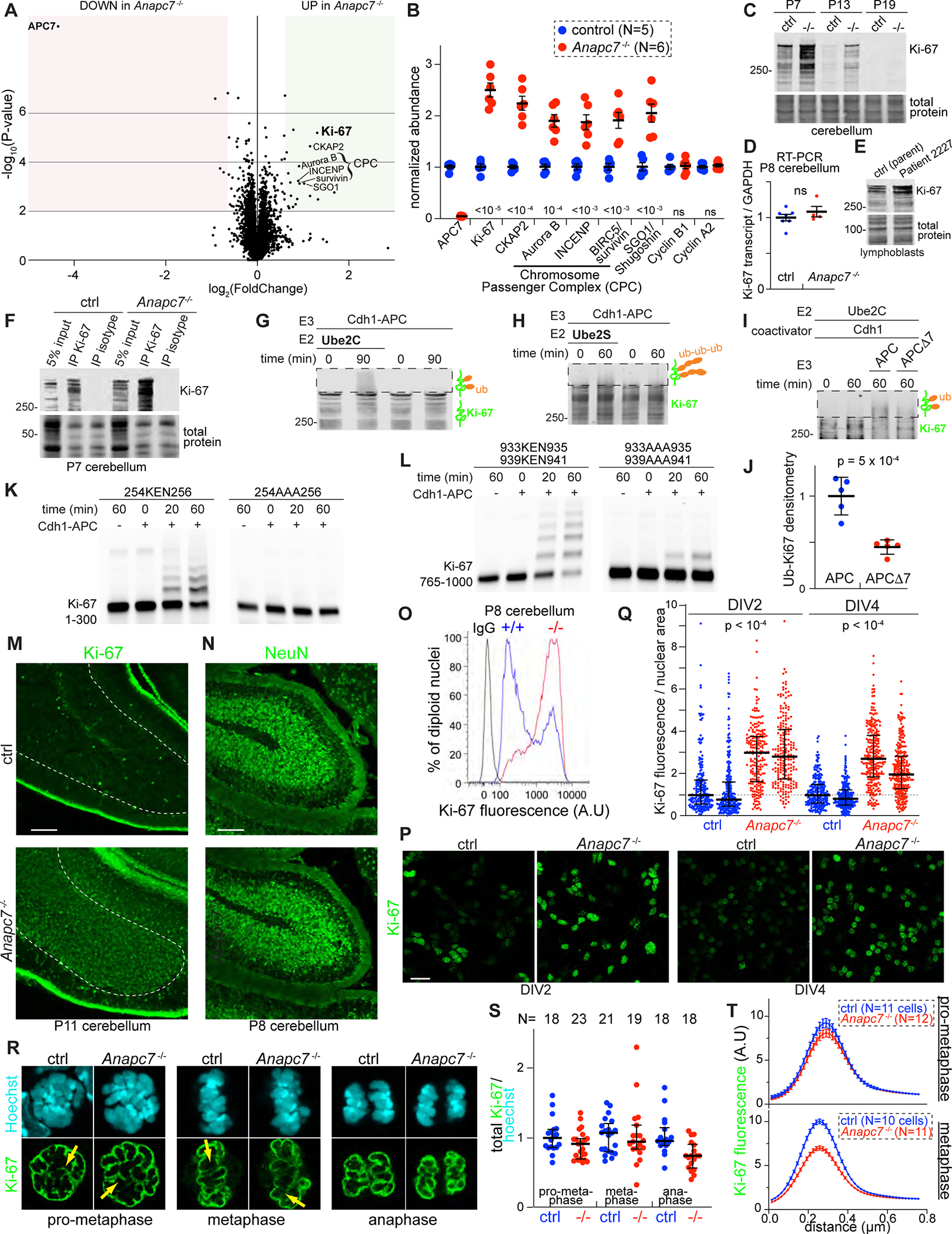
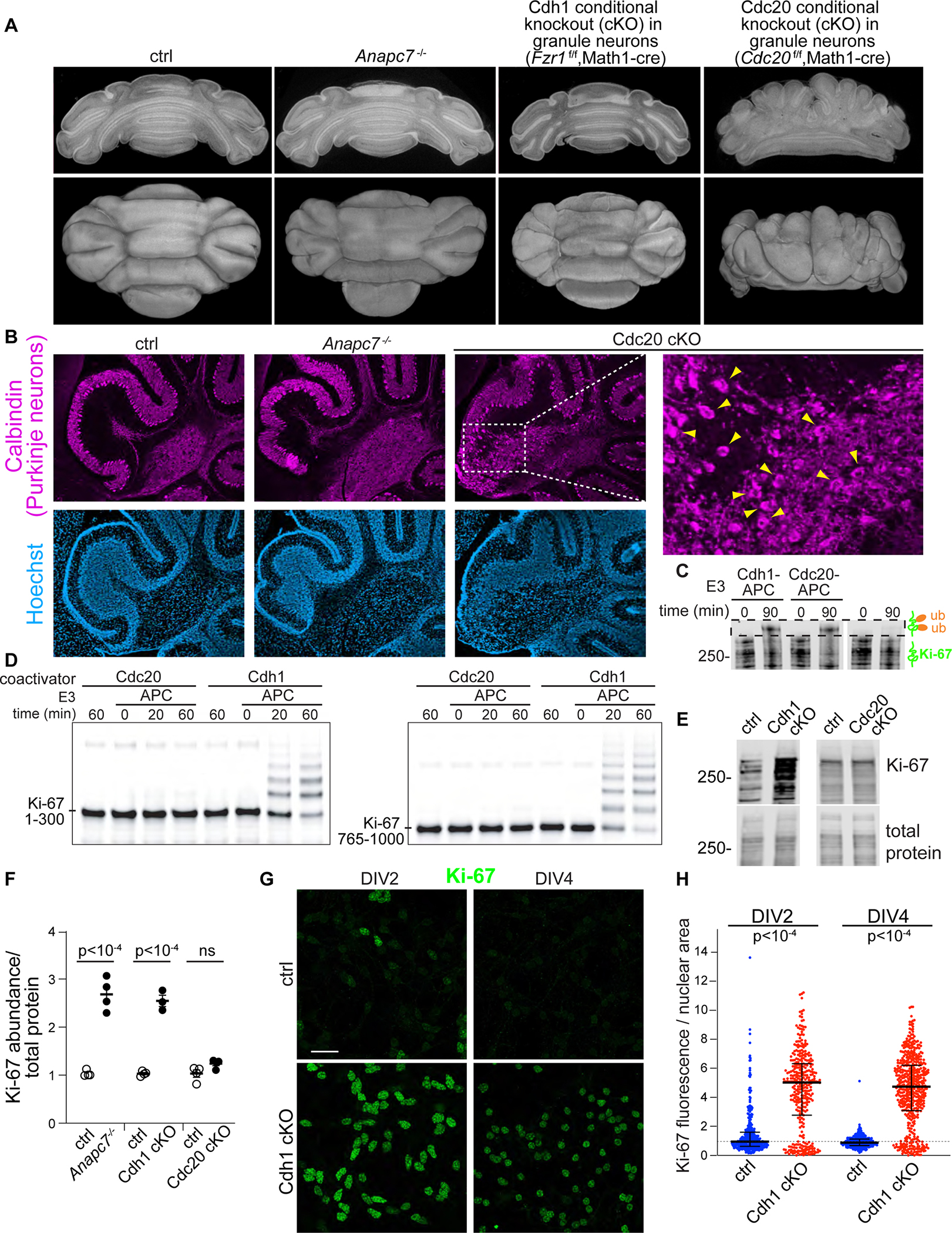
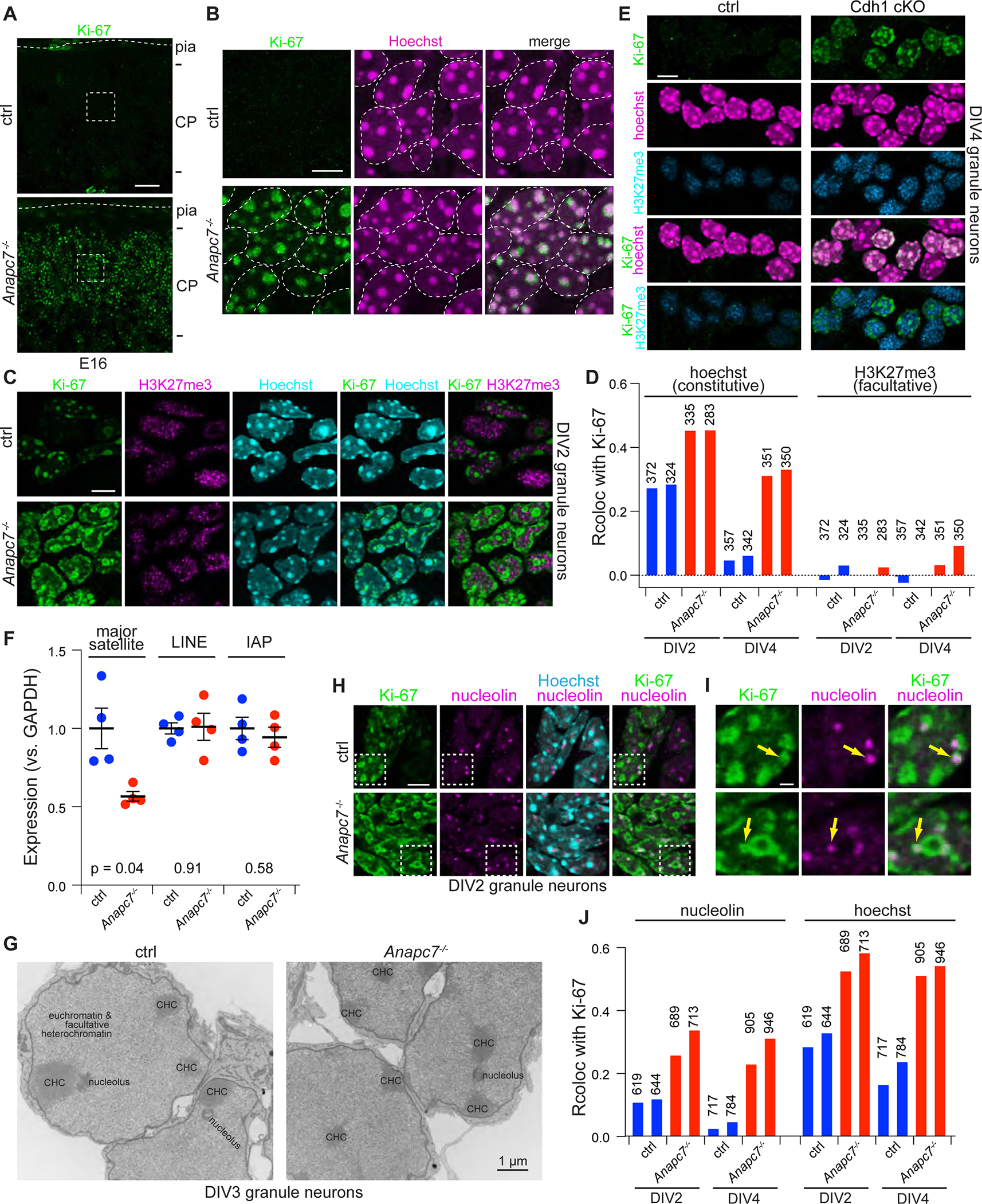
Neurodevelopmental cognitive disorders provide insights into mechanisms of human brain development. Here, we report an intellectual disability syndrome caused by the loss of APC7, a core component of the E3 ubiquitin ligase anaphase promoting complex (APC). In mechanistic studies, we uncover a critical role for APC7 during the recruitment and ubiquitination of APC substrates. In proteomics analyses of the brain from mice harboring the patient-specific APC7 mutation, we identify the chromatin-associated protein Ki-67 as an APC7-dependent substrate of the APC in neurons. Conditional knockout of the APC coactivator protein Cdh1, but not Cdc20, leads to the accumulation of Ki-67 protein in neurons in vivo, suggesting that APC7 is required for the function of Cdh1-APC in the brain. Deregulated neuronal Ki-67 upon APC7 loss localizes predominantly to constitutive heterochromatin. Our findings define an essential function for APC7 and Cdh1-APC in neuronal heterochromatin regulation, with implications for understanding human brain development and disease.

Keywords: APC7; Cdh1; Ki-67; anaphase-promoting complex; brain; chromatin; heterochromatin; neurodevelopment; ubiquitin; ubiquitin ligase.


Conflict of interest statement

Declaration of interests The authors declare no competing interests. A.B. is an employee of Roche. B.S. is on the scientific advisory board of BioTheryX and Interline Therapeutics, a shareholder of Interline Therapeutics, and a co-inventor of intellectual property licensed to Cinsano.

Figures

Figure 1:
Figure 1:. APC7 is not required for the conformation of the APC or its assembly. See also Figure S1.
A. Schematic of the APC. B. Published model of wild-type human APC (EMD-3386) showing the APC7 dimer (Chang et al., 2015). C. Empirically determined Cryo-EM structure of APCΔ7. APC subunits from published coordinates (PDB 4L9U) were rigid-body docked using Chimera (Pettersen et al., 2004). D. IP of APC1 from HAP1 cells followed by immunoblot (IB) for APC subunits. Flow through fractions reflect the remaining protein after antibody binding. E. IP of APC3 (TPR lobe) from HAP1 cells followed by IB for APC subunits. F. Published Cryo-EM structure of the APC holoenzyme (APC–Cdh1–Ube2C–substrate–Ubv) trapped by chemical crosslinking (Brown et al., 2016). G. Cryo-EM structure of APCΔ7–Cdh1–Ube2C–substrate–Ubv with Ube2C poised for ubiquitin transfer. Published coordinates (PDB 4L9U) were rigid-body docked using Chimera.
Figure 2:
Figure 2:. APC7 controls substrate recruitment and processive ubiquitination by Ube2C. See also Figure S2.
A. Time course of ubiquitination of securin by recombinant APC and APCΔ7. Boxed areas show reaction products. B. Kinetic evaluation of APCΔ7-dependent ubiquitination in response to escalating Cdh1. C. Kinetic analysis of substrate ubiquitination plotted using the Michaelis-Menten equation. Error bars SEM, N = 5. (*p = 0.02 by two-way ANOVA with Bonferroni test). D. In vitro ubiquitination assays using recombinant proteins. Boxed areas show products from reactions using the E2 enzymes Ube2C and Ube2S. E. The C-terminal peptide of Ube2S (Ube2SCTP) activates the APC to prime substrates during the initiation of ubiquitination. Shown are single encounter experiments with CycB-NTD (1K), a CycB-NTD variant with a single lysine for ubiquitin transfer. Use of methylated ubiquitin (meUb) ensures reactions terminate after priming. The E2-Ub sub mixture contained excess unlabeled substrate (Hsl1). Ube2SCTP−4A served as a negative control. F. Quantitation of ubiquitinated CycB-NTD (1K). Error bars SEM, N = 3 (p-value by one-way ANOVA with Tukey test). G. Securin ubiquitination at low (0.5 μM) and high (5 μM) substrate concentration. H. Quantitation of unmodified securin over time. Error bars SEM, N = 3. I. Ube2C-mediated processive ubiquitination in single and multiple encounter assays. Fluorescent securin detected during gel imaging is shown in green. J. APC7 is required for the function of the E2 Ube2C at multiple stages. During ubiquitin chain elongation by Ube2S, APC adopts a distinct catalytic architecture that does not require APC7.
Figure 3:
Figure 3:. APC7 is required for human and mouse brain development. See also Figures S3, S4 and S5.
A. 8054 bp deletion in the human ANAPC7 gene. B. APC7 IB in human lymphoblastoid cells. C. Patient 2137 with the ANAPC7 intellectual disability syndrome exhibits Pierre-Robin sequence. D. Stanford-Binet testing of IQ and subdomains. Age at testing is shown. E. APC7 IB in different mouse tissues. F. Recapitulation of the patient mutation in mice. G. RT-qPCR for APC7 in mouse cerebellum. The location of primers is shown in 3F. Error bars SEM, N = 4. H. APC7 in situ hybridization. Brown dots represent mRNA molecules. (EGL, external granule layer; ML, molecular layer; IGL, internal granule layer; VZ, ventricular zone). I. APC7 IB. J. In vitro ubiquitination assay of fluorescent APC substrates. The source of E3 was IP of APC7 from P5 brain. K. Ubiquitination of CycB-NTD by the APC isolated by APC3 IP from mouse brain at P5. L. Ubiquitination of CycB-NTD in response to increasing Ube2C. E3 (APC) was isolated by anti-APC3 IP from the brain at P5. M. Littermates at P5. N. Growth of mutant mice. Error bars SEM (*p < 0.001, mutant versus each of Anapc7+/− and Anapc7+/+, by mixed-effects ANOVA analysis and Tukey test). O. Total ambulations during open field test. Error bars SEM (*p = 0.01 by repeated measure ANOVA and Bonferroni test). P. Time rearing in the open field test. Error bars SEM (p-value by Kolmogorov-Smirnov test). Q. Total ambulatory distance in the elevated plus maze. Error bars SEM (p-value by Kolmogorov-Smirnov test). R. Assessment of contextual fear memory. Foot shock was applied on day 1 after habituation to the context environment. Freezing (conditioned response) was assessed on day 2 upon reintroduction to the context environment. Error bars SEM (p-value by one-way ANOVA with Tukey test). S. Long-term fear conditioning was assessed by reintroducing mice to the context environment after a one-week delay. Error bars SEM (*p = 0.01 by repeated measure ANOVA and Bonferroni test).
Figure 4:
Figure 4:. APC7 loss has little or no effect on mitosis. See also Figure S6.
A. APC7 IB in hTERT RPE-1 cells expressing Dox-inducible Cas9 and transfected with 4 CRISPR guide RNAs targeting APC7. B. Example images of hTERT RPE-1 cells expressing H2b-mRFP. Arrowhead indicates a lagging chromosome. C. Quantitation of the length of mitosis (NEBD to anaphase). The number of cells analyzed is shown. D. Quantitation of lagging chromosomes. The number of cells analyzed is shown. E. Flow-cytometry analysis of DNA content in dissociated P8 cerebellum stained with propidium iodide. F. H3S10ph immunofluorescence (IF). G. Cyclin B1 IF. H. Proliferating Cell Nuclear Antigen (PCNA) IF.
Figure 5:
Figure 5:. Identification of Ki-67 as an APC7-dependent substrate of the APC in neurons. See also Figure S7.
A. Volcano plot of protein abundance in P8 cerebellum as assessed by Tandem Mass Tag (TMT) proteomics (N=5 control, N=6 APC7 mutant). Shaded regions indicate a 1.5-fold change in abundance with p-value < 0.01 by two-tailed unpaired t-test. B. Normalized TMT ratios for relevant proteins (p-values by two-tailed unpaired t-test). C. Ki-67 IB in mouse cerebellum. D. Ki-67 RT-qPCR in P8 cerebellum (N=7 control, N=4 APC7 mutant). Not significant by two-tailed unpaired t-test. E. Ki-67 IB in human lymphoblasts. F. IP of Ki-67 from P8 cerebellum followed by Ki-67 IB. G. In vitro ubiquitination of Ki-67 by recombinant APC. Ki-67 was isolated by IP from wild-type P7 cerebellum. Boxed areas represent reaction products detected by Ki-67 IB. H. In vitro polyubiquitination of Ki-67 by recombinant Cdh1-APC and Ube2S. Ki-67 was isolated by IP from wild-type P7 cerebellum. I. In vitro ubiquitination assay using recombinant APC and APCΔ7. Ki-67 was isolated by IP from wild-type P7 cerebellum. J. Densitometry-based measurement of reaction products following ubiquitination of immunoprecipitated Ki-67 by APC and APCΔ7. Errors bars SEM (N = 5, p-value by two-tailed unpaired t-test). K. In vitro ubiquitination of amino acids 1–300 of human Ki-67 by recombinant APC. KEN to AAA substitution occurred at the indicated residues. L. In vitro ubiquitination of amino acids 765–1000 of human Ki-67 by recombinant APC. KEN to AAA substitutions occurred at the indicated residues M. Ki-67 IF in P11 cerebellum. N. NeuN IF in P11 cerebellum. O. Flow cytometry of diploid nuclei isolated from P8 cerebellum labeled with anti-Ki-67 or isotype antibody. P. Confocal IF of Ki-67 in primary mouse cerebellar granule neuron cultures. The day in vitro (DIV) is indicated. Q. Quantitation of Ki-67 in individual neuronal nuclei normalized to nuclear area. Two biological replicates are shown for each genotype and DIV. Error bars interquartile range (p-values by one-way ANOVA with Tukey test). R. Confocal optical sections of anti-Ki-67 IF in neuronal precursors during mitosis. Yellow arrows indicate regions sampled in Figure 5T. S. Quantitation of Ki-67 IF during mitosis. Ki-67 intensity was normalized to Hoechst. The number of cells analyzed is shown. Error bars SEM. T. Quantitation of the intensity of the chromosome periphery. For each cell, 3–5 segments were analyzed. Errors bars SEM.
Figure 6:
Figure 6:. APC7 operates in the context of Cdh1-APC to drive Ki-67 degradation in neurons.
A. Electron tomography of P8 cerebellum. B. Calbindin IF in sagittal sections of P12 cerebellum. An expanded region from the Cdc20f/f,Math1-cre is shown (PCL, Purkinje cell layer). C. In vitro ubiquitination of full-length Ki-67 by recombinant Cdh1-APC and Cdc20-APC. Ki-67 was immunoprecipitated from wild-type P7 cerebellum and detected by Ki-67 IB. D. In vitro ubiquitination of human Ki-67 amino acids 1–300 (left) and 765–1000 (right) by Cdh1-APC and Cdc20-APC. E. Ki-67 IB in P12 cerebellum. F. Densitometry quantitation of Ki-67 IB. Mutants were normalized and compared to littermates. Error bars SEM (N = 4, p-value by two-tailed unpaired t-test). G. IF of Ki-67 in primary cerebellar granule neuron cultures. H. Quantitation of Ki-67 IF normalized to nuclear area in cultured neurons on the indicated DIV. Error bars interquartile range (p-values by Kruskal-Wallis test for non-parametric data followed by Dunn test).
Figure 7:
Figure 7:. The APC regulates Ki-67 protein turnover in heterochromatin in neurons.
A. Ki-67 IF in the cortical plate (CP) at E16. B. Expanded area from part A. Outlines delimit nuclei. C. IF of Ki-67 and H3K27me3 in primary cerebellar granule neurons on DIV2. D. Quantitation of colocalization between Ki-67 and Hoechst versus Ki-67 and H3K27me3. The number of neurons analyzed is shown. E. Ki-67, H3K27me3 and Hoechst in DIV4 cultured granule neurons from Cdh1 cKO mice. F. RT-qPCR analysis of repeat expression in P11 cerebellum. Error bars SEM (p-values by two-tailed Welch’s t-test) G. Transmission EM of cultured neurons at DIV3. Constitutive heterochromatin appears electron dense and resides within lamin-associated and nucleolar-associated domains. H. Fluorescence imaging of Ki-67, nucleoli and Hoechst in cultured neurons. I. Magnified areas from 7H, arrows demonstrate colocalization of Ki-67 and nucleolin. J. Quantitation of colocalization between Ki-67 and nucleolin, versus Ki-67 and Hoechst in cultured neurons. The number of cells analyzed is shown.

References

    1. Adams DJ, Quail MA, Cox T, Van Der Weyden L, Gorick BD, Su Q, Chan WI, Davies R, Bonfield JK, Law F, et al. (2005). A genome-wide, end-sequenced 129Sv BAC library resource for targeting vector construction. Genomics 86, 753–758. - PubMed
    1. Avagliano L, Parenti I, Grazioli P, Di Fede E, Parodi C, Mariani M, Kaiser FJ, Selicorni A, Gervasini C, and Massa V (2020). Chromatinopathies: A focus on Cornelia de Lange syndrome. Clin. Genet. 97, 3–11. - PubMed
    1. Basak O, van de Born M, Korving J, Beumer J, van der Elst S, van Es JH, and Clevers H (2014). Mapping early fate determination in Lgr5+ crypt stem cells using a novel Ki67-RFP allele. EMBO J. 33, 2057–2068. - PMC - PubMed
    1. Bilimoria PM, and Bonni A (2008). Cultures of cerebellar granule neurons. Cold Spring Harb. Protoc. 3, 1–8. - PMC - PubMed
    1. Bodnar AG, Ouellette M, Frolkis M, Holt SE, Chiu C, Morin GB, Harley CB, Shay JW, Lichtsteiner S, and Wright WE (1998). Extension of Life-Span by Introduction of Telomerase into Normal Human Cells. 279. - PubMed

Publication types

MeSH terms