. 2021 Nov 25;10(12):1885.
doi: 10.3390/antiox10121885.

Radioprotective and Radiomitigative Effects of Melatonin in Tissues with Different Proliferative Activity

Affiliations

Radioprotective and Radiomitigative Effects of Melatonin in Tissues with Different Proliferative Activity

Serazhutdin A Abdullaev et al. Antioxidants (Basel). .

Abstract

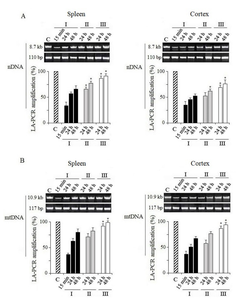
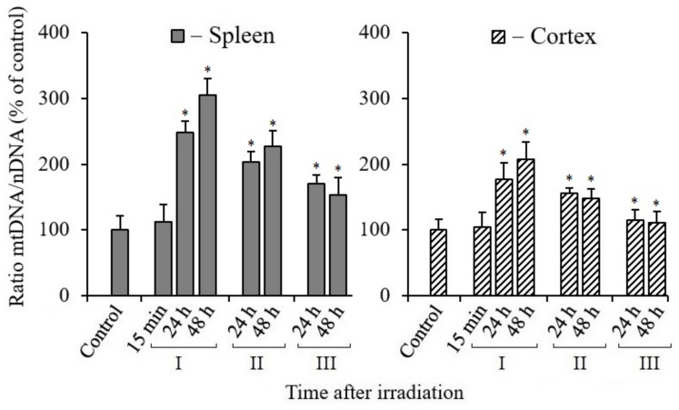
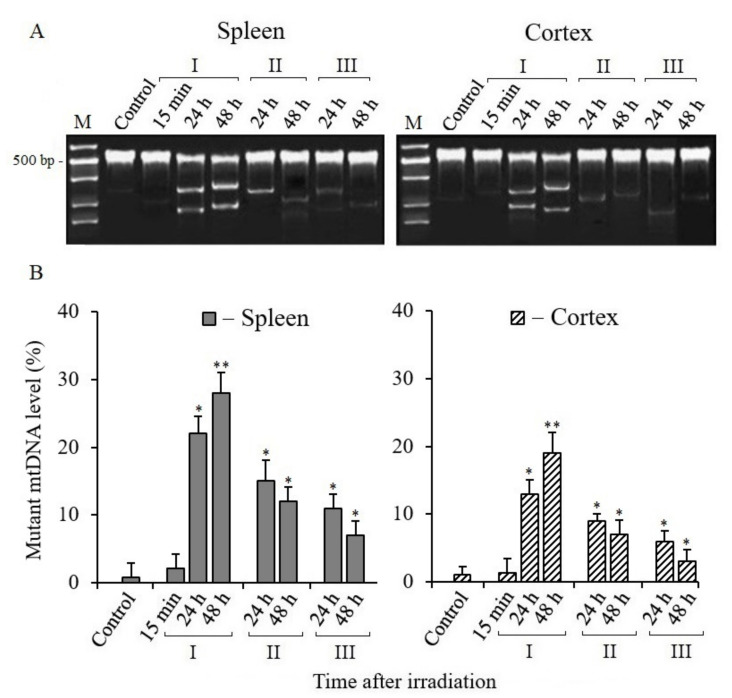
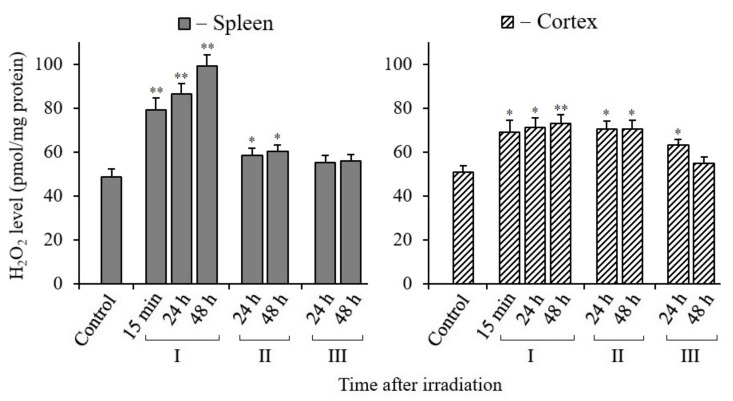
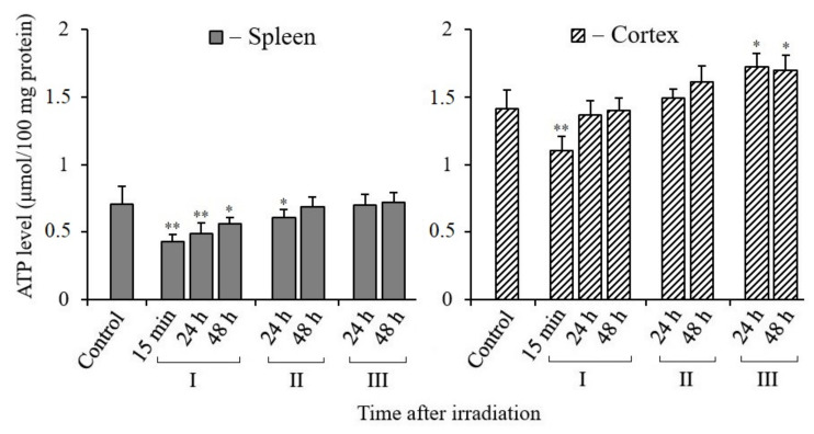
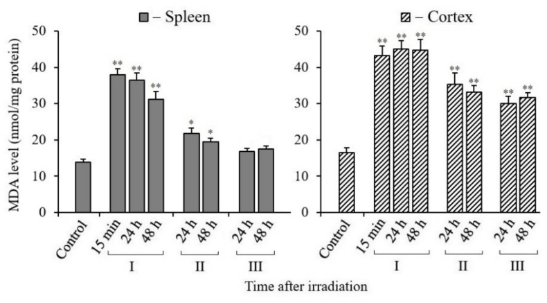
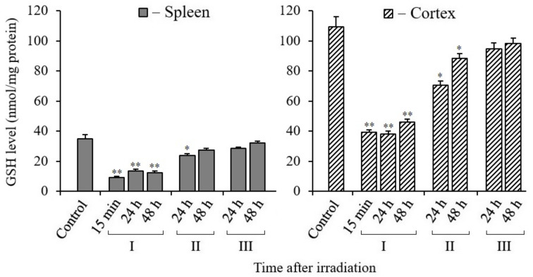
We used various markers to analyze damage to mouse tissues (spleen and cerebral cortex) which have different proliferative activity and sensitivity to ionizing radiation (IR). We also assessed the degree of modulation of damages that occurs when melatonin is administered to mice prior to and after their X-ray irradiation. The data from this study showed that lesions in nuclear DNA (nDNA) were repaired more actively in the spleen than in the cerebral cortex of mice irradiated and treated with melatonin (N-acetyl-5-methoxytryptamine). Mitochondrial biogenesis involving mitochondrial DNA (mtDNA) synthesis was activated in both tissues of irradiated mice. A significant proportion of the newly synthesized mtDNA molecules were mutant copies that increase oxidative stress. Melatonin reduced the number of mutant mtDNA copies and the level of H2O2 in both tissues of the irradiated mice. Melatonin promoted the restoration of ATP levels in the tissues of irradiated mice. In the mouse tissues after exposure to X-ray, the level of malondialdehyde (MDA) increased and melatonin was able to reduce it. The MDA concentration was higher in the cerebral cortex tissue than that in the spleen tissue of the mouse. In mouse tissues following irradiation, the glutathione (GSH) level was low. The spleen GSH content was more than twice as low as that in the cerebral cortex. Melatonin helped restore the GSH levels in the mouse tissues. Although the spleen and cerebral cortex tissues of mice differ in the baseline values of the analyzed markers, the radioprotective and radiomitigative potential of melatonin was observed in both tissues.

Keywords: ATP; GSH; H2O2; MDA; melatonin; mitigation; mtDNA-mutations; nDNA-repair; oxidation stress; protection; radiation.


Conflict of interest statement

All authors declare no conflict of interest.

Figures

Figure 1
Figure 1
Analysis of damage and repair of nuclear DNA and recovery of mitochondrial DNA. Long fragments of nDNA (8.7 kb) and mtDNA (10.9 kb) were measured. These data were normalized by the measured levels of the short fragment of nDNA (110 bp) and mtDNA (117 bp), obtained using the same DNA sample. (A) Quantitative analysis of the LA-QPCR amplicons of nDNA extracted from spleen and cerebral cortex. (B) Quantitative analysis of the LA-QPCR amplicons of mtDNA extracted from spleen and cerebral cortex. Data are presented in % to control (C). Here and in other figures: the dose of X-ray irradiation of mice was 5 Gy and MEL was administered to mice before and after irradiation as a single dose of 125 mg/kg. Electropherogram samples of synthesized amplicons are presented above the histograms. The numbers (15 min, 24 h, 48 h) above and below indicate the time after irradiation. I—mice without MEL administration; II—MEL administration before irradiation; III—MEL administration after irradiation. The data are presented as mean ± SEM of 5–6 independent experiments. Statistical significance was set at * p < 0.05.
Figure 2
Figure 2
Ratio of mtDNA/nDNA in the tissues of the spleen and cerebral cortex of mice after their irradiation. The y-axis shows the percentage (%) of the change in mtDNA to nDNA ratio relative to control. The numbers (15 min, 24 h, 48 h) on X-axis indicate the time after irradiation. I—mice without MEL administration; II—MEL administration before irradiation; III—MEL administration after irradiation. The data are presented as mean ± SEM of 5–6 independent experiments. Statistical significance was set at * p < 0.05.
Figure 3
Figure 3
Detection of mtDNA mutant copies of spleen and cerebral cortex tissues in mice 15 min, 24, and 48 h after X-ray irradiation. (A) Electrophoresis of cleavage products obtained by Surveyor nuclease digestion of heteroduplexes of mtDNA PCR amplicons from spleen and cerebral cortex tissues. (B) Percentage of Surveyor nuclease cleaved heteroduplexes of PCR amplicons of mtDNA (ND3 gene, 534 bp). I—mice without MEL administration; II—MEL administration before irradiation; III—MEL administration after irradiation. The data are presented as mean ± SEM of 5–6 independent experiments. Statistical significance was set at * p < 0.05, ** p < 0.01.
Figure 4
Figure 4
Changes in the H2O2 content in spleen and cerebral cortex tissues of mice 15 min, 24, and 48 h after their exposure to X-rays. I—mice groups without MEL administration; II—MEL administration before irradiation; III—MEL administration after irradiation. The data are presented as mean ± SEM of 5–6 independent experiments. Statistical significance was set at * p < 0.05; ** p < 0.01.
Figure 5
Figure 5
Changes in the ATP content in spleen and cerebral cortex tissues of mice 15 min, 24, and 48 h after their irradiation. I—mice groups without MEL administration; II—MEL administration before irradiation; III—MEL administration after irradiation. The data are presented as mean ± SEM of 5–6 independent experiments. Statistical significance was set at * p < 0.05; ** p < 0.01.
Figure 6
Figure 6
Changes in the MDA content in spleen and cerebral cortex tissues of mice 15 min, 24, and 48 h after their exposure to X-rays. I—mice without MEL administration; II—MEL administration before irradiation; III—MEL administration after irradiation. The data are presented as mean ± SEM of 5–6 independent experiments. Statistical significance was set at * p < 0.05, ** p < 0.01.
Figure 7
Figure 7
Changes in the GSH in spleen and cerebral cortex tissues of mice 15 min, 24, and 48 h after their irradiation. I—mice without MEL administration; II—MEL administration before irradiation; III—MEL administration after irradiation. The data are presented as mean ± SEM of 5–6 independent experiments. Statistical significance was set at * p < 0.05, ** p < 0.01.

References

    1. Karri J., Lachman L., Hanania A., Marathe A., Singh M., Zacharias N., Orhurhu V., Gulati A., Abd-Elsayed A. Radiotherapy-specific chronic pain syndromes in the cancer population: An evidence-based narrative review. Adv. Ther. 2021;38:1425–1446. doi: 10.1007/s12325-021-01640-x. - DOI - PubMed
    1. Chancellor J.C., Scott G.B., Sutton J.P. Space radiation: The number one risk to astronaut health beyond low Earth orbit. Life. 2014;4:491–510. doi: 10.3390/life4030491. - DOI - PMC - PubMed
    1. Hall E.J., Giaccia A.J. Radiobiology for the Radiologist. 8th ed. Wolters Kluwer; Philadelphia, PA, USA: 2019.
    1. Rosen E.M., Day R., Singh V.K. New approaches to radiation protection. Front. Oncol. 2015;4:381. doi: 10.3389/fonc.2014.00381. - DOI - PMC - PubMed
    1. Reiter R.J., Tan D.X., Herman T.S., Thomas C.R., Jr. Melatonin as a radioprotective agent: A review. Int. J. Radiat. Oncol. Biol. Phys. 2004;59:639–653. - PubMed

LinkOut - more resources