Review
. 2021 Dec 9;11(12):2318.
doi: 10.3390/diagnostics11122318.

Pulmonary Vasculitides: A Radiological Review Emphasizing Parenchymal HRCT Features

Affiliations
Review

Pulmonary Vasculitides: A Radiological Review Emphasizing Parenchymal HRCT Features

Stefano Palmucci et al. Diagnostics (Basel). .

Abstract

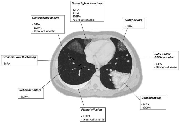
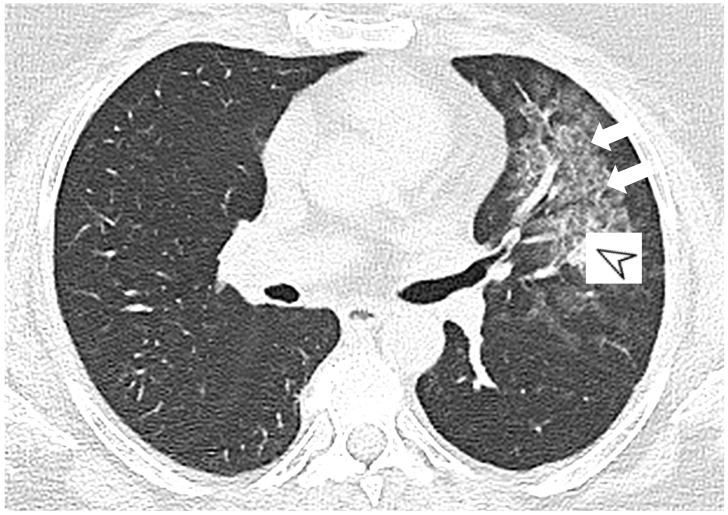
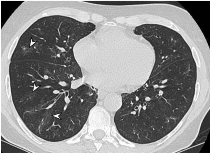
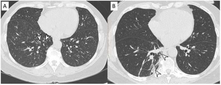
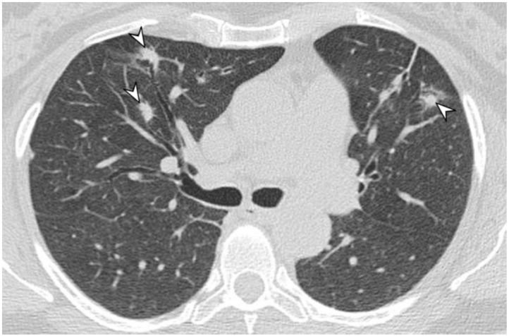
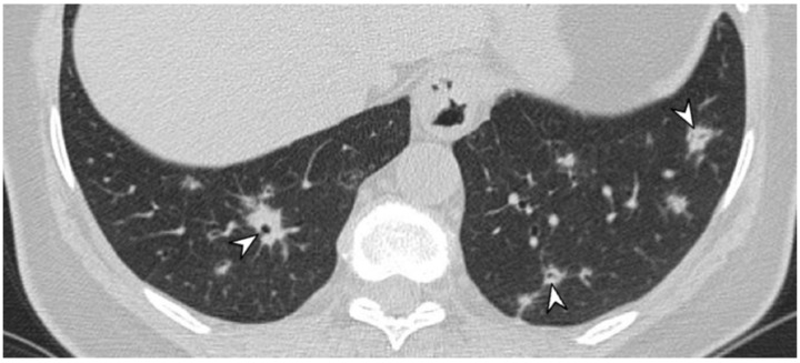
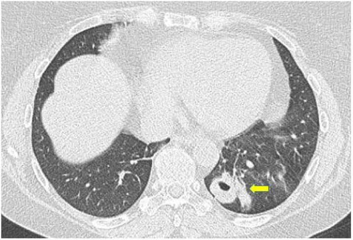
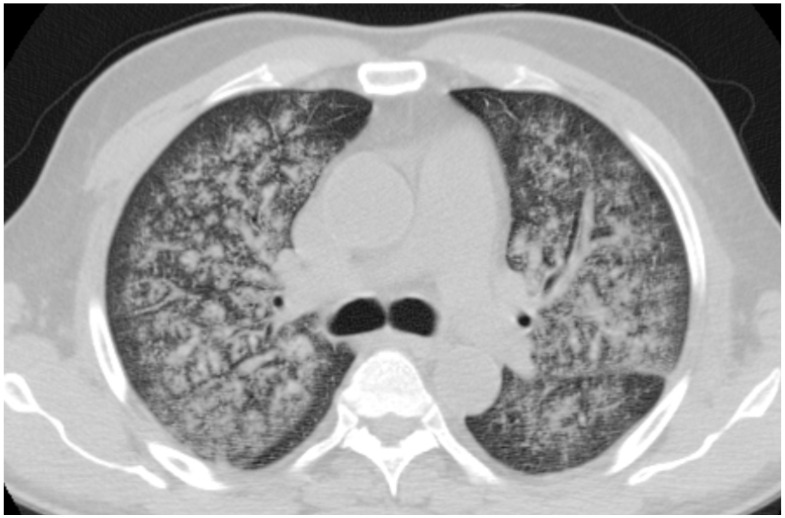
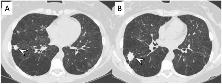
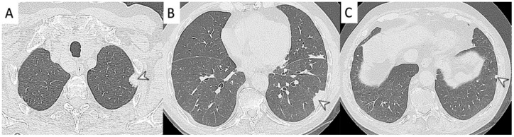
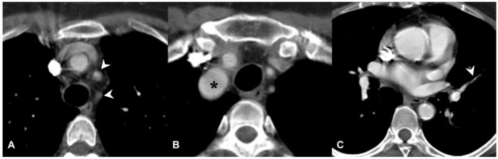
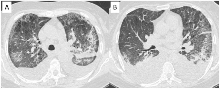
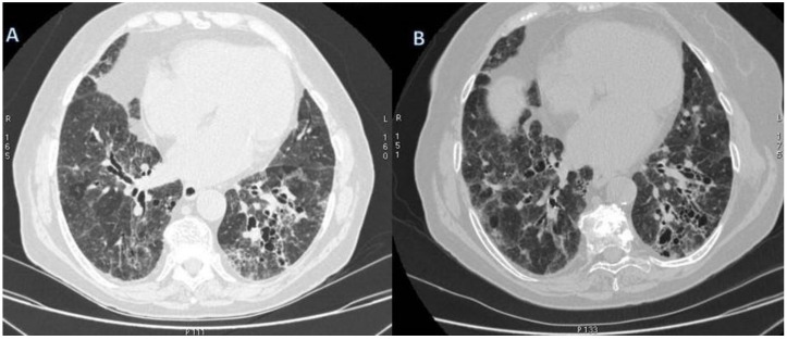
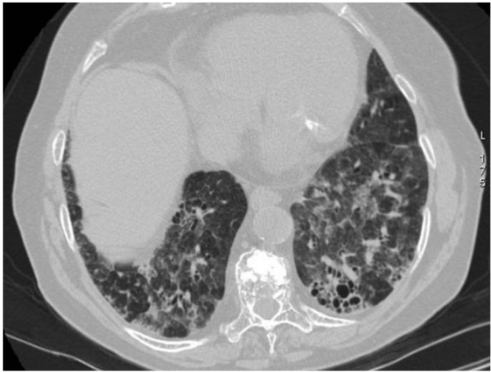
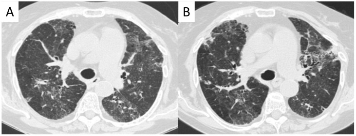
Vasculitides represent a heterogeneous group of immune-mediated disorders, characterized by a systemic inflammatory destructive process of the blood vessels resulting either in ischemia or hemorrhage. The organ involved and vessel size influence the pattern of presentation of the pathology. The lung is commonly involved in systemic vasculitides, with heterogeneous clinical, radiological, and histopathological presentations. Primary vasculitides most commonly associated with lung parenchymal involvement include small-vessel antineutrophil cytoplasmic autoantibody (ANCA)-associated vasculitides, such as granulomatosis with polyangiitis (GPA), eosinophilic granulomatosis with polyangiitis (EGPA), and microscopic polyangiitis (MPA). Several studies have reported cases of interstitial lung diseases (ILDs) associated with systemic vasculitis, particularly those positive for ANCA associated vasculitis/vasculitidis: AAV. We have selected from our case series different radiological features of pulmonary vasculitis (i.e., solitary or multiple nodules, cavitary lesions, nodules with centrilobular or peribronchial distribution, airspace consolidations, "crazy paving" appearance, interstitial disease), including cases with interstitial lung alterations. Therefore, the aim of this review is to describe the typical clinical manifestations of vasculitides and their main radiologic features (especially AAV).

Keywords: antibodies; antineutrophil cytoplasmic; interstitial; lung diseases; multidetector computed tomography; vasculitis.


Conflict of interest statement

The authors declare no conflict of interest.

Figures

Figure 1
Figure 1
A schematic drawn of parenchymal HRCT features in most common primitive vasculitides whit pulmonary involvement.
Figure 2
Figure 2
A 52-year-old female with MPA vasculitis. Axial high-resolution CT shows GGO in the left upper lobe (white arrows), due to alverolar hemorrhage; a peribronchial opacity is also depicted in the pulmonary parenchyma (white arrowhead).
Figure 3
Figure 3
A 57-year-old female with p-ANCA associated vasculitidis. Axial high-resolution CT shows patchy ground-glass opacities (arrowheads), predominantly located in the right lower lobe, sparing subpleural regions.
Figure 4
Figure 4
Microscopic Polyangiitis (MPA). Axial CT scan shows micronodules with centrilobular distribution and diffuse bronchial wall thickening (white arrowheads in (A)). After treatment, 2 years later, axial CT scan shows the appearance of (focal organizing pattern) focal consolidation (white arrowheads in (B)), as clearly depicted in the right lower lobe.
Figure 5
Figure 5
A female patient, with diagnosis of Wegener’s disease. CT image shows multiple bilateral lung nodules (arrowheads).
Figure 6
Figure 6
A patient with diagnosis of Wegener’s disease. CT image shows multiple bilateral lung nodules (arrowheads), with air bronchogram sign.
Figure 7
Figure 7
Axial high resolution CT scan in a female patient with Granulomatosis with polyangiitis shows nodule with cavitation presenting thick walls and irregular margins (yellow arrow).
Figure 8
Figure 8
A 64-year-old, male, with fever, weakness, hemoptysis and hematuria. Diagnosis of granulomatosis with polyangiitis. Axial CT shows diffuse hemorrhagic alveolitis diffuse alveolar hemorrhage.
Figure 9
Figure 9
Peripheral nodular consolidations (arrowheads in (A,B)) located in the right lower lung of a female patient with eosinophilic granulomatosis with polyangiitis. Other pulmonary HRCT abnormalities, represented by emphysematous changes and not related to vasculitis, are also shown.
Figure 10
Figure 10
Peripheral consolidations (arrowheads in (AC)), located in the left lower lung of a female patient with eosinophilic granulomatosis with polyangiitis.
Figure 11
Figure 11
Wall thickening in a patient with Takayasu arteritis. Occlusions of left subclavian artery and common carotid artery (white arrowheads) are depicted in the figure (A); dilation of brachiocephalic trunk is also evident in (B) (black asterisk). Stenosis of pulmonary segmental artery could be also appreciated in figure (C) (white arrowhead), due to granulomatous inflammation of the arterial wall.
Figure 12
Figure 12
Cryoglobulinemic vasculitis in a patient with chronic renal failure and dyspnoea. Axial HRCT images show diffuse areas of ground-glass attenuation, due to alveolar hemorrhage, with interlobular and intralobular septal thickening. Pleural effusions are also shown in the figures (A,B).
Figure 13
Figure 13
A 76-year-old female patient with GPA. Axial high-resolution CT shows irregular bronchial dilatation and interstitial lung alterations with interlobular and intralobular septal thickening and reticulations, predominantly located in the lower lobe (A). After six-months follow-up interstitial and bronchial are still recognizable (B).
Figure 14
Figure 14
CT scan shows subpleural reticulation and architectural distortion in the lower lobes in an 81-year-old female with AAV. Small, rounded cysts with thick walls are distributed in concentric layers in the subpleural region of the lower left lobe (honeycombing).
Figure 15
Figure 15
An 80-year-old female patient with granulomatosis with polyangiitis (GPA). HRCT shows irregular bronchial wall thickening, bronchial dilatation, and interstitial lung alterations with interlobular septal thickening and GGOs. It is possibly to appreciate areas of different attenuation of lung parenchyma, resembling the appearance of the so called “mosaic attenuation” (A). Follow-up CT (B), acquired one year later, confirms the presence of these alterations.

References

    1. Jennette J.C., Falk R.J., Andrassy K., Bacon A.P., Churg J., Wolfgang L.G., Christiaan Hagen E., Hoffman G.S., Hunder G.G., Kallenberg C.G.M., et al. Nomenclature of systemic vasculitides: The proposal of an international consensus conference. Arthritis Rheum. 1994;37:187–192. doi: 10.1002/art.1780370206. - DOI - PubMed
    1. Jennette J.C., Falk R.J., Bacon A.P., Basu N., Cid M.C., Ferrario F., Flores-Suarez L.F., Gross W.L., Guillevin L., Hagen E.C., et al. 2012 revised International Chapel Hill Consensus Conference nomenclature of vasculitides. Arthritis Rheum. 2013;65:1–11. doi: 10.1002/art.37715. - DOI - PubMed
    1. Brown K.K. Pulmonary vasculitis. Proc. Am. Thorac. Soc. 2006;3:48–57. doi: 10.1513/pats.200511-120JH. - DOI - PMC - PubMed
    1. Maffessanti M., Dalpiaz G. Diffuse Lung Disease—Clinical Features, Pathology, HRCT. Springer; Berlin/Heidelberg, Germany: 2004.
    1. Alba M.A., Flores-Suárez L.F., Henderson A.G., Xiao H., Hu P., Nachman P.H., Falk R.J., Jennetteet J.C. Interstitial lung disease in ANCA vasculitis. Autoimmun. Rev. 2017;16:722–729. doi: 10.1016/j.autrev.2017.05.008. - DOI - PMC - PubMed

LinkOut - more resources