. 2021 Nov 24;10(12):3281.
doi: 10.3390/cells10123281.

Gas6/TAM Signalling Negatively Regulates Inflammatory Induction of GM-CSF in Mouse Brain Microglia

Affiliations

Gas6/TAM Signalling Negatively Regulates Inflammatory Induction of GM-CSF in Mouse Brain Microglia

Shannon E Gilchrist et al. Cells. .

Abstract

Microglia and astrocytes are the main CNS glial cells responsible for the neuroinflammatory response, where they release a plethora of cytokines into the CNS inflammatory milieu. The TAM (Tyro3, Axl, Mer) receptors and their main ligand Gas6 are regulators of this response, however, the underlying mechanisms remain to be determined. We investigated the ability of Gas6 to modulate the CNS glial inflammatory response to lipopolysaccharide (LPS), a strong pro-inflammatory agent, through a qPCR array that explored Toll-like receptor signalling pathway-associated genes in primary cultured mouse microglia. We identified the Csf2 gene, encoding granulocyte-macrophage colony-stimulating factor (GM-CSF), as a major Gas6 target gene whose induction by LPS was markedly blunted by Gas6. Both the Csf2 gene induction and the suppressive effect of Gas6 on this were emulated through measurement of GM-CSF protein release by cells. We found distinct profiles of GM-CSF induction in different glial cell types, with microglia being most responsive during inflammation. Also, Gas6 markedly inhibited the LPS-stimulated nuclear translocation of NF-κB p65 protein in microglia. These results illustrate microglia as a major resident CNS cellular source of GM-CSF as part of the neuroinflammatory response, and that Gas6/TAM signalling inhibits this response through suppression of NF-κB signalling.

Keywords: GM-CSF; Gas6; NF-κB; TAM receptors; astrocytes; glial cells; lipopolysaccharide; microglia; neuroinflammation; primary culture.


Conflict of interest statement

The authors declare no conflict of interest.

Figures

Figure 1
Figure 1
A TLR signalling gene array identified the Csf2 gene as markedly affected by Gas6/TAM signalling in microglial cells undergoing inflammatory stimulation. An RT2 Profiler Gene Array for the TLR signalling pathway was used to measure the expression of 84 genes of interest in pure mouse microglial cultures. Microglia were treated with LPS (10 ng/mL; blue bars) for 8 h in the presence or absence of Gas6 co-incubation (1.6 μg/mL; red bars) added 1 h before and present thereafter. Results are presented as: (A) genes upregulated more than 10-fold after LPS stimulation, (B) genes downregulated more than 2-fold with LPS or (C) genes that were mostly unchanged with LPS treatment. Data is displayed as fold regulation (the negative inverse of fold change for values less than one) of gene expression and is from an exploratory experiment in one mouse culture. Green boxes highlight the genes for which Gas6 presence markedly altered the LPS-induced expression changes.
Figure 2
Figure 2
Gas6 inhibits the inflammatory LPS-induced upregulation of Csf2 mRNA in microglia. RT-qPCR was used to measure the fold change in gene expression of: (A) Csf2 and (B) Cd80 in microglial cultures in response to 8 h LPS (10 ng/mL) exposure in the presence or absence of Gas6 (1.6 μg/mL), which was added 1 h before. Data displayed is the fold change (2−ΔΔCt) normalised to non-stimulated cells (value of 1) with each pairwise change illustrated with a connected line for each separate culture and bars displaying mean ± SEM (n = 6–7 independent experiments on separate cultures). Data was analysed by Wilcoxon signed-rank test; * p < 0.05.
Figure 3
Figure 3
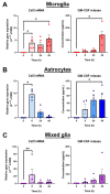
Microglia are the main resident CNS glial source of GM-CSF. Time course of GM-CSF upregulation by LPS (10 ng/mL) in: (A) pure microglial cultures, (B) pure astrocyte cultures and (C) mixed glial cultures, using RT-qPCR and ELISA to measure gene expression and protein release into the cell media, respectively. Gene expression is displayed as relative gene expression (2−ΔCt) and protein concentration is in pg/mL. Statistical analysis was determined using Friedman’s tests with p < 0.05 (n = 4–5 independent experiments on separate cultures). * p < 0.05, ** p < 0.01.
Figure 4
Figure 4
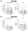
Gas6 supresses the LPS-upregulated GM-CSF protein release by microglia. RT-qPCR and ELISA were used to measure the gene expression and protein release of GM-CSF from: (A) pure microglial cultures or (B) mixed glial cultures after 48-h LPS treatment (10 ng/mL) with or without 1-h pre-treatment with Gas6 (1.6µg/mL), the Gas6 remaining throughout. Gene expression data shows fold change (2−ΔΔCt) and protein release data displays GM-CSF protein concentration (pg/mL). Non-treated microglia had a protein concentration of 0 pg/mL and mixed glia had baseline levels of 0–2 pg/mL (data not shown). Statistical significance was determined using Friedman’s tests for ELISA data or Wilcoxon signed-rank tests for RT-qPCR data with p < 0.05 (n = 4–10 independent experiments on separate cultures). * p < 0.05.
Figure 5
Figure 5
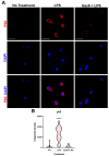
Gas6 inhibits LPS-induced nuclear translocation of the NF-κB p65 subunit in microglia. Pure microglial cell cultures were treated with LPS (10 ng/mL) for 30 min with or without 1 h pre-incubation with Gas6 (1.6 µg/mL), which then remained throughout. (A) Cells underwent immunofluorescence staining with anti-p65 primary antibody with AlexaFluor647 anti-mouse secondary antibody and DAPI nuclear counterstaining. Scale bar = 100 µm. (B) p65 staining within the nuclear area of each cell was quantified for each treatment group. Data is shown in a violin plot with median and interquartile ranges visible (n > 30 cells). Data was statistically analysed using one-way ANOVA; **** p < 0.0001 vs. both other conditions.

References

    1. Frischer J.M., Bramow S., Dal-Bianco A., Lucchinetti C.F., Rauschka H., Schmidbauer M., Laursen H., Sorensen P.S., Lassmann H. The relation between inflammation and neurodegeneration in multiple sclerosis brains. Brain. 2009;132:1175–1189. doi: 10.1093/brain/awp070. - DOI - PMC - PubMed
    1. Kwon H.S., Koh S.H. Neuroinflammation in neurodegenerative disorders: The roles of microglia and astrocytes. Transl. Neurodegener. 2020;9:1–12. doi: 10.1186/s40035-020-00221-2. - DOI - PMC - PubMed
    1. Guzman-Martinez L., Maccioni R.B., Andrade V., Navarrete L.P., Pastor M.G., Ramos-Escobar N. Neuroinflammation as a common feature of neurodegenerative disorders. Front. Pharmacol. 2019;10:1008. doi: 10.3389/fphar.2019.01008. - DOI - PMC - PubMed
    1. Sofroniew M.V., Vinters H.V. Astrocytes: Biology and pathology. Acta Neuropathol. 2010;119:7–35. doi: 10.1007/s00401-009-0619-8. - DOI - PMC - PubMed
    1. Nimmerjahn A., Kirchhoff F., Helmchen F. Resting Microglial Cells Are Highly Dynamic Surveillants of Brain Parenchyma in Vivo. Science. 2005;308:1314–1319. doi: 10.1126/science.1110647. - DOI - PubMed

Publication types

MeSH terms

Substances