Review
. 2021 Nov 25;10(12):3308.
doi: 10.3390/cells10123308.

Translational Stroke Research Review: Using the Mouse to Model Human Futile Recanalization and Reperfusion Injury in Ischemic Brain Tissue

Affiliations
Review

Translational Stroke Research Review: Using the Mouse to Model Human Futile Recanalization and Reperfusion Injury in Ischemic Brain Tissue

Emilia Conti et al. Cells. .

Abstract

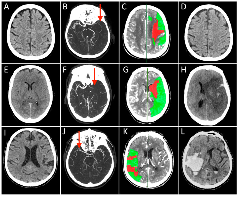
The approach to reperfusion therapies in stroke patients is rapidly evolving, but there is still no explanation why a substantial proportion of patients have a poor clinical prognosis despite successful flow restoration. This issue of futile recanalization is explained here by three clinical cases, which, despite complete recanalization, have very different outcomes. Preclinical research is particularly suited to characterize the highly dynamic changes in acute ischemic stroke and identify potential treatment targets useful for clinical translation. This review surveys the efforts taken so far to achieve mouse models capable of investigating the neurovascular underpinnings of futile recanalization. We highlight the translational potential of targeting tissue reperfusion in fully recanalized mouse models and of investigating the underlying pathophysiological mechanisms from subcellular to tissue scale. We suggest that stroke preclinical research should increasingly drive forward a continuous and circular dialogue with clinical research. When the preclinical and the clinical stroke research are consistent, translational success will follow.

Keywords: brain; futile recanalization; ischemic stroke; mechanical thrombectomy; reperfusion; translational stroke research.


Conflict of interest statement

The authors declare no conflict of interest. The funders had no role in the design of the study; in the collection, analyses, or interpretation of data; in the writing of the manuscript, or in the decision to publish the results.

Figures

Figure 1
Figure 1
Each row represents a single patient’s images derived in different modalities and timepoints (Case 1 presented in (AD), Case 2 in (EH), and Case 3 in (IL)). The first three columns on the left show the Non-Contrast-CT (NCCT), multiphase CT Angiography (CTA), and CT Perfusion (CTP) performed at hospital arrival, while the last column on the right displays the NCCT acquired at 24 h after stroke. On presenting NCCT (A,E,I) no early ischemic changes can be seen in the brain tissue in Case 2 and 3 (ASPECT score = 10), while a tissue swelling was detected in the right insular lobe in Case 1 (ASPECT score = 9, not shown). The mCTA images identified proximal (M1 segment) Middle Cerebral Artery (MCA) occlusion in the left hemisphere in (B,F), and contralateral side in (J) (red arrows). CTP showed in all cases a small infarct core corresponding to CBV lesion (red) and a large ischemic penumbra consisting of the difference between MTT and CBV lesions (green), representing the expected “salvageable” tissue after recanalization. In these patients, CBV lesion volume ≤ 50% of MTT lesion size. All patients were treated with combined IVT and MT, obtaining a complete recanalization, but the 24 h NCCT showed three different conditions: no ischemic lesion visible (D), complete MCA territory infarct associated with a massive cerebral oedema (H), and a vast hemorrhagic transformation of the ischemic lesion.
Figure 2
Figure 2
Schematic representation of intraluminal suture of middle cerebral artery occlusion (MCAO) stroke model, created with Biorender.com (accessed on 1 September 2021). On the right pros and cons table of the model.
Figure 3
Figure 3
Schematic representation of Endothelin-1 stroke model, created with Biorender.com (accessed on 1 September 2021). On the right pros and cons table of the model.
Figure 4
Figure 4
Schematic representation of photothrombotic stroke model, created with Biorender.com (accessed on 1 September 2021). On the right pros and cons table of the model.
Figure 5
Figure 5
Hemorrhage and edema evaluation in preclinical research: (A) Evaluation of Evans Blue extravasation at different time points from the induction of photothrombosis. Modified from Stoll et al., 2008. (B) Representative images of T2-MRI scans (+1.1 mm to −2.9 mm relative to bregma) in a mouse with small hematoma and a mouse with large hematoma at 6 h after induction of intracerebral hemorrhage. The boundary between hematoma and surrounding tissues is indicated by a solid white line. Modified from Matsushita et al., 2013. (C) Upper left: red arrows depict the optical beam scan pattern for three-dimensional 3-D imaging of the sample; upper right: 3-D volume of in vivo mouse brain rendered from OCT volumetric scan; lower left: sagittal OCT intensity image of in vivo mouse brain and corresponding (lower right) attenuation image. Scale bar = 0.5 mm. Modified from Rodriguez et al., 2014. (D) Depth projection images illustrate the transient BBB disruption induced by microbubbles and focused ultrasound at 0.6 MPa (scale bar: 100 μm). Modified from Nhan et al. 2013.
Figure 6
Figure 6
(A) Two-photon imaging of local changes in the dendritic structure before, during, and after MCAO. In the left panel, intact dendritic structures were observed; in the middle panel, extensive dendritic blebbing was observed; in the right panel, a significant recovery of dendritic structures after reperfusion was observed. Modified by Li and Murphy 2008, Copyright (2008) Society for Neuroscience. (B) Time-lapse imaging of apical dendrites showed the retraction of a dendritic spine. Modified by Brown et al., 2007, Copyright (2007) Society for Neuroscience. (C) Time-lapse images of maximum intensity z-projections (from 20 to 60 μm) before (left) and after (right) the laser-induced ischemic hemorrhage. The figures shown in green are the GFP-labeled neurons in a GFP-M mouse, and in red are the vascular networks labeled with Texas-red dextran dye. The tip of the yellow lightning symbol represents the laser irradiation point. The first image was acquired just before the laser irradiation. Scale bar, 20 μm. Modified from Allegra Mascaro et al., 2010. (D) Image sequences of cortical activation as assessed by calcium imaging during pulling of the handle by the contralateral forelimb of CTRL (top), STROKE (bottom) Thy1-GCaMP6f mice in the M-Platform. A small area located in the motor-sensory region reproducibly lit up in CTRL mice, while a large area covering most of the cortical surface of the injured hemisphere was activated in STROKE mice 1 month after stroke. A–P, anterior posterior, M-L, medio-lateral, M1, primary motor area, V1, primary visual area, S1, primary sensory area, Rs, Retro splenial area, BF, barrel field. The black dashed lines define the lesion borders. The black dot indicates bregma. Scale bar 1 mm. Modified from Allegra Mascaro et al., 2019.

References

    1. Nie X., Pu Y., Zhang Z., Liu X., Duan W., Liu L. Futile Recanalization after Endovascular Therapy in Acute Ischemic Stroke. Biomed. Res. Int. 2018;2018:5879548. doi: 10.1155/2018/5879548. - DOI - PMC - PubMed
    1. Stoll G., Pham M. Beyond recanalization—A call for action in acute stroke. Nat. Rev. Neurol. 2020;16:591–592. doi: 10.1038/s41582-020-00417-0. - DOI - PubMed
    1. Ter Schiphorst A., Charron S., Hassen W.B., Provost C., Naggara O., Benzakoun J., Seners P., Turc G., Baron J.C., Oppenheim C. Tissue no-reflow despite full recanalization following thrombectomy for anterior circulation stroke with proximal occlusion: A clinical study. J. Cereb. Blood Flow Metab. 2021;41:253–266. doi: 10.1177/0271678X20954929. - DOI - PMC - PubMed
    1. Hacke W., Schwab S., Horn M., Spranger M., De Georgia M., von Kummer R. ‘Malignant’ middle cerebral artery territory infarction: Clinical course and prognostic signs. Arch. Neurol. 1996;53:309–315. doi: 10.1001/archneur.1996.00550040037012. - DOI - PubMed
    1. Brogan M.E., Manno E.M. Treatment of malignant brain edema and increased intracranial pressure after stroke. Curr. Treat. Opt. Neurol. 2015;17:327. doi: 10.1007/s11940-014-0327-0. - DOI - PubMed

Publication types