Review
. 2021 Nov 27;10(12):3330.
doi: 10.3390/cells10123330.

Molecular Signaling to Preserve Mitochondrial Integrity against Ischemic Stress in the Heart: Rescue or Remove Mitochondria in Danger

Affiliations
Review

Molecular Signaling to Preserve Mitochondrial Integrity against Ischemic Stress in the Heart: Rescue or Remove Mitochondria in Danger

Justin D Yu et al. Cells. .

Abstract

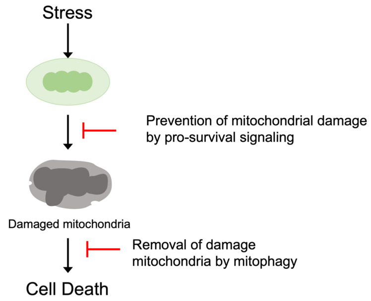
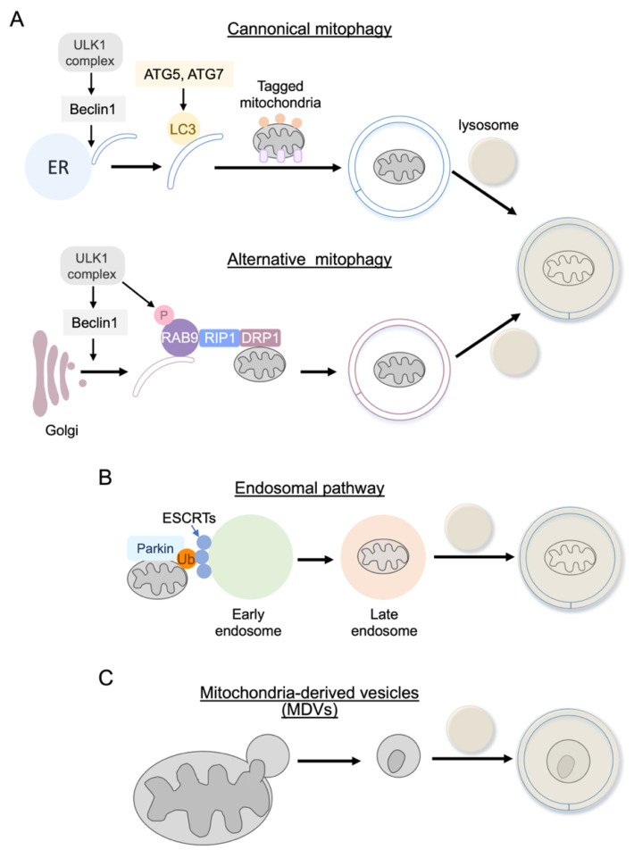
Cardiovascular diseases are one of the leading causes of death and global health problems worldwide, and ischemic heart disease is the most common cause of heart failure (HF). The heart is a high-energy demanding organ, and myocardial energy reserves are limited. Mitochondria are the powerhouses of the cell, but under stress conditions, they become damaged, release necrotic and apoptotic factors, and contribute to cell death. Loss of cardiomyocytes plays a significant role in ischemic heart disease. In response to stress, protective signaling pathways are activated to limit mitochondrial deterioration and protect the heart. To prevent mitochondrial death pathways, damaged mitochondria are removed by mitochondrial autophagy (mitophagy). Mitochondrial quality control mediated by mitophagy is functionally linked to mitochondrial dynamics. This review provides a current understanding of the signaling mechanisms by which the integrity of mitochondria is preserved in the heart against ischemic stress.

Keywords: BCL-2 family proteins; cell death; heart; ischemia; mPTP; mitochondria; mitophagy; pro-survival signaling.


Conflict of interest statement

The authors declare no conflict of interest.

Figures

Figure 1
Figure 1
Prevention of the mitochondrial death pathway. Pro-survival signaling pathways antagonize the induction of the mitochondrial death pathway. Damaged mitochondria are also eliminated by mitophagy to preserve mitochondrial integrity and inhibit cell death.
Figure 2
Figure 2
The BCL-2 family proteins and the mitochondrial permeability transition pore (mPTP). (A) BCL-2 family proteins regulate mitochondrial outer membrane permeabilization to induce apoptotic cell death, which is positively and negatively regulated by intracellular signaling molecules. (B) mPTP is a large conductance channel that is activated by Ca2+ overloading and ROS, inducing the rupture of mitochondrial membranes and eventual cell death. Although the molecular constituents of the mPTP continue to be disputed, it has been shown that the opening of the mPTP is regulated by intracellular signaling molecules.
Figure 3
Figure 3
The PINK1/Parkin-mediated mitophagy. (A) In healthy cells, PINK1, a mitochondrial serine/threonine kinase, is cleaved by an inner-mitochondrial membrane protease, and the cleaved PINK1 is then retro-translocated to the cytosol where it undergoes proteasomal degradation. (B) Under stress conditions, dissipation of mitochondrial membrane potential leads to the inhibition of PINK1 cleavage and supports the accumulation of full-length PINK1 at the MOM. PINK1 phosphorylates basal MOM ubiquitin which drives Parkin recruitment to the mitochondria and further phosphorylates Parkin to stabilize the binding of Parkin to ubiquitin. In addition, PINK1 phosphorylates mitofusin-2 (MFN2), a mitochondria fusion protein, which also serves as a receptor for Parkin at MOM. Ubiquitinated mitochondria are recognized by the autophagosome membrane through the autophagy receptor proteins, which interact with LC3 on the autophagosome membrane. (C) Signaling molecules regulating the PINK1/Parkin pathway.
Figure 4
Figure 4
Ubiquitin independent mitophagy mechanisms. BNIP3, NIX/BNIP3L, FUNDC1, BCL2L13, and cardiolipin function as LC3 receptors, targeting damaged mitochondria for autophagosomal engulfment and clearance.
Figure 5
Figure 5
Alternative (non-canonical) autophagy. (A) Rab9-dependent mitophagy is not dependent on ATG5/ATG7 and LC3, which are required for canonical autophagy, including PINK1/Parkin-mediated mitophagy. Rab9-dependent autophagy is mediated by ULK1, Rab9, Rip1, and dynamin-related protein 1 (Drp1) in the heart. (B) Endosomal pathway. Mitochondria ubiquitinated by Parkin are also eliminated by the endosome pathway. A ubiquitinated mitochondrion is captured by ESCRT complexes on the Rab5 positive early endosome and, after its maturation, the endosome fuses with the lysosome for degradation. (C) Mitochondrial-derived vesicles (MDVs). Mitochondria labeled by the PINK1/Parkin pathway are also subjected to MDVs-dependent elimination.

References

    1. Benjamin E.J., Muntner P., Alonso A., Bittencourt M.S., Callaway C.W., Carson A.P., Chamberlain A.M., Chang A.R., Cheng S., Das S.R., et al. Heart Disease and Stroke Statistics-2019 Update: A Report From the American Heart Association. Circulation. 2019;139:e56–e528. doi: 10.1161/CIR.0000000000000659. - DOI - PubMed
    1. Jennings R.B. Historical perspective on the pathology of myocardial ischemia/reperfusion injury. Circ. Res. 2013;113:428–438. doi: 10.1161/CIRCRESAHA.113.300987. - DOI - PubMed
    1. Baines C.P., Kaiser R.A., Purcell N.H., Blair N.S., Osinska H., Hambleton M.A., Brunskill E.W., Sayen M.R., Gottlieb R.A., Dorn G.W., et al. Loss of cyclophilin D reveals a critical role for mitochondrial permeability transition in cell death. Nature. 2005;434:658–662. doi: 10.1038/nature03434. - DOI - PubMed
    1. Kwong J.Q., Molkentin J.D. Physiological and pathological roles of the mitochondrial permeability transition pore in the heart. Cell Metab. 2015;21:206–214. doi: 10.1016/j.cmet.2014.12.001. - DOI - PMC - PubMed
    1. Bonora M., Wieckowski M.R., Sinclair D.A., Kroemer G., Pinton P., Galluzzi L. Targeting mitochondria for cardiovascular disorders: Therapeutic potential and obstacles. Nat. Rev. Cardiol. 2019;16:33–55. doi: 10.1038/s41569-018-0074-0. - DOI - PMC - PubMed

Publication types