. 2021 Dec 9;10(12):3472.
doi: 10.3390/cells10123472.

Long-Term Treatment with Atypical Antipsychotic Iloperidone Modulates Cytochrome P450 2D (CYP2D) Expression and Activity in the Liver and Brain via Different Mechanisms

Affiliations

Long-Term Treatment with Atypical Antipsychotic Iloperidone Modulates Cytochrome P450 2D (CYP2D) Expression and Activity in the Liver and Brain via Different Mechanisms

Przemysław J Danek et al. Cells. .

Erratum in

Abstract

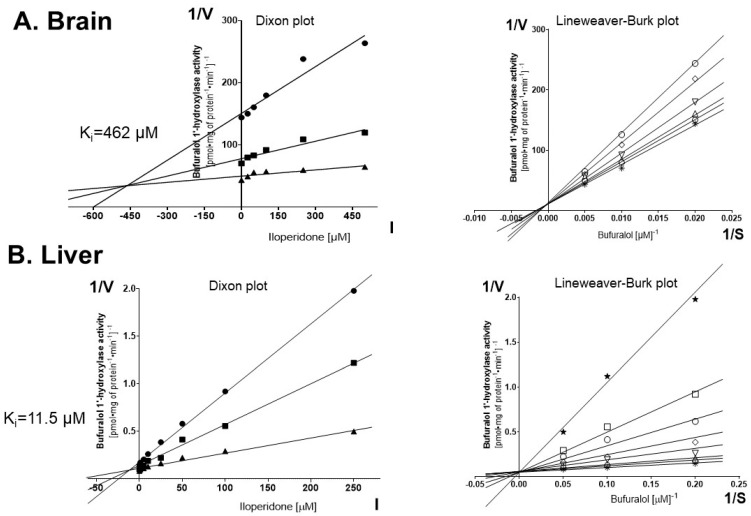
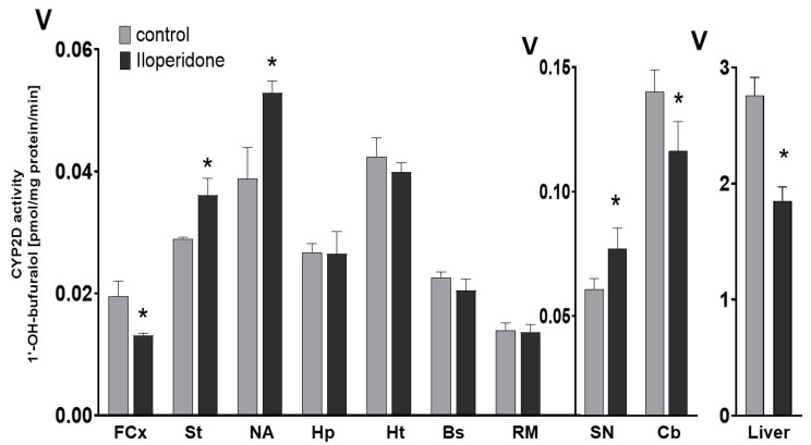
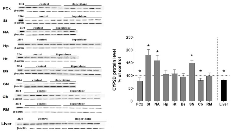
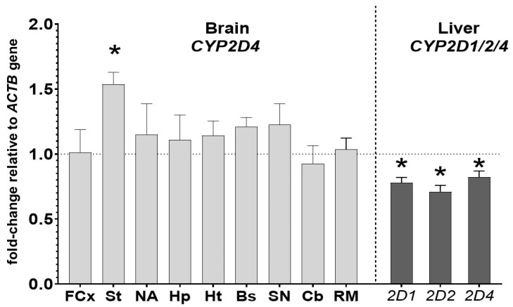
CYP2D enzymes engage in the synthesis of endogenous neuroactive substances (dopamine, serotonin) and in the metabolism of neurosteroids. The present work investigates the effect of iloperidone on CYP2D enzyme expression and activity in rat brains and livers. Iloperidone exerted a weak direct inhibitory effect on CYP2D activity in vitro in the liver and brain microsomes (Ki = 11.5 μM and Ki = 462 μM, respectively). However, a two-week treatment with iloperidone (1 mg/kg ip.) produced a significant decrease in the activity of liver CYP2D, which correlated positively with the reduced CYP2D1, CYP2D2 and CYP2D4 protein and mRNA levels. Like in the liver, iloperidone reduced CYP2D activity and protein levels in the frontal cortex and cerebellum but enhanced these levels in the nucleus accumbens, striatum and substantia nigra. Chronic iloperidone did not change the brain CYP2D4 mRNA levels, except in the striatum, where they were significantly increased. In conclusion, by affecting CYP2D activity in the brain, iloperidone may modify its pharmacological effect, via influencing the rate of dopamine and serotonin synthesis or the metabolism of neurosteroids. By elevating the CYP2D expression/activity in the substantia nigra and striatum (i.e., in the dopaminergic nigrostriatal pathway), iloperidone may attenuate extrapyramidal symptoms, while by decreasing the CYP2D activity and metabolism of neurosteroiods in the frontal cortex and cerebellum, iloperidone can have beneficial effects in the treatment of schizophrenia. In the liver, pharmacokinetic interactions involving chronic iloperidone and CYP2D substrates are likely to occur.

Keywords: brain; cytochrome P450 2D expression/activity; iloperidone; liver; prolonged administration.


Conflict of interest statement

The authors declare no conflict of interest.

Figures

Figure 1
Figure 1
The effects of iloperidone added in vitro to pooled brain or liver microsomes (n = 10) on the activity of CYP2D determined as the rate of bufuralol 1′-hydroxylation. Each point shows the mean value of two independent analyses. (A) Brain. Kinetic parameters: Km = 492.1 μM, Vmax = 0.076 pmol∙mg protein−1∙min−1, Ki = 462 µM. Dixon plot: bufuralol concentrations of 50 µM (●), 100 µM (■) and 200 µM (▲). Lineweaver-Burk plot: control—no asenapine (✱); the asenapine concentrations of 25 µM (⎔), 50 µM (△), 100 µM (▽), 250 µM (◊) and 500 µM (○). (B) Liver. Kinetic parameters: Km = 8.26 μM, Vmax = 15.45 pmol∙mg protein−1∙min−1, Ki = 11.5 µM. Dixon plot: bufuralol concentrations of 5 µM (●), 10 µM (■) and 20 µM (▲). Lineweaver–Burk plot: control—no asenapine (✱); the asenapine concentrations of 1 µM (⎔), 5 µM (△), 10 µM (▽), 25 µM (◊), 50 µM (○), 100 µM (◻) and 250 µM (★). V, velocity of the reaction; I, the inhibitor (iloperidone) concentration; S, the substrate (bufuralol) concentration.
Figure 2
Figure 2
The influence of the two-week treatment with iloperidone on the CYP2D activity measured in microsomes derived from the selected brain structures or liver. The presented values are the means ± S.E.M. of 12 samples (from 12 animals) for the cerebellum, remainder of brain and the liver, of 6 samples (each sample contained 2 pooled brain structures from 2 animals) for the frontal cortex, brain stem, striatum and hippocampus, and of 4 samples (each sample contained 3 pooled brain structures from 3 animals) for the hypothalamus, nucleus accumbens and substantia nigra. Student’s t-test: * p < 0.05 vs. control group. FCx—the frontal cortex, St—the striatum, NA—the nucleus accumbens, Hp—the hippocampus, Ht—the hypothalamus, Bs—the brain stem, SN—the substantia nigra, Cb—the cerebellum and RM—the remainder.
Figure 3
Figure 3
The effect of two-week treatment with iloperidone on the CYP2D protein levels measured in microsomes derived from the selected brain structures or liver. The presented values are the means ± S.E.M. of 12 samples (from 12 animals) for the cerebellum, remainder of brain and the liver, of 6 samples (each sample contained 2 pooled brain structures from 2 animals) for the frontal cortex, brain stem, striatum and hippocampus or of 4 samples (each sample contained 3 pooled brain structured from 3 animals) for the hypothalamus, nucleus accumbens and substantia nigra. The representative CYP2D protein bands of the Western blot analysis are shown. Brain or liver microsomal protein (10 µg) was subjected to Western blot analysis. cDNA expressed CYP2D4 protein (Bactosomes) and cDNA expressed CYP2D6 protein (Supersomes) was used as a positive control. Student’s t-test: * p < 0.05 vs. control group. FCx—the frontal cortex, St—the striatum, NA—the nucleus accumbens, Hp—the hippocampus, Ht—the hypothalamus, Bs—the brain stem, SN—the substantia nigra, Cb—the cerebellum and RM—the remainder.
Figure 4
Figure 4
The influence of two-week treatment with iloperidone on the mRNA levels of the CYP2D4 gene in the selected brain structures and on the mRNA levels of the CYP2D1, CYP2D2 and CYP2D4 genes in the liver. The results are expressed as the fold change compared to the ACTB housekeeping gene. The presented values are the mean fold change quantified by the comparative delta-delta Ct method for the control and iloperidone-treated rats (mean ± S.E.M. of 12 samples (from 12 animals) for the cerebellum, remainder of brain and the liver, of 6 samples (each sample contained 2 pooled brain structures from 2 animals) for the frontal cortex, brain stem, striatum and hippocampus or of 4 samples (each sample contained 3 pooled brain structured from 3 animals) for the hypothalamus, nucleus accumbens and substantia nigra). Student’s t-test: * p < 0.05 vs. control group. FCx—the frontal cortex, St—the striatum, NA—the nucleus accumbens, Hp—the hippocampus, Ht—the hypothalamus, Bs—the brain stem, SN—the substantia nigra, Cb—the cerebellum and RM—the remainder.

References

    1. Winship I.R., Dursun S.M., Baker G.B., Balista P.A., Kandratavicius L., Maia-de-Oliveira J.P., Hallak J., Howland J.G. An Overview of Animal Models Related to Schizophrenia. Can. J. Psychiatry. 2019;64:5–17. doi: 10.1177/0706743718773728. - DOI - PMC - PubMed
    1. Caccia S., Pasina L., Nobili A. New Atypical Antipsychotics for Schizophrenia: Iloperidone. Drug Des. Devel. Ther. 2010;4:33–48. doi: 10.2147/DDDT.S6443. - DOI - PMC - PubMed
    1. Tarazi F.I., Stahl S.M. Iloperidone, Asenapine and Lurasidone: A Primer on Their Current Status. Expert Opin. Pharmacother. 2012;13:1911–1922. doi: 10.1517/14656566.2012.712114. - DOI - PubMed
    1. Hertel P., Fagerquist M.V., Svensson T.H. Enhanced Cortical Dopamine Output and Antipsychotic-like Effects of Raclopride by Alpha2 Adrenoceptor Blockade. Science. 1999;286:105–107. doi: 10.1126/science.286.5437.105. - DOI - PubMed
    1. Meltzer H.Y., Li Z., Kaneda Y., Ichikawa J. Serotonin Receptors: Their Key Role in Drugs to Treat Schizophrenia. Prog. Neuropsychopharmacol. Biol. Psychiatry. 2003;27:1159–1172. doi: 10.1016/j.pnpbp.2003.09.010. - DOI - PubMed

Publication types

MeSH terms

LinkOut - more resources