. 2021 Dec 10;10(12):3491.
doi: 10.3390/cells10123491.

Intra-Tumoral Nerve-Tracing in a Novel Syngeneic Model of High-Grade Serous Ovarian Carcinoma

Affiliations

Intra-Tumoral Nerve-Tracing in a Novel Syngeneic Model of High-Grade Serous Ovarian Carcinoma

Jeffrey L Barr et al. Cells. .

Abstract

Dense tumor innervation is associated with enhanced cancer progression and poor prognosis. We observed innervation in breast, prostate, pancreatic, lung, liver, ovarian, and colon cancers. Defining innervation in high-grade serous ovarian carcinoma (HGSOC) was a focus since sensory innervation was observed whereas the normal tissue contains predominantly sympathetic input. The origin, specific nerve type, and the mechanisms promoting innervation and driving nerve-cancer cell communications in ovarian cancer remain largely unknown. The technique of neuro-tracing enhances the study of tumor innervation by offering a means for identification and mapping of nerve sources that may directly and indirectly affect the tumor microenvironment. Here, we establish a murine model of HGSOC and utilize image-guided microinjections of retrograde neuro-tracer to label tumor-infiltrating peripheral neurons, mapping their source and circuitry. We show that regional sensory neurons innervate HGSOC tumors. Interestingly, the axons within the tumor trace back to local dorsal root ganglia as well as jugular-nodose ganglia. Further manipulations of these tumor projecting neurons may define the neuronal contributions in tumor growth, invasion, metastasis, and responses to therapeutics.

Keywords: innervation; nerve-tracing; ovarian cancer; ultrasound.


Conflict of interest statement

Ronny Drapkin is a member of the scientific advisory boards for Repare Therapeutics, Inc. and VOC Health, and Paola D. Vermeer has a patent pending on EphrinB1 inhibitors for tumor control. Daniel W. Vermeer has a patent under licensing agreement with NantHealth for an HPV vaccine.

Figures

Figure 1
Figure 1
Representative serial sections of three HGSOC cases with serial sections histologically stained by H&E and immunohistochemically stained for β-III tubulin. In the first example (A,B) tumor islands (T) are surrounded by stroma. β-III tubulin positive twigs are found coursing through out the stroma. The boxed area is shown in higher magnification (insets) where β-III tubulin positive twigs are in close proximity to tumor cells. Arrows point out additional areas where β-III tubulin positive twigs are in close proximity to another tumor island. In the second case (C,D), tumor and stroma are clearly defined and β-III tubulin positive twigs are found throughout the stroma. The boxed area depicts higher magnification of these regions. In the third representative case (E,F), small tumor islands (T) are surrounded by stroma. The boxed regions are shown in higher magnification and illustrate the complexity of these sprouted twigs. Scale bar, 300 µm. n = 75 cases were analyzed.
Figure 2
Figure 2
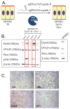
(A) Graphic illustration depicting methodology used to generate an MOSEC line deleted for Trp53 and Pten. CRISPR-Cas9 was used to delete Trp53 and Pten in early passage primary MOSECs. (B) Western blot analysis of CRISPR-Cas9 mediated knockout of Trp53 and Pten. Five clones were evaluated for protein expression, including Pax8, Ovgp1, Pten, phospho-AKT, and β-actin. Loss of Pten protein was associated with acquisition of phospho-AKT in clones 2 and 4. MOSEC represents the parental murine oviductal secretory epithelial cells that was used for genome editing to generate the Trp53; Pten double knockout cell lines. (C) Morphology and immunophenotype of Trp53; Pten double knockout tumors. Top panel: H&E of tumors (20× and 40×) show a morphology consistent with a high-grade carcinoma. Lower panel: immunohistochemistry for Pax8 and WT1 (20×) shows that tumors retain lineage markers associated with high-grade serous carcinomas.
Figure 3
Figure 3
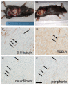
Trp53-/- Pten-/- syngeneic ovarian tumors grow in the peritoneal cavity (A) as well as subcutaneously (B). Immunohistochemical staining of subcutaneous Trp53 -/- Pten -/- tumors demonstrates they harbor β-III tubulin (C), TRPV1 (D), neurofilament (E), peripherin (F) positive nerve twigs (arrows). Scale bar, 50 µm.
Figure 4
Figure 4
Representative ultrasound images of (A) transverse view of intraperitoneal tumor in lateral lower right quadrant of the abdomen showing skin line and fat under the skin. (B) Placement of the needle within the tumor for injection. Dotted line highlights tumor.
Figure 5
Figure 5
(A) Representative 4× confocal image of Trp53 -/- Pten -/- tumor at the site of WGA-A568 (red) injection. (B) Tracer positive (red) intra-tumoral neuronal twigs distal to the injection site. Scale bar, 500 µm.
Figure 6
Figure 6
Representative 10× confocal images of whole mount DRG containing labeled neurons innervating HGSOC. (A) Labeled thoracic DRG neurons following WGA injections into abdominal HGSOC. (B) Labeled neurons of the jugular–nodose ganglia containing sensory peripheral neurons of the vagus nerve. (C) Labeled neurons of lumbar DRG (L4 spinal segment) following tracer injection into subcutaneous tumor in hind limb. (D) Labeling in a thoracic DRG following ip. injection of WGA into non-tumor bearing animal. Scale bar, 250 μm.
Figure 7
Figure 7
Cartoon of spinal cord and associated DRG. Panels (AK) contain fluorescent images of WGA positive DRG from each of the thoracic segments (Scale bar = 250 μm, Cartoon was created using Motifolio Illustration Neuroscience Toolkit, https://www.motifolio.com. accessed on 1 September 2021).
Figure 8
Figure 8
Representative confocal images of whole mount DRG containing WGA labeled (red) neurons innervating HGSOC tumor with TRPV1 (green, (AC)), VIP (green, (DF)) or TH (green, (GI)) staining. Expression of TRPV1 was observed in tracer labeled thoracic DRG neurons (A, arrows) whereas no TH or VIP staining was observed in tracer labeled neurons (D,G). Scale bar = 100 μm.

References

    1. Zheng L., Cui C., Shi O., Lu X., Li Y.-K., Wang W., Li Y., Wang Q. Incidence and mortality of ovarian cancer at the global, regional, and national levels, 1990–2017. Gynecol. Oncol. 2020;159:239–247. doi: 10.1016/j.ygyno.2020.07.008. - DOI - PubMed
    1. Wild C., Weiderpass E., Stewart B., editors. World Cancer Report: Cancer Research for Cancer Prevention. IARC; Lyon, France: 2020.
    1. Jacobs I., Menon U. Progress and Challenges in Screening for Early Detection of Ovarian Cancer. Mol. Cell. Proteom. 2004;3:355–366. doi: 10.1074/mcp.R400006-MCP200. - DOI - PubMed
    1. Badgwell D., Bast R.C., Jr. Early detection of ovarian cancer. Dis. Markers. 2007;23:397–410. doi: 10.1155/2007/309382. - DOI - PMC - PubMed
    1. James N.E., Woodman M., DiSilvestro P.A., Ribeiro J.R. The Perfect Combination: Enhancing Patient Response to PD-1-Based Therapies in Epithelial Ovarian Cancer. Cancers. 2020;12:2150. doi: 10.3390/cancers12082150. - DOI - PMC - PubMed

Publication types

MeSH terms

Substances