. 2021 Dec 11;10(12):3501.
doi: 10.3390/cells10123501.

Protein Kinase A (PRKA) Activity Is Regulated by the Proteasome at the Onset of Human Sperm Capacitation

Affiliations

Protein Kinase A (PRKA) Activity Is Regulated by the Proteasome at the Onset of Human Sperm Capacitation

Héctor Zapata-Carmona et al. Cells. .

Abstract

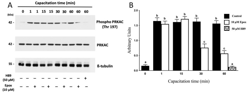
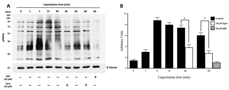
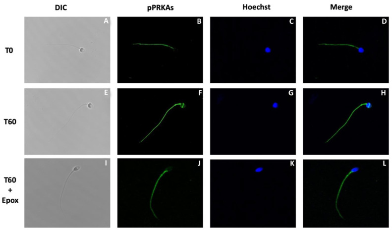
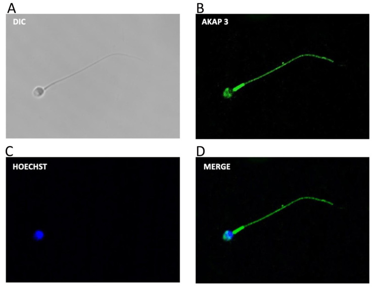
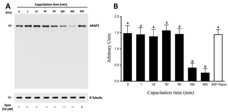
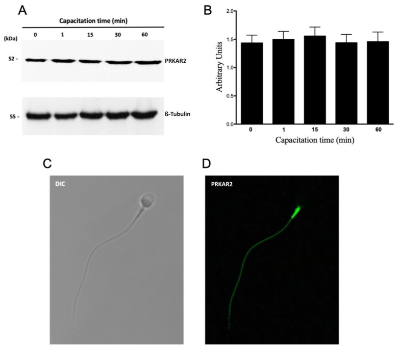
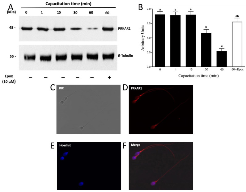
The proteasome increases its activity at the onset of sperm capacitation due to the action of the SACY/PRKACA pathway; this increase is required for capacitation to progress. PRKA activity also increases and remains high during capacitation. However, intracellular levels of cAMP decrease in this process. Our goal was to evaluate the role of the proteasome in regulating PRKA activity once capacitation has started. Viable human sperm were incubated in the presence and absence of epoxomicin or with 0.1% DMSO. The activity of PRKA; the phosphorylation pattern of PRKA substrates (pPRKAs); and the expression of PRKAR1, PRKAR2, and AKAP3 were evaluated by Western blot. The localization of pPRKAs, PRKAR1, PRKAR2, and AKAP3 was evaluated by immunofluorescence. Treatment with epoxomicin changed the localization and phosphorylation pattern and decreased the percentage of pPRKAs-positive sperm. PRKA activity significantly increased at 1 min of capacitation and remained high throughout the incubation. However, epoxomicin treatment significantly decreased PRKA activity after 30 min. In addition, PRKAR1 and AKAP3 were degraded by the proteasome but with a different temporal kinetic. Our results suggest that PRKAR1 is the target of PRKA regulation by the proteasome.

Keywords: AKAP3; PRKA regulatory subunits; capacitation; proteasome; protein kinase A; sperm.


Conflict of interest statement

The authors declare no conflict of interest.

Figures

Figure 1
Figure 1
Effect of proteasome inhibition on the phosphorylation of PRKAC in Thr197 during human sperm capacitation. Human sperm were incubated for different times with 0.1% DMSO, 10 µM epoxomicin, or with 50 μM H89. Time 0 (0 min) corresponds to sperm incubated in non-capacitating conditions. (A) Western blot of PRKA activity evaluated using the anti-phospho Thr197 antibody against the catalytic subunit of PRKA (PKA-C). (B) For the densitometric analysis, total PRKAC/ß-tubulin levels were used as loading control. Bars represent the mean ± S.E.M of five different experiments. Different letters indicate statistically significant differences (p < 0.01) between groups.
Figure 2
Figure 2
Effect of proteasome inhibition on the phosphorylation of PRKA substrates (pPRKAs) during human sperm capacitation. Human sperm were incubated for different times with 0.1% DMSO, 10 µM epoxomicin, or with 50 μM H89. The time 0 (0 min) corresponds to sperm incubated in non-capacitating conditions. (A) Phosphorylated PRKA substrates (pPRKAs) were evaluated by Western blot. (B) For the densitometric analysis, β-tubulin was used as loading control. Bars represent mean ± S.E.M. of seven different experiments. a indicates p < 0.001 vs. corresponding control.
Figure 3
Figure 3
Effect of proteasome inhibition on the localization of phosphorylated PRKA substrates (pPRKAs) during sperm capacitation. Human sperm were incubated for 0 (T0) and 60 (T60) min with 0.1% DMSO or for 60 min with 10 µM epoxomicin (T60 + Epox). Cells were then fixed and labeled with a primary anti-PRKA phosphosubstrates antibody (B,F,J; green) and with Hoechst (C,G,K; blue). DIC: differential interference contrast (A,E,I). Merge: merged image of phosphorylated PRPKA substrates and Hoechst (D,H,L; green and blue).
Figure 4
Figure 4
Subcellular location of AKAP3 during human sperm capacitation. Sperm suspensions were incubated under capacitating conditions for 60 min. Then, the cells were fixed and labeled with a primary anti-AKAP3 antibody (B, green) and with Hoechst (C), (blue). DIC: differential interference contrast (A). Merge: merged image of AKAP3 and Hoechst ((D); green and blue).
Figure 5
Figure 5
The degradation of AKAP3 is dependent on the sperm proteasome. Total protein extracts were obtained from sperm incubated at different times with 0.1% DMSO or for 300 min with 10 μM epoxomicin (Epox). (A) The level of AKAP3 was detected by Western blot using an anti-AKAP3 antibody. (B) For the densitometric analysis, β-tubulin was used as a loading control. Bars represent the mean ± S.E.M of five different experiments. Different letters indicate statistically significant differences (p < 0.01) between the groups.
Figure 6
Figure 6
Protein levels and subcellular localization of PRKAR2 during human sperm capacitation. (A) Total protein extracts were obtained from sperm incubated for 0, 1, 15, 30, or 60 min. The level of PRKAR2 was detected by Western blot using an anti-PRKAR2 antibody. (B) For the densitometric analysis, β-tubulin was used as a loading control. Bars represent the mean ± S.E.M of six different experiments. (C,D) For subcellular localization, the sperm were incubated under capacitated conditions for 1 min. Then, the cells were fixed and labeled with a primary anti-PRKAR2 antibody (D, green). DIC: differential interference contrast (C).
Figure 7
Figure 7
The degradation of PRKAR1 is dependent on the sperm proteasome. Total protein extracts were obtained from sperm incubated at different times with 0.1% DMSO or for 60 min with 10 µM epoxomicin (Epox). (A) The level of PRKAR1 was detected by Western blot using an anti-PRKAR1 antibody. (B) For the densitometric analysis, β-tubulin was used as a loading control. Bars represent the mean ± S.E.M of seven different experiments. Different letters indicate statistically significant differences (p < 0.01) between groups. (CF) For subcellular localization of PRKAR1, sperm were incubated under capacitation conditions for 1 min. Then, the cells were fixed and labeled with a primary anti-PRKARI antibody (D, red) and with Hoechst (E, blue). DIC: differential interference contrast (A). Merge: merged image of PRKAR1 and Hoechst (F; red and blue).

References

    1. Schwartz A.L., Ciechanover A. Targeting proteins for destruction by the ubiquitin system: Implications for human pathobiology. Annu. Rev. Pharmacol. Toxicol. 2009;49:73–96. doi: 10.1146/annurev.pharmtox.051208.165340. - DOI - PubMed
    1. Meyer-Schwesinger C. The ubiquitin–proteasome system in kidney physiology and disease. Nat. Rev. Nephrol. 2019;15:393–411. doi: 10.1038/s41581-019-0148-1. - DOI - PubMed
    1. Schlesinger D.H., Goldstein G., Niall H.D. The complete amino acid sequence of ubiquitin, an adenylate cyclase stimulating polypeptide probably universal in living cells. Biochemistry. 1975;14:2214–2218. doi: 10.1021/bi00681a026. - DOI - PubMed
    1. Bebington C., Doherty F.J., Fleming S.D. The possible biological and reproductive functions of ubiquitin. Hum. Reprod. Update. 2001;7:102–111. doi: 10.1093/humupd/7.1.102. - DOI - PubMed
    1. Ciechanover, a Intracellular protein degradation: From a vague idea thru the lysosome and the ubiquitin-proteasome system and onto human diseases and drug targeting. Cell Death Differ. 2005;12:1178–1190. doi: 10.1038/sj.cdd.4401692. - DOI - PubMed

Publication types

MeSH terms

Substances

LinkOut - more resources