Review
. 2021 Dec 14;10(12):3528.
doi: 10.3390/cells10123528.

Role of Mitochondrial Protein Import in Age-Related Neurodegenerative and Cardiovascular Diseases

Affiliations
Review

Role of Mitochondrial Protein Import in Age-Related Neurodegenerative and Cardiovascular Diseases

Andrey Bogorodskiy et al. Cells. .

Abstract

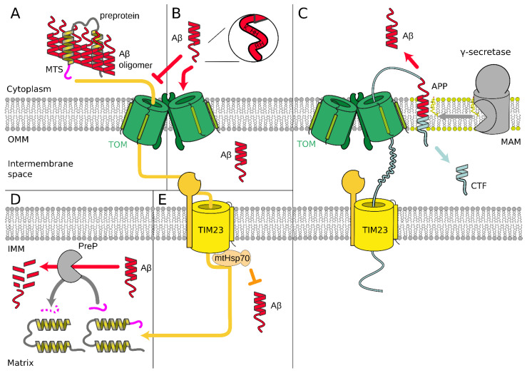
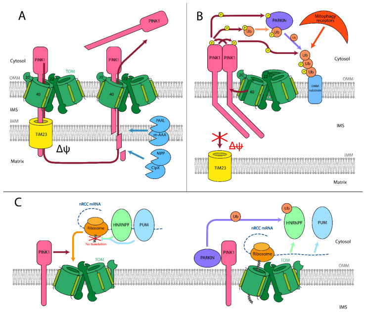
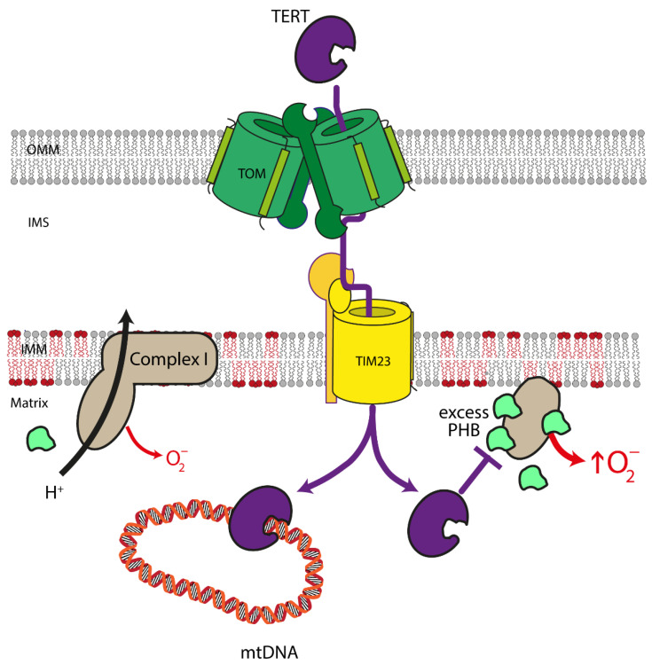
Mitochondria play a critical role in providing energy, maintaining cellular metabolism, and regulating cell survival and death. To carry out these crucial functions, mitochondria employ more than 1500 proteins, distributed between two membranes and two aqueous compartments. An extensive network of dedicated proteins is engaged in importing and sorting these nuclear-encoded proteins into their designated mitochondrial compartments. Defects in this fundamental system are related to a variety of pathologies, particularly engaging the most energy-demanding tissues. In this review, we summarize the state-of-the-art knowledge about the mitochondrial protein import machinery and describe the known interrelation of its failure with age-related neurodegenerative and cardiovascular diseases.

Keywords: Alzheimer’s disease; Parkinson’s disease; TERT; age-related diseases; cardiolipin; cardiovascular disease; mitochondria; mitochondrial protein import.


Conflict of interest statement

The authors declare no conflict of interest.

Figures

Figure 3
Figure 3
(A) Aβ peptides form cytosolic Aβ–preprotein coaggregates that inhibit the initial steps of the mitochondrial preprotein transfer through TIM/TOM complex [98]. (B) Positively charged amino acid residues of Aβ, localized closely at the N-terminal cation binding domain [115], are possibly sufficient for recognition by the TOM complex. Aβ species possibly could be imported into the mitochondria through the TOM complex by the same mechanism as proteins with the signal peptide of mitochondrial localization [116]. (C) Amyloid beta precursor protein (APP) in mitochondria-associated membranes (MAM) [108] is degraded by mitochondrial γ-secretase [112], producing Aβ [96,108,111]. APP possibly blocks TIM/TOM complex by arresting translocation [95,96,97]. (D) Mitochondrial presequence peptidase (PreP) is able to degrade Aβ [118]. Aβ possibly inhibits PreP function of degrading presequences and processing protein intermediates after translocation through TIM/TOM complex [119]. (E) Overexpression of mortalin (mtHsp70) suppresses Aβ-mediated mitochondrial fragmentation and cell death [124].
Figure 1
Figure 1
Overview of mitochondrial protein import in mammalian cells. Proteins are recognized by TOMM70A/TOMM20/TOMM22 and are imported either co- or post-translationally through the TOM complex, containing TOMM40, TOMM22, TOMM5, TOMM6, and TOMM7. Hydrophobic proteins employ Hsp70/90 complex with participation of TOMM34 to prevent misfolding in the cytoplasm. Inside the intermembrane space, depending on the nature and destination of the precursor protein, proteins are delivered to different compartments. β-barrels of the outer membrane are inserted into the outer mitochondrial membrane by SAM complex. Intermembrane space proteins with cysteine motifs are oxidized to the mature form by the MIA40/ALR system. Metabolite carriers are inserted into IMM by TIM22 complex, composed of TIMM22, TIMM29, and acylglycerol kinase (AGK). Other IMM and matrix proteins are inserted/transported by TIM23 complex. Primary TIMM23 pore is associated with TIMM50 (recognizes signals and interacts with TOM complex), TIMM44 with associated mtHsp70 (forming presequence-associated motor helping matrix protein transfer), or TIMM21 for protein release into IMM. Mitochondrial-encoded proteins are inserted into the IMM by OXA1L insertase from the matrix side.
Figure 2
Figure 2
Role of mitochondrial protein import in Parkinson’s disease. (A) PINK1/PARKIN mitochondrial quality control model. In healthy mitochondria, PINK1 is transported through both TOM and TIM complexes, utilizing the IMM electrostatic potential. Afterward, it is cleaved inside the matrix and on the IMM by mitochondrial proteases. The remaining part of PINK1 is dislocated into the cytosol, where it is degraded. This process allows keeping PINK1 levels on the OMM negligible. (B) In unhealthy mitochondria, PINK1 is unable to be transported through the TIM complex. It is ejected from TOMM40 laterally into the OMM, and the ejection is facilitated by TOMM7. PINK1 remains in connection with TOMM20. In that state, it accumulates on the OMM and starts forming homodimers within an HMW PINK1 complex. In dimeric form, PINK1 phosphorylates its dimeric partner and starts to phosphorylate PARKIN and ubiquitin. Phosphorylated PARKIN and ubiquitin form complex and ubiquitinate targets on the OMM. PINK1 phosphorylates ubiquitin on such targets, which attracts mitophagy receptors, starting the mitophagy process. (C) Proposed nRCC translation regulation mechanism. While the nRCC mRNA is in the cytosol and away from the mitochondria, PUM and HNRNPF are attached to it and serve as translation repressors. PINK1 attracts mRCC mRNA to the TOM complex on the outer mitochondrial membrane. Then, PARKIN removes the translation repressors HNRNPF and PUM from the mRNA. For HNRNPF protein, this process involves ubiquitination by PARKIN. When the repressors are released, co-translational synthesis through the TOM complex starts.
Figure 4
Figure 4
Mitochondrial functions of TERT in the cardiovascular system. In the mitochondria, Telomerase Reverse Transcriptase (TERT) binds to mitochondrial DNA (mtDNA) and protects it against damage. In addition, mitochondrial TERT improves the stoichiometry of the different subunits of complex I of the electron transport chain. Moreover, an increase in mitochondrial TERT reduces the levels of Prohibitin (PHB) in these organelles, which—when in excess—stabilizes free matrix arm subunits of complex I of the electron transport chain resulting in increased production of mitochondrial superoxide. Consequently, mitochondrial TERT improves complex I composition and activity, decreases mitochondrial reactive oxygen levels, and, thereby, contributes to enhanced mitochondrial functionality.

References

    1. Pfanner N., Warscheid B., Wiedemann N. Mitochondrial Proteins: From Biogenesis to Functional Networks. Nat. Rev. Mol. Cell Biol. 2019;20:267–284. doi: 10.1038/s41580-018-0092-0. - DOI - PMC - PubMed
    1. Harman D. The Free Radical Theory of Aging. Antioxid. Redox Signal. 2003;5:557–561. doi: 10.1089/152308603770310202. - DOI - PubMed
    1. Park C.B., Larsson N.-G. Mitochondrial DNA Mutations in Disease and Aging. J. Cell Biol. 2011;193:809–818. doi: 10.1083/jcb.201010024. - DOI - PMC - PubMed
    1. Paradies G., Ruggiero F.M., Petrosillo G., Quagliariello E. Age-Dependent Decline in the Cytochrome c Oxidase Activity in Rat Heart Mitochondria: Role of Cardiolipin. FEBS Lett. 1997;406:136–138. doi: 10.1016/S0014-5793(97)00264-0. - DOI - PubMed
    1. Garcia-Euiz C., Mari M., Coiell A., Morales A., Caballero F., Montero J., Terrones O., Basañes G., Fernandez-Checa J.C. Mitochondrial Cholesterol in Health and Disease. Histol. Histopathol. 2009;24:117–132. doi: 10.14670/HH-24.117. - DOI - PubMed

Publication types

Substances