. 2021 Dec 18;10(12):3581.
doi: 10.3390/cells10123581.

Neutrophil-Derived Extracellular Vesicles Activate Platelets after Pneumolysin Exposure

Affiliations

Neutrophil-Derived Extracellular Vesicles Activate Platelets after Pneumolysin Exposure

Eleftheria Letsiou et al. Cells. .

Abstract

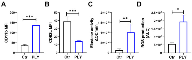
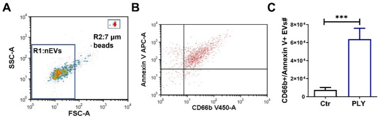
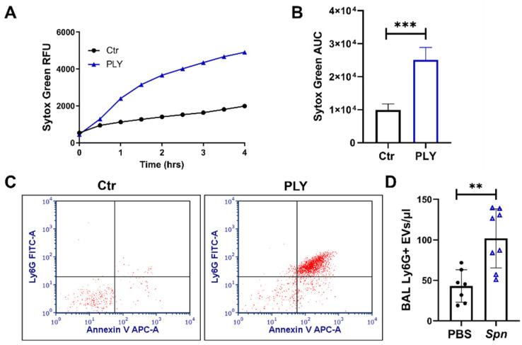
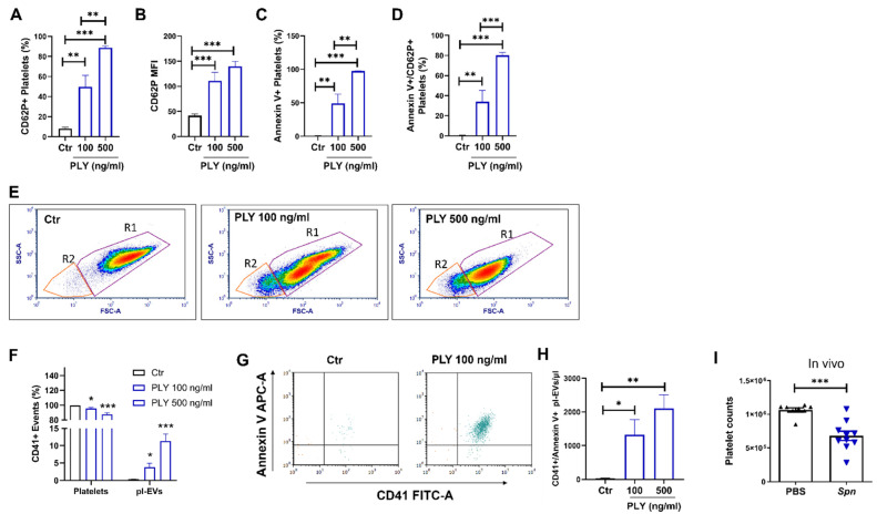
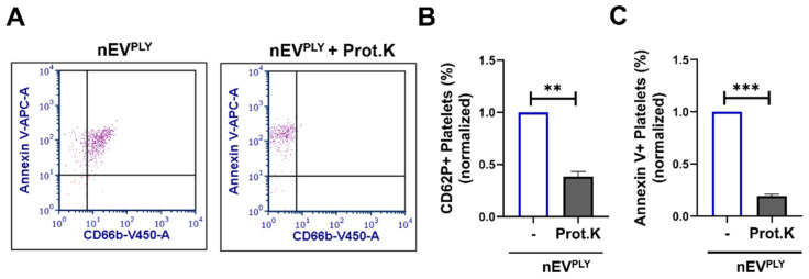
Pneumolysin (PLY) is a pore-forming toxin of Streptococcus pneumoniae that contributes substantially to the inflammatory processes underlying pneumococcal pneumonia and lung injury. Host responses against S. pneumoniae are regulated in part by neutrophils and platelets, both individually and in cooperative interaction. Previous studies have shown that PLY can target both neutrophils and platelets, however, the mechanisms by which PLY directly affects these cells and alters their interactions are not completely understood. In this study, we characterize the effects of PLY on neutrophils and platelets and explore the mechanisms by which PLY may induce neutrophil-platelet interactions. In vitro studies demonstrated that PLY causes the formation of neutrophil extracellular traps (NETs) and the release of extracellular vesicles (EVs) from both human and murine neutrophils. In vivo, neutrophil EV (nEV) levels were increased in mice infected with S. pneumoniae. In platelets, treatment with PLY induced the cell surface expression of P-selectin (CD62P) and binding to annexin V and caused a significant release of platelet EVs (pl-EVs). Moreover, PLY-induced nEVs but not NETs promoted platelet activation. The pretreatment of nEVs with proteinase K inhibited platelet activation, indicating that the surface proteins of nEVs play a role in this process. Our findings demonstrate that PLY activates neutrophils and platelets to release EVs and support an important role for neutrophil EVs in modulating platelet functions in pneumococcal infections.

Keywords: NETs; Streptococcus pneumoniae; lung injury; microvesicles; pneumonia.


Conflict of interest statement

The authors declare no conflict of interest.

Figures

Figure 1
Figure 1
Pneumolysin (PLY) induces neutrophil activation. Human neutrophils were treated with PLY (100 ng/mL, 30 min) and cell surface expression (MFI) of (A) CD11b or (B) CD62L was assessed using flow cytometry. (C) Elastase activity was determined in cell culture supernatants. (D) ROS production was measured using a luminol based kinetic assay. Bars represent area under the curve (AUC) values calculated from kinetic curves. n = 5–9. * p < 0.05, ** p < 0.01, *** p < 0.001, t-test.
Figure 2
Figure 2
Pneumolysin triggers the formation of neutrophil extracellular traps (NETs). (A,B) NET formation was quantified using the cell-impermeant nucleic acid dye, SYTOX Green, in human neutrophils treated with PLY (100 or 500 ng/mL). (A) NET production (SYTOX Green fluorescence) was monitored over a period of 4 h (representative graph), (B) Bars represent the area under the curve (AUC) of Sytox Green fluorescence for Ctr and PLY-treated cells calculated from the kinetic curves. n = 12, * p < 0.05, ** p < 0.01, one-way ANOVA. (C) NET production in neutrophils pre-treated with U0126 (25 μM) and then challenged with PLY (100 ng/mL). AUC for each condition was calculated from the kinetic curves (Sytox Green assay). Each line represents neutrophils from the same donor differentially treated. n = 4. * p < 0.05, paired t-test. (D) Neutrophils were treated with PLY, fixed, and stained with the nuclear stain DAPI. Depicted are representative pseudocolor images. Red represents intact nuclei. White staining indicates extracellular DNA. Arrows indicate extracellular DNA structures. Pictures were taken at 40x magnification. Scale bar: 50 μm. n = 3. (E) Elastase activity was measured in isolated NETs from PLY-treated or control neutrophils. n = 7–9. * p < 0.05, *** p < 0.001, one-way ANOVA.
Figure 3
Figure 3
Pneumolysin induces extracellular vesicle release from human neutrophils. Human neutrophils were treated with PLY (100 ng/mL, 1h) or vehicle (Ctr). Neutrophil-EVs (nEVs) were isolated from the cell culture supernatant and analyzed by flow cytometry. (A) Representative dot plot of isolated human nEVs. Gate 1 (R1) includes events less than 1 μm; 7 μm counting beads were included in each sample for nEV quantification (gate 2; R2). (B) Representative dot plot showing nEVs after staining with annexin V and CD66b (double-positive events are within the upper right quadrant), and (C) Quantification of nEVs released from Ctr and PLY-treated neutrophils. n = 9. *** p < 0.001, paired t-test.
Figure 4
Figure 4
Neutrophil extracellular trap and EV production in murine neutrophils. (A,B) NET formation was quantified using the cell-impermeant nucleic acid dye Sytox Green in murine neutrophils treated with PLY (100 ng/mL) or HBSS (Ctr). (A) NET production (Sytox Green fluorescence) was monitored over a period of 4 h (representative graph). (B) Bars represent the area under the curve (AUC) for Ctr and PLY-treated neutrophils calculated from the kinetic curves. n = 9, *** p < 0.001, paired t-test. (C) Representative dot blots of nEVs isolated from murine neutrophils treated with vehicle (Ctr) or PLY (100 ng/mL) and stained with annexin V-APC and Ly6G-FITC. Double-positive EVs are within the upper right quadrant. n = 3. (D) Quantification of nEVs (Ly6G+) in the BAL of mice 48 h after S. pneumoniae (Spn) infection. n = 7–8 mice/group. ** p < 0.01, t-test.
Figure 5
Figure 5
Pneumolysin activates platelets and induces the release extracellular vesicles (pl-Evs). (AH) Human platelets (diluted PRP) were stimulated with PLY (100 or 500 ng/mL, 30 min), stained with CD62P (PerCP) antibody or annexin V (APC), and then analyzed by flow cytometry. (A) Percentage of CD62P positive platelets, (B) Median fluorescence intensity (MFI) of CD62P, (C) Percentage of annexin V positive platelets, and (D) Percentage of CD62P/annexin V double-positive platelets. (E) Representative FSC versus SSC dot plots of resting platelets (Ctr) or platelets treated with PLY (100 or 500 ng/mL). R1 gate includes platelets and R2 gate includes pl-EVs. (F) Quantification of the percentage of platelets and pl-EVs for each condition. (G) Representative dot plots of pl-EVs (gate R2; (E)) and (H) corresponding quantification of EVs from Ctr or PLY-treated platelets (CD41/annexin V double positive). (I) Platelet counts in whole blood of mice infected with S. pneumoniae (Spn) (48 h). * p < 0.05, ** p < 0.01, *** p < 0.001. (AH) n = 7–9, one-way ANOVA, (F) comparisons are vs. corresponding controls, (I) n = 8–10 mice per condition, Mann–Whitney test.
Figure 6
Figure 6
Effects of neutrophil extracellular traps (NETs) and vesicles (nEVs) on platelets. Human platelets were stimulated with nEVs (A–C) or NETs (D–F) from control or PLY-stimulated neutrophils, and CD62P/annexin V expression was assessed by flow cytometry. (A) Percentage of CD62P positive platelets, (B) median fluorescence intensity (MFI) of CD62P in platelets, and (C) percentage of annexin V positive platelets treated with nEVs (derived from Ctr- or PLY-treated neutrophils; nEVCtr, nEVPLY). (D) Percentage of CD62P positive platelets, (E) MFI of CD62P in platelets, and (F) percentage of annexin V positive platelets treated with NETs (from Ctr- or PLY-treated neutrophils; NETCtr, NETPLY). n = 5–10. * p < 0.05, ** p < 0.01, *** p < 0.001, Kruskal–Wallis test.
Figure 7
Figure 7
Neutrophil extracellular vesicle (nEV) surface proteins mediate platelet activation. Human platelets were stimulated with nEVs (pre-treated with proteinase K or not) from PLY-stimulated neutrophils (nEVPLY). nEVs and platelets were analyzed by flow cytometry. (A) Staining of nEVPLY with CD66b (Pacific Blue) and annexin V (APC) shows that proteinase K treatment effectively removes proteins from EVs but not lipids (annexin V binds to PS). Representative dot plots are depicted. Percentage of (B) CD62P positive or (C) annexin V positive platelets treated with nEVPLY (proteinase K pre-treated or not). n = 3. ** p < 0.01, *** p < 0.001, Welch’s t-test.

References

    1. Dockrell D.H., Whyte M.K.B., Mitchell T.J. Pneumococcal pneumonia: Mechanisms of infection and resolution. Chest. 2012;142:482–491. doi: 10.1378/chest.12-0210. - DOI - PMC - PubMed
    1. Müller-Redetzky H., Lienau J., Suttorp N., Witzenrath M. Therapeutic strategies in pneumonia: Going beyond antibiotics. Eur. Respir. Rev. 2015;24:516–524. doi: 10.1183/16000617.0034-2015. - DOI - PMC - PubMed
    1. Feldman C., Anderson R. Pneumococcal virulence factors in community-acquired pneumonia. Curr. Opin. Pulm. Med. 2020;26:222–231. doi: 10.1097/MCP.0000000000000674. - DOI - PubMed
    1. Anderson R., Feldman C. Pneumolysin as a potential therapeutic target in severe pneumococcal disease. J. Infect. 2017;74:527–544. doi: 10.1016/j.jinf.2017.03.005. - DOI - PubMed
    1. Nishimoto A.T., Rosch J.W., Tuomanen E.I. Pneumolysin: Pathogenesis and Therapeutic Target. Front. Microbiol. 2020;11:1543. doi: 10.3389/fmicb.2020.01543. - DOI - PMC - PubMed

Publication types

MeSH terms

Substances

LinkOut - more resources