. 2021 Nov 30;11(12):3413.
doi: 10.3390/ani11123413.

Histological and Ultrastructural Description of Benign Adipocytic Tumors in Farmed Striped Sea Bream (Lythognathus mormyrus)

Affiliations

Histological and Ultrastructural Description of Benign Adipocytic Tumors in Farmed Striped Sea Bream (Lythognathus mormyrus)

Massimo Orioles et al. Animals (Basel). .

Abstract

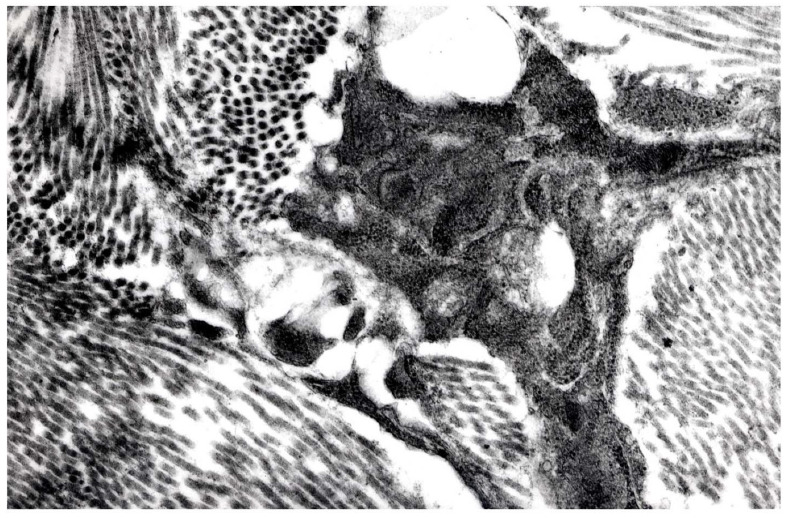
Cutaneous neoplasms affecting wild striped bream (Lythognathus mormyrus) have been recorded after their introduction in a marine aquaculture farm in the Adriatic Sea. The tumors were evident on 24% of the reared fish, showing single or multiple nodules, with a diameter ranging between 0.5-4.0 cm. Histologically, all the neoplastic lesions were located in the stratum spongiosum of the dermis and were surrounded by a thin capsule of connective tissue. The tumors were predominantly composed of adipocytes grouped and surrounded by a thin net of fibroblasts and collagen fibers. In some lipomas a mixture of adipocytes and uniform spindle cells were also observed. Fibroblasts and collagen fibers, or spindle cells, showing few mitotic figures were mainly observed in other nodules. Three of the tumors showed bands of cells with elongated nuclei. Five neoplasms differed from the classic spindle cell lipoma due to the presence of scattered giant cells. These cells presented acidophilic abundant cytoplasm with multiple hyperchromatic nuclei showing a concentric "floret-like" arrangement. The tumors were further characterized by ultrastructural observations that allowed ruling out the presence of virus-like particles within the lesions. Histological features of the masses lead to the identification of four prevalent patterns of neoplasms: lipoma, fibrolipoma, spindle cell lipoma (SCL), and atypical spindle cell-like lipoma (ASCL). The different neoplasms could arise from the transformation of mesenchymal cells of dermal origin. To the author's knowledge, this is the first report describing key differential histological and ultrastructural features of these neoplasms in striped sea bream.

Keywords: atypical spindle cell-like lipoma; fibrolipoma; lipoma; spindle cell lipoma; striped sea bream; tumors.


Conflict of interest statement

The authors declare no conflict of interest.

Figures

Figure 1
Figure 1
Cut section (a) and lateral (b) and dorsal (c) view of the masses on striped sea bream showing the exophytic growth and a well-circumscribed appearance, mainly present on fish flank. Multiple masses are present on the same fish (bar = 1 cm).
Figure 2
Figure 2
(a,b) LIPOMA. (a,b). Histological sections of a lipoma. (a) shows the lipoma arising from the dermis. The epidermal layer is mostly unaffected H&E (bar = 500 µm). The capsule is present, and infiltration of lymphocytes can be seen in the subcapsular area. (b) Higher magnification of (a). The nodule is composed of adipocytes, generally well separated from each other by a thin cytoplasm layer. The large central vacuole displaces the nuclei to the cellular periphery. Cells are almost uniform in size and shape. H&E (bar = 100 µm).
Figure 3
Figure 3
(ac). FIBROLIPOMA. (ac). Histological section of a fibrolipoma. (a) shows the prevalence of fibroblasts and the collagen fibers especially in the center of the neoplasia. Groups of infiltrating adipocytes and adipoblasts are located among the connective component. The capsule is present, and infiltration of lymphocytes (black arrows) is present in the subcapsular area. H&E (bar = 500 µm). (b) Higher magnification of the (a) showing groups of adipocytes in between the fibroblastic component H&E (bar = 100 µm). (c) shows a stroma composed by a mixture of collagenous and reticular fibers which stains blue with Masson’s trichromic and are arranged in wavy bundles. Masson’s stain (bar = 200 µm).
Figure 4
Figure 4
(a,b) SPINDLE CELL LIPOMA (SCL). (a,b). Histological section of a spindle cells lipoma. (a) shows a net of fibroblasts and collagen fibers surrounding mature adipocytes. Spindle cells are often located in one side of the nodule, while the other side reveals mainly adipocytes. H&E (bar = 500 µm). (b) shows how spindle cells tend to be well aligned or sometimes assuming an irregular distribution pattern. These cells tend to have a mild cellular pleomorphism. H&E (bar = 50 µm).
Figure 5
Figure 5
(a,b). ATYPICAL SPINDLE CELL-LIKE LIPOMA (ASCL). (a,b). Histological section of an atypical spindle cell-like lipoma. These neoplasms display a prevalent aspect of spindle cell lipoma, for the abundance of spindle-shaped and fibroblastic components. The structure consists of poorly defined whorls and interlacing bundles of fibroblasts (b) H&E (bar = 100 µm). (b) Higher magnification of the (a). The image shows a mixture of adipocytes and uniform spindle cells. These are associated with bundles of collagen. These tumors differ from the spindle-shaped lipoma due to the presence of scattered bizarre multinucleated giant cells (black arrow) H&E (bar = 50 µm).
Figure 6
Figure 6
(a,b) Tumor n. 6 (SPINDLE CELL LIPOMA). (a) Lipoblasts with intracytoplasmatic lipidic droplets of various size (12,000×), (b) Spindle cells: the nucleus presents folded and corrugate profile (8700×).
Figure 7
Figure 7
Tumor n. 12 (ATYPICAL SPINDLE CELL-LIKE LIPOMA). Pleomorphic cells with irregular borders within abundant collagen matrix (14,000×).

References

    1. Roberts R.J., editor. Fish Pathology. 4th ed. WB Saunders Co; London, UK: 2012. Neoplasia of teleosts; pp. 167–185.
    1. Vergneau-Grosset C., Nadeau M.E., Groff J.M. Fish Oncology: Diseases, diagnostics and therapeutics. Vet. Clin. N. Am. Exot. Anim. Pract. 2017;20:21–56. doi: 10.1016/j.cvex.2016.07.002. - DOI - PubMed
    1. Romano L.A., Pedrosa V.F. Neoplasias in Fish: Review of the Last 20 Years. A Look from the Pathology. Annu. Res. Rev. Biol. 2020;35:134–153. doi: 10.9734/arrb/2020/v35i1230319. - DOI
    1. Hendrick M.J., Mahaffey E.A., Moore F.M., Vos J.H., Walder E.J. Histological Classification of Mesenchymal Tumors of Skin and Soft Tissues of Domestic Animals. Volume 2 Armed Forces Institute of Pathology; Washington, DC, USA: 1998. (2nd Series).
    1. Hendrick M.J. Mesenchymal Tumors of the Skin and Soft Tissues. In: Meuten D.J., editor. Tumors in Domestic Animals. 5th ed. Iowa State Press; Ames, IA, USA: 2016. pp. 142–165.