. 2021 Dec 2;11(12):1812.
doi: 10.3390/biom11121812.

The Nuts and Bolts of SARS-CoV-2 Spike Receptor-Binding Domain Heterologous Expression

Affiliations

The Nuts and Bolts of SARS-CoV-2 Spike Receptor-Binding Domain Heterologous Expression

Mariano Maffei et al. Biomolecules. .

Abstract

COVID-19 is a highly infectious disease caused by a newly emerged coronavirus (SARS-CoV-2) that has rapidly progressed into a pandemic. This unprecedent emergency has stressed the significance of developing effective therapeutics to fight the current and future outbreaks. The receptor-binding domain (RBD) of the SARS-CoV-2 surface Spike protein is the main target for vaccines and represents a helpful "tool" to produce neutralizing antibodies or diagnostic kits. In this work, we provide a detailed characterization of the native RBD produced in three major model systems: Escherichia coli, insect and HEK-293 cells. Circular dichroism, gel filtration chromatography and thermal denaturation experiments indicated that recombinant SARS-CoV-2 RBD proteins are stable and correctly folded. In addition, their functionality and receptor-binding ability were further evaluated through ELISA, flow cytometry assays and bio-layer interferometry.

Keywords: COVID-19; SARS-CoV-2; heterologous expression; protein production; receptor-binding domain; spike protein.


Conflict of interest statement

Mariano Maffei, Grazia Vitagliano, Shaila Sellathurai, Federica Bucci, Alessia Muzi and Valerio Chiarini are employees of the companies Evvivax and Takis. Mirco Compagnone is an employee at NeoMatrix. Luigi Fedele is a former employee of the company Takis. Giuseppe Roscilli and Emanuele Marra are co-founders of the companies Takis and Evvivax. The other authors declare no conflict of interest.

Figures

Figure 1
Figure 1
SARS-CoV-2 RBD production in E. coli, insect and mammalian cells. (a) Schematic representation of the RBD protein constructs expressed in E. coli (left), insect cells (middle) and mammalian HEK-293 cells (right). (b) Diagram summarizing the RBD recombinant expression from E. coli (left), insect cells (middle) and mammalian HEK-293 cells (right) and the subsequent purification. (c) SDS-PAGE (left panel) and Western blot analysis (right panels) of E. coli-purified RBD protein. (d) SDS-PAGE (left panel) and Western blot analysis (right panels) of RBD fragment produced in Hi-5 insect cells and mammalian HEK-293. L = molecular weight ladder. (e) Theoretical molecular masses calculated according to RBD amino acid composition (above) and RBD production yields (below).
Figure 2
Figure 2
Biochemical characterization of recombinant RBD. (a) Gel filtration chromatographic profiles. Protein separation was performed at room temperature using a Superdex 200 Increase 10/300 GL with 40 µg of RBD produced in HEK-293 (black), 47 µg of RBD produced in insect (blue) and 40 µg of RBD produced in E. coli (green), each in 50 mM Tris·HCl and 150 mM NaCl (pH 8.3). (b) Far-UV CD spectra of RBD produced in HEK-293 (black), Insect (blue) and E. coli (green) cells. All spectra were collected at 20 °C, using a 0.1 cm path length quartz cuvette. (c) The histogram reports the distribution of the secondary structure content determined for the RBD proteins (at least three independent CD experiments (means ± standard deviation)), in comparison with the secondary structure composition of RBD reported by Lan et al. (dark grey bars, Literature) [33]. (d) Thermal denaturation profiles of RBD E. coli (green), Insect (blue) and HEK-293 (black), continuously monitored by far-UV CD at 222 nm over the range 293–350 K. Data were fitted using a two-state model. The estimated thermodynamic parameters derived from these analyses are presented in Table S1.
Figure 3
Figure 3
ELISA assays. (a) Serum from immunized rat with COVID-eVax was used to compare different concentrations (1, 3 and 5 μg/mL) of the RBD expressed in E. coli (green), insect (blue) and HEK-293 cells (black). The y-axis represents the optical density (OD) measured at 405 nm, while the x-axis accounts for RBD concentrations and serum dilution factors (1:1000, 1:10,000 and 1:50,000). Bars indicate standard deviations. Dashed line = cut-off value. (b) Commercial antibody against the S1 subunit of SARS-CoV-2 Spike was used to compare different concentrations (1, 3 and 5 μg/mL) of RBD produced in E. coli (green), insect (blue) and HEK-293 cells (black). Optical density (OD) was measured at 450 nm and bars indicate standard deviations. Dashed line = cut-off value.
Figure 4
Figure 4
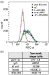
Flow cytometry assays. RBD-Vero E6 cells binding experiment. (a) Gray curve, Vero E6 cells alone; red curve, Vero E6 cells incubated with only secondary antibody; green curve, Vero E6 cells incubated with E. coli-RBD; blue curve, Vero E6 cells incubated with Insect-RBD; black curve, Vero E6 cells incubated with HEK-293-RBD. Incubation with RBD was followed by anti-RBD primary antibody and secondary antibody. (b) Intensity of the staining measured as geometric mean (median fluorescence intensity, MFI) value.
Figure 5
Figure 5
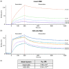
BLI measurements. (a) BLI profiles accounting for the binding of insect-RBD and (b) HEK-293-RBD to ACE2-hFc. After a baseline, the sensorgram starts with the association (0–300 s) of the RBD to the ACE2-loaded sensor, followed by the dissociation phase (900 s). (c) Kd measured values.

References

    1. Lai C.C., Shih T.P., Ko W.C., Tang H.J., Hsueh P.R. Severe acute respiratory syndrome coronavirus 2 (SARS-CoV-2) and coronavirus disease-2019 (COVID-19): The epidemic and the challenges. Int. J. Antimicrob. Agents. 2020;55:105924. doi: 10.1016/j.ijantimicag.2020.105924. - DOI - PMC - PubMed
    1. World Health Organization (WHO) Coronavirus. 2021. [(accessed on 27 October 2021)]. Available online: https://covid19.who.int.
    1. Tai W., He L., Zhang X., Pu J., Voronin D., Jiang S., Zhou Y., Du L. Characterization of the receptor-binding domain (RBD) of 2019 novel coronavirus: Implication for development of RBD protein as a viral attachment inhibitor and vaccine. Cell. Mol. Immunol. 2020;17:613–620. doi: 10.1038/s41423-020-0400-4. - DOI - PMC - PubMed
    1. Wang M.-Y., Zhao R., Gao L.-J., Gao X.-F., Wang D.-P., Cao J.-M. SARS-CoV-2: Structure, Biology, and Structure-Based Therapeutics Development. Front. Cell. Infect. Microbiol. 2020;10:587269. doi: 10.3389/fcimb.2020.587269. - DOI - PMC - PubMed
    1. Yang Y., Du L. SARS-CoV-2 spike protein: A key target for eliciting persistent neutralizing antibodies. Signal Transduct. Target. Ther. 2021;6:1–3. doi: 10.1038/s41392-021-00523-5. - DOI - PMC - PubMed

Publication types

MeSH terms

Substances