Review
. 2021 Dec 9;11(12):1849.
doi: 10.3390/biom11121849.

The Annexin A2/S100A10 Complex: The Mutualistic Symbiosis of Two Distinct Proteins

Affiliations
Review

The Annexin A2/S100A10 Complex: The Mutualistic Symbiosis of Two Distinct Proteins

Alamelu Bharadwaj et al. Biomolecules. .

Abstract

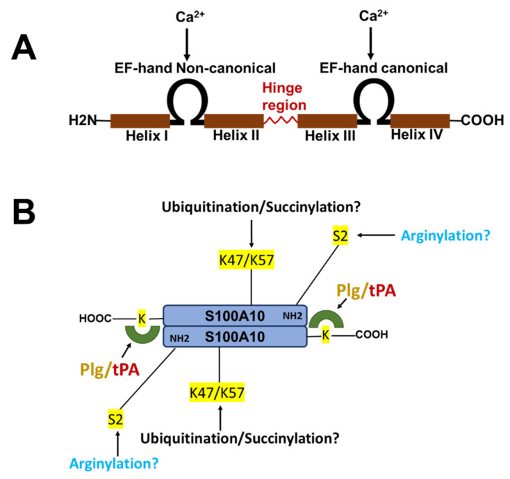
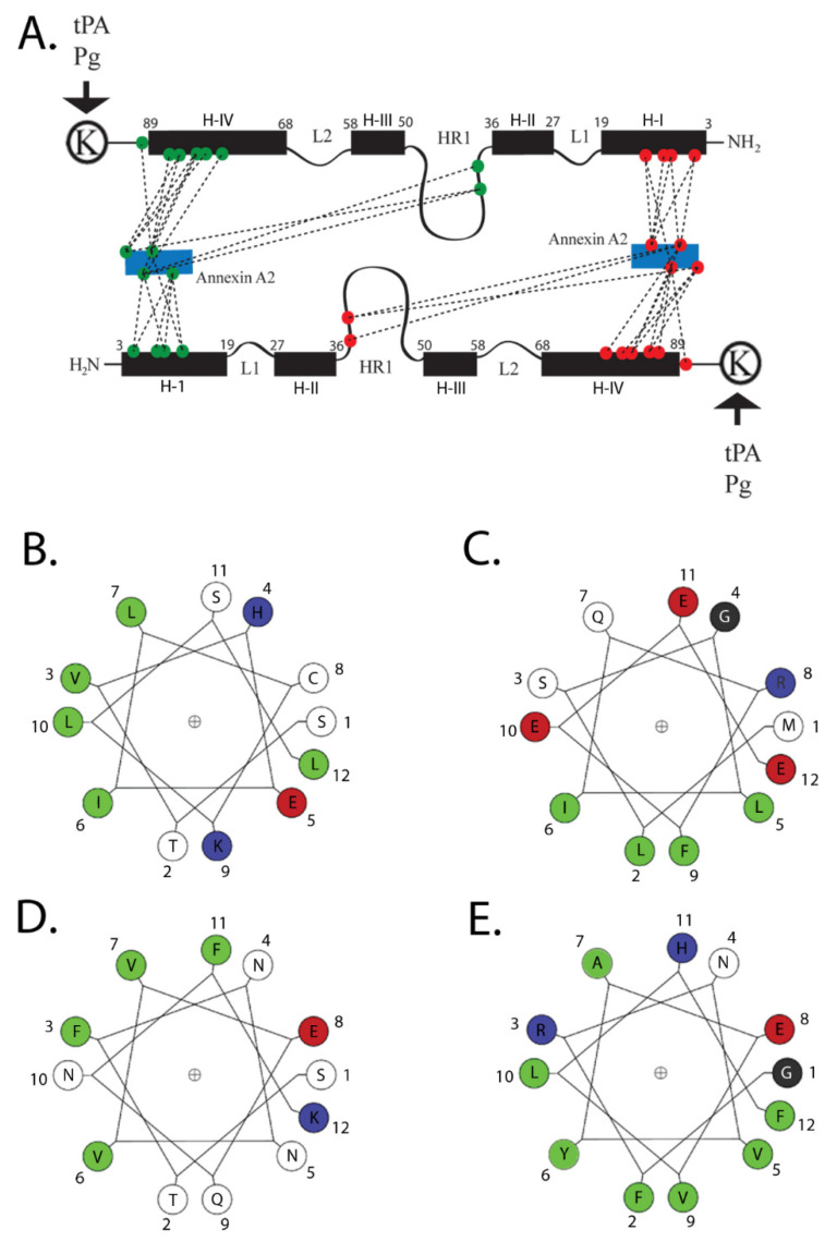
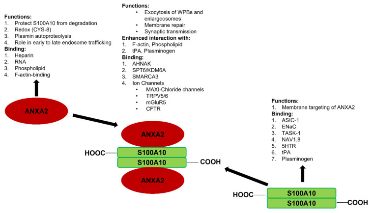
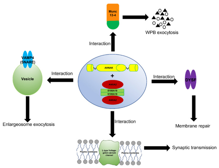
Mutualistic symbiosis refers to the symbiotic relationship between individuals of different species in which both individuals benefit from the association. S100A10, a member of the S100 family of Ca2+-binding proteins, exists as a tight dimer and binds two annexin A2 molecules. This association forms the annexin A2/S100A10 complex known as AIIt, and modifies the distinct functions of both proteins. Annexin A2 is a Ca2+-binding protein that binds F-actin, phospholipid, RNA, and specific polysaccharides such as heparin. S100A10 does not bind Ca2+, but binds tPA, plasminogen, certain plasma membrane ion channels, neurotransmitter receptors, and the structural scaffold protein, AHNAK. S100A10 relies on annexin A2 for its intracellular survival: in the absence of annexin A2, it is rapidly destroyed by ubiquitin-dependent and independent proteasomal degradation. Annexin A2 requires S100A10 to increase its affinity for Ca2+, facilitating its participation in Ca2+-dependent processes such as membrane binding. S100A10 binds tissue plasminogen activator and plasminogen, and promotes plasminogen activation to plasmin, which is a process stimulated by annexin A2. In contrast, annexin A2 acts as a plasmin reductase and facilitates the autoproteolytic destruction of plasmin. This review examines the relationship between annexin A2 and S100A10, and how their mutualistic symbiosis affects the function of both proteins.

Keywords: S100A10; annexin A2; ion channels; plasmin; plasminogen.


Conflict of interest statement

The authors declare no conflict of interest.

Figures

Figure 1
Figure 1
Structure of S100 proteins. (A) The structure of a typical S100 protein. (B) The structure and key regulatory sites of S100A10. Shown are the sites for ubiquitylation, arginylation and succinylation.
Figure 2
Figure 2
Structure of AIIt. The numbering of the amino acids represents the position of the amino acids in the wheel. Colors are—green, hydrophobic; red, acidic; blue, basic; black, neutral. A cartoon of the association of S100A10 with its two primary ligands, ANXA2 and plasminogen, is presented in (A). The figure illustrates the structure of S100A10 and the association of S100A10 with the amino-terminus of ANXA2 and with plasminogen. Each S100A10 monomer is composed of four α-helical domains: H-I, H-II, H-III and H-IV. Separating the H-I and H-II helical regions is a loop, L1. The H-III and H-IV are separated by a second loop (L2). The H-II and H-III are connected by a flexible linker or hinge region (HR1). The points of interaction between the amino-terminus of ANXA2 and S100A10 are quite extensive, and four hydrophobic amino acids of the amino terminus of ANXA2 (V3, I6, L7 and L10) form seven points of contact with helix H-I of one monomer, two points of contact with the hinge region, and nine points of contact with helix H-IV of the other monomer, for a total of nineteen points of contact with S100A10. (B) The helical wheel projections for the S100A10-binding site for ANXA2. (C) Bluetongue virus, NS3; (D) DLC1; and (E) TASK-1 are presented. The S100A10-binding region of these ligands consists of an amphipathic α-helix in which hydrophobic residues form a binding site on one side of the helix. The program for helical wheel projections was obtained from http://lbqp.unb.br › NetWheels/, accessed on 5 November 2021. The numbering of the amino acids represents the position of the amino acids in the wheel. This cartoon and figure legend was originally published in Madureira, O’Connell, Surette, Miller, and Waisman 2012, ‘The Biochemistry and Regulation of S100A10: A Multifunctional Plasminogen Receptor Involved in Oncogenesis. J Biomed Biotechnol 2012:353687’.
Figure 3
Figure 3
Synopsis of the mutualistic symbiotic relationship between S100A10 and ANXA2. Annexin A2 is a monomeric Ca2+-binding protein that binds F-actin, phospholipid, RNA, and specific polysaccharides, such as heparin. S100A10 does not bind Ca2+, but binds tPA, plasminogen, certain plasma membrane ion channels, and neurotransmitter receptors. ANXA2 and S100A10 form a tight 2:2 heterotetrameric complex, which is called AIIt. The formation of AIIt results in the enhancement of many of the functions of each subunit. For example, ANXA2 enhances the ability of S100A10 to bind plasminogen and facilitate the tPA-dependent conversion of plasminogen to plasmin, whereas S100A10 enhances the interaction of ANXA2 with phospholipids. The formation of AIIt permits the interaction of the complex with new binding partners such as certain ion channels, dysferlin, and the structural scaffold protein AHNAK.
Figure 4
Figure 4
Schematic representation of the function of the AIIt-AHNAK complex. The formation of the ANXA2/S100A10/AHNAK (2:2:1) complex forms a key hub for the regulation of several important biological functions such as the exocytosis of enlargesomes, the regulation of the L-type voltage-gated calcium channel (VGCC), and possibly the MUNC-13-dependent regulation of the exocytosis of Weibel–Palade bodies (WPB).

References

    1. Krieg P., Schuppler M., Koesters R., Mincheva A., Lichter P., Marks F. Repetin (Rptn), a New Member of the “Fused Gene” Subgroup within the S100 Gene Family Encoding a Murine Epidermal Differentiation Protein. Genomics. 1997;43:339–348. doi: 10.1006/geno.1997.4818. - DOI - PubMed
    1. Donato R. S100: A Multigenic Family of Calcium-Modulated Proteins of the EF-Hand Type with Intracellular and Extracellular Functional Roles. INTJ Biochem. Biol. 2001;33:637–668. doi: 10.1016/S1357-2725(01)00046-2. - DOI - PubMed
    1. Gonzalez L.L., Garrie K., Turner M.D. Role of S100 Proteins in Health and Disease. Biochim. Biophys. Acta Mol. Cell Res. 2020;1867:118677. doi: 10.1016/j.bbamcr.2020.118677. - DOI - PubMed
    1. Mischke D., Korge B.P., Marenholz I., Volz A., Ziegler A. Genes Encoding Structural Proteins of Epidermal Cornification and S100 Calcium-Binding Proteins Form a Gene Complex (“epidermal Differentiation Complex”) on Human Chromosome 1q21. J. Investig. Dermatol. 1996;106:989–992. doi: 10.1111/1523-1747.ep12338501. - DOI - PubMed
    1. Marenholz I., Heizmann C.W., Fritz G. S100 Proteins in Mouse and Man: From Evolution to Function and Pathology (Including an Update of the Nomenclature) Biochem. Biophys. Res. Commun. 2004;322:1111–1122. doi: 10.1016/j.bbrc.2004.07.096. - DOI - PubMed

Publication types

Grants and funding

LinkOut - more resources