. 2021 Dec 17;11(12):1892.
doi: 10.3390/biom11121892.

Gut Metabolite Trimethylamine N-Oxide Protects INS-1 β-Cell and Rat Islet Function under Diabetic Glucolipotoxic Conditions

Affiliations

Gut Metabolite Trimethylamine N-Oxide Protects INS-1 β-Cell and Rat Islet Function under Diabetic Glucolipotoxic Conditions

Emily S Krueger et al. Biomolecules. .

Abstract

Serum accumulation of the gut microbial metabolite trimethylamine N-oxide (TMAO) is associated with high caloric intake and type 2 diabetes (T2D). Impaired pancreatic β-cell function is a hallmark of diet-induced T2D, which is linked to hyperglycemia and hyperlipidemia. While TMAO production via the gut microbiome-liver axis is well defined, its molecular effects on metabolic tissues are unclear, since studies in various tissues show deleterious and beneficial TMAO effects. We investigated the molecular effects of TMAO on functional β-cell mass. We hypothesized that TMAO may damage functional β-cell mass by inhibiting β-cell viability, survival, proliferation, or function to promote T2D pathogenesis. We treated INS-1 832/13 β-cells and primary rat islets with physiological TMAO concentrations and compared functional β-cell mass under healthy standard cell culture (SCC) and T2D-like glucolipotoxic (GLT) conditions. GLT significantly impeded β-cell mass and function by inducing oxidative and endoplasmic reticulum (ER) stress. TMAO normalized GLT-mediated damage in β-cells and primary islet function. Acute 40µM TMAO recovered insulin production, insulin granule formation, and insulin secretion by upregulating the IRE1α unfolded protein response to GLT-induced ER and oxidative stress. These novel results demonstrate that TMAO protects β-cell function and suggest that TMAO may play a beneficial molecular role in diet-induced T2D conditions.

Keywords: beta cell; glucolipotoxicity (GLT); glucose stimulated insulin secretion (GSIS); islet; type 2 diabetes (T2D); unfolded protein response (UPR).


Conflict of interest statement

The authors declare no conflict of interest. The funders had no role in the design of the study; in the collection, analyses, or interpretation of data; in the writing of the manuscript, or in the decision to publish the results.

Figures

Figure 1
Figure 1
Trimethylamine N-oxide (TMAO) does not alter glucolipotoxicity (GLT)-mediated reduction of INS-1 β-cell mass. (A) Tetrazolium salt MTT measured mitochondrial viability of INS-1 β-cells cultured with or without TMAO for 24 h in standard cell culture (SCC), and (B) in GLT. (C) Flow cytometric measurement of dead and dying β-cell populations as measured by Annexin V and 7-aminoactinomycin D staining of β-cells cultured with or without TMAO for 24 h in SCC, and (D) in GLT. (E) β-cell proliferation measured by [3H]-thymidine incorporation of β-cells cultured with or without TMAO for 24 h in SCC, and (F) in GLT. (A,B,E,F) Values are normalized to SCC no treatment (NT) controls and (C,D) values reported as percent. (E,F) Proliferation data were normalized to cell protein content. All values represent the mean of biological triplicates (n = 3). Error bars indicate the standard error. * Indicates significant one-way ANOVA showing GLT and TMAO effects with p-values * <0.05, *** <0.001, **** <0.0001, or not significant (ns).
Figure 2
Figure 2
TMAO normalizes GLT-damaged INS-1 β-cells and primary rat islet function. (A) Insulin secretion under low (solid bars) vs. high glucose (striped bars) for INS-1 β-cells cultured with or without TMAO for 24 h in SCC condition, and (B) corresponding insulin content. (C) Insulin secretion from β-cells cultured with or without TMAO for 24 h in GLT condition, and (D) corresponding insulin content. (E) Insulin secretion from primary rat islets cultured with or without TMAO for 24 h in SCC, and (F) corresponding insulin content. (G) Insulin secretion from islets cultured with or without TMAO for 24 h in GLT, and (H) corresponding insulin content. All values are normalized to SCC NT controls and represent the mean of biological replicates (n = 3 or 4). (A,C,E,D) # Indicates significant paired two-way ANOVA comparing low vs. high glucose. (AH) Error bars indicate the standard error. * Indicates significant one-way ANOVA showing GLT and TMAO effects, where p-values < 0.1 given, * <0.05, ** <0.01, **** <0.0001, or ns.
Figure 3
Figure 3
TMAO recovers the decrease in INS-1 β-cell insulin granule number induced by GLT. (A) Representative scanning transmission electron microscopy images of INS-1 β-cells cultured with or without 40 µM TMAO for 24 h in SCC and GLT. White arrows identify examples of insulin granules. (B) Quantification of insulin granules/cytosol area. All values represent the mean of biological triplicates (n = 3). Error bars indicate the standard error. * Indicates significant one-way ANOVA showing GLT and TMAO effects, where p-values *** <0.001.
Figure 4
Figure 4
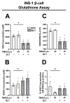
TMAO does not alter GLT-mediated changes to GSH or redox potential in INS-1 β-cells. INS-1 β-cells cultured with or without 40 µM TMAO for 24 h in SCC and GLT were measured for (A) Glutathione (GSH) concentration, (B) Glutathione disulfide (GSSG) concentration, (C) GSH/GSSG redox state, and (D) Protein S-glutathionylation (Pr-SSG) concentration. All values represent the mean of biological triplicates (n = 3). Error bars indicate the standard error. * Indicates significant one-way ANOVA showing GLT effects, where p-values * <0.05 or ns.
Figure 5
Figure 5
TMAO normalizes GLT-mediated reduction of the IRE1α unfolded protein response (UPR) in INS-1 β-cells. INS-1 β-cells cultured with or without 40 µM TMAO for 24 in SCC and GLT were measured for changes to UPR pathway proteins by Western blotting and mRNA by qPCR. Samples were probed and quantified for p-PERK (170kd) and PERK (140kd) (AC), ATF4 (39kd) (D,E), IRE1α (130kd) (F,G), sXBP1 (40kd) and uXBP1 (29kd) (JM). mRNA of sXBP1 and total XBP1 were measured by qPCR (H,I). Abbreviations: quantitative polymerase chain reaction (qPCR), phospho-protein kinase RNA-like ER kinase (p-PERK), activating transcription factor (ATF4), inositol-requiring enzyme 1α (IRE1α) (A,B), spliced and unspliced X-box bind protein 1 (sXBP1, uXBP1). All Western blot values are normalized to the loading control tubulin and represent the mean of biological triplicates (n = 3). qPCR data represent the mean of 6 to 8 replicates (n = 6–8). Error bars indicate the standard error. * Indicates significant one-way ANOVA showing GLT and TMAO effects, where p-values <0.1 given, * <0.05, ** <0.01, *** <0.001, **** <0.0001, or ns.

References

    1. Kones R., Rumana U. Cardiometabolic diseases of civilization: History and maturation of an evolving global threat. An update and call to action. Ann. Med. 2017;49:260–274. doi: 10.1080/07853890.2016.1271957. - DOI - PubMed
    1. Stanhope K.L., Goran M.I., Bosy-Westphal A., King J.C., Schmidt L.A., Schwarz J.M., Stice E., Sylvetsky A.C., Turnbaugh P.J., Bray G.A., et al. Pathways and mechanisms linking dietary components to cardiometabolic disease: Thinking beyond calories. Obes. Rev. 2018;19:1205–1235. doi: 10.1111/obr.12699. - DOI - PMC - PubMed
    1. Kopp W. How Western Diet and Lifestyle Drive the Pandemic of Obesity And Civilization Diseases. Diabetes Metab. Syndr. Obes. Targets Ther. 2019;12:2221–2236. doi: 10.2147/DMSO.S216791. - DOI - PMC - PubMed
    1. Shan Z., Sun T., Huang H., Chen S., Chen L., Luo C., Yang W., Yang X., Yao P., Cheng J., et al. Association between microbiota-dependent metabolite trimethylamine-N-oxide and type 2 diabetes. Am. J. Clin. Nutr. 2017;106:888–894. doi: 10.3945/ajcn.117.157107. - DOI - PubMed
    1. Svingen G.F., Schartum-Hansen H., Pedersen E.R., Ueland P.M., Tell G.S., Mellgren G., Njolstad P.R., Seifert R., Strand E., Karlsson T., et al. Prospective Associations of Systemic and Urinary Choline Metabolites with Incident Type 2 Diabetes. Clin. Chem. 2016;62:755–765. doi: 10.1373/clinchem.2015.250761. - DOI - PubMed

Publication types

MeSH terms