. 2021 Nov 24;9(12):1758.
doi: 10.3390/biomedicines9121758.

Combined Therapy of Vitamin D3-Tolerogenic Dendritic Cells and Interferon-β in a Preclinical Model of Multiple Sclerosis

Affiliations

Combined Therapy of Vitamin D3-Tolerogenic Dendritic Cells and Interferon-β in a Preclinical Model of Multiple Sclerosis

Bibiana Quirant-Sánchez et al. Biomedicines. .

Abstract

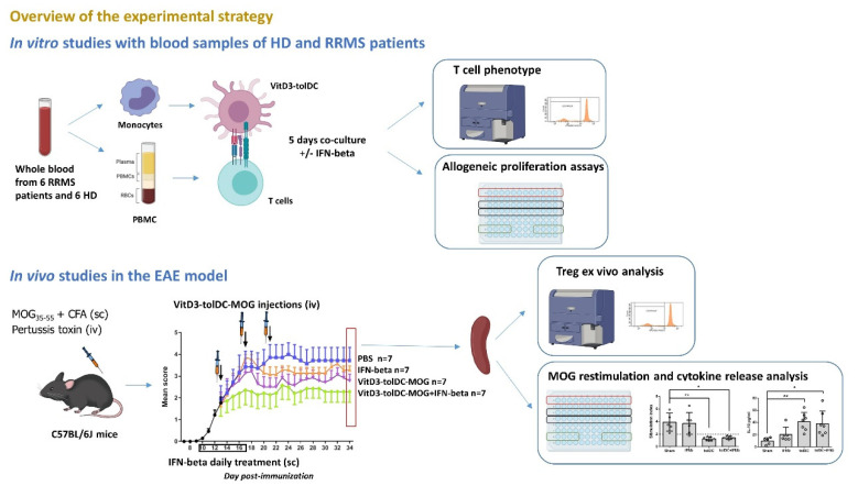
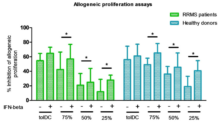
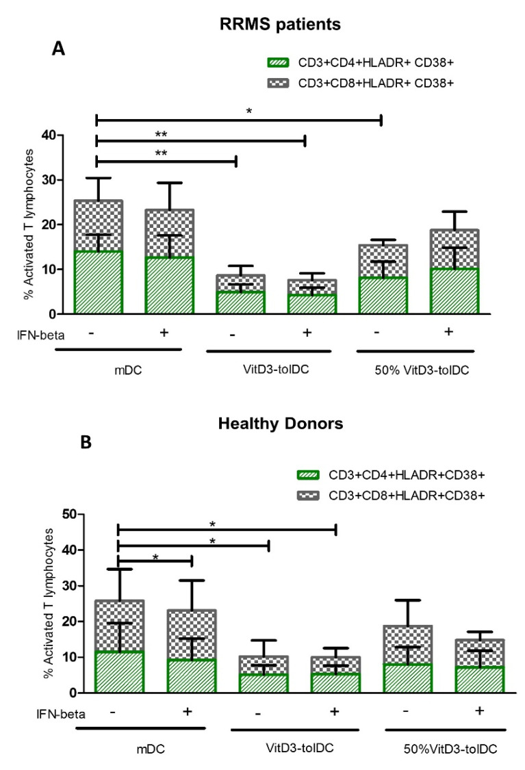
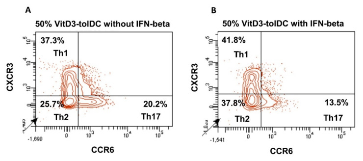
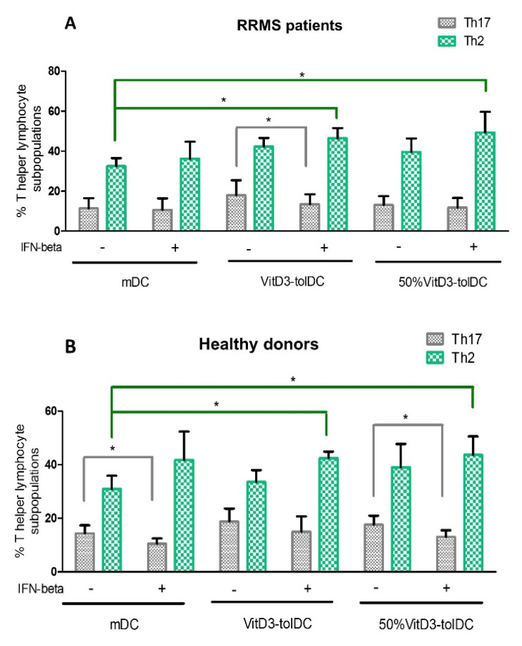
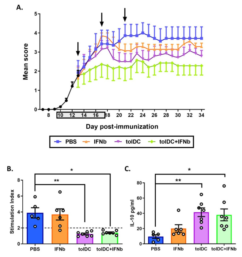
Autologous antigen-specific therapies based on tolerogenic dendritic cells (tolDC) offer the possibility to treat autoimmune diseases by restoring homeostasis and targeting specifically autoreactive responses. Here, we explore the hypothesis that systemic inflammation occurring in autoimmune diseases, such as multiple sclerosis (MS), can generate a disease-specific environment able to alter the functionality of tolDC. In this context in fact, a combined therapy of tolDC with an immunomodulatory treatment could potentiate the beneficial effect of this antigen-specific cell therapy. For this purpose, we analyzed the efficacy of a combined therapy based on the use of vitamin D3 (VitD3)-tolDC plus interferon beta (IFN-beta) in MS. VitD3-tolDC were generated from healthy donors and MS patients and co-cultured with allogeneic peripheral blood mononuclear cells, in the presence or absence of IFN-beta. In vitro, VitD3-tolDC treatment reduced the percentage of activated T cells and allogeneic proliferation, whereas VitD3-tolDC+IFN-beta treatment enhanced the suppressive ability of VitD3-tolDC and, additionally, induced a shift towards a Th2 profile. To determine the clinical benefit of the combined therapy, C57BL/6-experimental autoimmune encephalomyelitis (EAE)-induced mice were treated with antigen-specific VitD3-tolDC and/or IFN-beta. Treatment of EAE mice with combined therapy ameliorated the disease course compared to each monotherapy. These results suggest that a combined therapy based on antigen-specific VitD3-tolDC and IFN-beta may represent a promising strategy for MS patients.

Keywords: antigen-specific therapies; combined therapy; immunomodulatory; multiple sclerosis.


Conflict of interest statement

The authors declare no conflict of interest.

Figures

Figure 1
Figure 1
Overview of the experimental strategy. Two experimental approaches were used to investigate the effect of the combined VitD3-tolDC plus IFN-beta therapy in MS. In vitro studies: we performed in vitro co-cultures of VitD3-tolDC plus PBMC of RRMS patients and of HD (n = 6/group) in presence/absence of IFN-beta. After 5 days of culture, the phenotype of cultured T cells and the induction of allogeneic proliferation by different concentrations of VitD3-tolDC in presence/absence of IFN-beta were analyzed (see Methods for a more detailed explanation); in vivo studies: EAE was induced to C57BL/6J mice. Mice were treated subcutaneously (sc) with IFN-beta (from day 10 to 17 post-immunization, (pi)) and /or intravenously (iv) with antigen-specific VitD3-tolDC (VitD3-tolDC-MOG) (on days 13, 17 and 21 pi) (n = 7/group). Clinical scores were recorded daily. Splenocytes of mice (n = 7/group) were analyzed to evaluate antigen-specific proliferation (ex-vivo restimulation with MOG antigen), secretion of cytokines and percentage of regulatory T cells (Treg). * p < 0.05, ** p < 0.01.
Figure 2
Figure 2
Dose-dependent reduction of allogeneic proliferation of PBMC, cultured with different percentages of VitD3-tolDC in presence/absence of IFN-beta. Suppressive ability of VitD3-tolDC on allogeneic PBMC from RRMS patients (green) and HD (blue) is represented as the percentage of proliferation compared to mature DC (mDC) stimulus. Different percentages of VitD3-tolDC and mDC are represented: 100% (100% VitD3-tolDC), 75% (75% VitD3-tolDC + 25% mDC), 50% (50% VitD3-tolDC + 50% mDC) and 25% (25% VitD3-tolDC +75% mDC) in presence/absence of IFN-beta. Error bars correspond to SEM; * p < 0.05.
Figure 3
Figure 3
VitD3-tolDC reduce the activation of T lymphocytes in RRMS patients and HD. The percentage of activated CD4 + lymphocytes (CD3+CD4+HLA-DR+CD38+) (green) and CD8+ lymphocytes (CD3+CD8+HLA-DR+CD38+) (gray) in co-cultures of PBMC with mature DC (mDC), (100% VitD3-tolDC) or (50%VitD3-tolDC), in presence/absence of IFN-beta was analyzed in relapsing-remitting multiple sclerosis (RRMS) patients (A) and healthy donors (HD) (B) (n = 6 each group). Data represent the relative percentage of each lymphocyte subpopulation n relation to total percentages of CD4+ or CD8+ T cells. Error bars correspond to SEM; * p < 0.05, ** p < 0.01.
Figure 4
Figure 4
Immunomodulatory effect of VitD3-tolDC with/without IFN-beta on T lymphocytes. Representative example of changes in the relative percentages of Th1, Th2 and Th17 lymphocytes in relation to total CD4+ T lymphocytes, after 5 days co-culture of 50% VitD3-tolDC in absence (A) or presence (B) of IFN-beta.
Figure 5
Figure 5
Effect of VitD3-tolDC in presence/absence of IFN-beta on the differentiation of Th2 and Th17 cells. Percentage of Th2 (CD3+ CD4+ CXCR3- CCR6-) lymphocytes (green) and Th17 (CD3+ CD4+ CXCR3- CCR6+) lymphocytes (gray) from (A) relapsing-remitting multiple sclerosis (RRMS) patients (n = 6) and (B) healthy donors (HD) (n = 6). The percentages of Th2 and Th17 lymphocyte subpopulations were analyzed on PBMC co-cultured with mature DC (mDC), 100%VitD3-tolDC or 50%VitD3-tolDC, in presence/absence of IFN-beta after 5 days of culture. Data represent the relative percentage of each lymphocyte subpopulation in relation to total CD4+ T cells. Error bars correspond to SEM; * p < 0.05.
Figure 6
Figure 6
Combined therapy of antigen-specific VitD3-tolDC-MOG+IFN-beta ameliorates clinical signs of EAE. (A) Representation of daily mean clinical score of mice treated with vehicle (PBS) (blue), IFN-beta (IFNb) [5000IU] (orange), VitD3-tolDC-MOG [1 × 10⁶ cells] (tolDC) (violet) or VitD3-tolDC-MOG+IFN-beta (tolDC+IFNb) (green) (n = 7/group) for 34 days of follow-up. Gray square in the X axis and arrows indicate daily treatment period with IFN-beta (from day 10 to 17 post-immunization, pi) and VitD3-tolDC-MOG administration (days 13, 17 and 21 pi), respectively. (B) Analysis of antigen-specific T cell reactivity to MOG35-55 in splenocytes from mice treated with vehicle (PBS), IFN-beta (IFNb), VitD3-tolDC-MOG (tolDC) or VitD3-tolDC-MOG+IFN-beta (tolDC+IFNb) on day 34 pi. (C) Level of IL-10 in the supernatant of re-stimulated splenocytes with MOG35-55 antigen from each group of mice. Errors bars correspond to SEM. * p < 0.05, ** p < 0.01.

References

    1. Theofilopoulos A.N., Kono D.H., Baccala R. The multiple pathways to autoimmunity. Nat. Immunol. 2017;18:716–724. doi: 10.1038/ni.3731. - DOI - PMC - PubMed
    1. Mansilla M.J., Presas-Rodríguez S., Teniente-Serra A., González-Larreategui I., Quirant-Sánchez B., Fondelli F., Djedovic N., Iwaszkiewicz-Grześ D., Chwojnicki K., Miljković Đ., et al. Paving the way towards an effective treatment for multiple sclerosis: Advances in cell therapy. Cell. Mol. Immunol. 2021;18:1353–1374. doi: 10.1038/s41423-020-00618-z. - DOI - PMC - PubMed
    1. Goverman J. Autoimmune T cell responses in the central nervous system. Nat. Rev. Immunol. 2009;9:393–407. doi: 10.1038/nri2550. - DOI - PMC - PubMed
    1. Machado-Santos J., Saji E., Tröscher A., Paunovic M., Liblau R., Gabriely G., Bien C.G., Bauer J., Lassmann H. The compartmentalized inflammatory response in the multiple sclerosis brain is composed of tissue-resident CD8+ T lymphocytes and B cells. Brain. 2018;141:2066–2082. doi: 10.1093/brain/awy151. - DOI - PMC - PubMed
    1. Xie Z.-X., Zhang H., Wu X.-J., Zhu J., Ma D.-H., Jin T. Role of the Immunogenic and Tolerogenic Subsets of Dendritic Cells in Multiple Sclerosis. Mediat. Inflamm. 2015;2015:1–20. doi: 10.1155/2015/513295. - DOI - PMC - PubMed

LinkOut - more resources