. 2021 Dec 8;13(24):6182.
doi: 10.3390/cancers13246182.

Cervical Cancer Development: Implications of HPV16 E6E7-NFX1-123 Regulated Genes

Affiliations

Cervical Cancer Development: Implications of HPV16 E6E7-NFX1-123 Regulated Genes

Kevin M Quist et al. Cancers (Basel). .

Abstract

High-risk human papillomavirus (HR HPV) causes nearly all cervical cancers, half of which are due to HPV type 16 (HPV16). HPV16 oncoprotein E6 (16E6) binds to NFX1-123, and dysregulates gene expression, but their clinical implications are unknown. Additionally, HPV16 E7's role has not been studied in concert with NFX1-123 and 16E6. HR HPVs express both oncogenes, and transformation requires their expression, so we sought to investigate the effect of E7 on gene expression. This study's goal was to define gene expression profiles across cervical precancer and cancer stages, identify genes correlating with disease progression, assess patient survival, and validate findings in cell models. We analyzed NCBI GEO datasets containing transcriptomic data linked with cervical cancer stage and utilized LASSO analysis to identify cancer-driving genes. Keratinocytes expressing 16E6 and 16E7 (16E6E7) and exogenous NFX1-123 were tested for LASSO-identified gene expression. Ten out of nineteen genes correlated with disease progression, including CEBPD, NOTCH1, and KRT16, and affected survival. 16E6E7 in keratinocytes increased CEBPD, KRT16, and SLPI, and decreased NOTCH1. Exogenous NFX1-123 in 16E6E7 keratinocytes resulted in significantly increased CEBPD and NOTCH1, and reduced SLPI. This work demonstrates the clinical relevance of CEBPD, NOTCH1, KRT16, and SLPI, and shows the regulatory effects of 16E6E7 and NFX1-123.

Keywords: E6; E7; HPV16; NFX1-123; cervical cancer; keratinocytes.


Conflict of interest statement

The authors declare no conflict of interest.

Figures

Figure 1
Figure 1
The expression of genes involved in RNA binding correlates with NFX1 expression in cervical cancer. (A) Plot showing the distribution of genes in the TCGA database graphed based on the extent to which their expression correlates with NFX1 on the x-axis (Spearman correlation coefficient) and the significance of this correlation (p-value). Those with a p-value less than 0.05 are shown in blue and considered genes with expression that correlates with NFX1 expression. All other genes are represented as black dots. (B) A gene ontology analysis was performed on the genes that significantly correlated with NFX1 expression (using GOrilla software). The output of this analysis is shown. Gene ontology terms are shown in boxes. The color of the box denotes the statistical significance of the enrichment, with darker colors represented greater significance. Arrows indicate increasing specificity of the gene ontology terms.
Figure 2
Figure 2
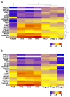
Heat maps showing the expression of NFX1-123-responsive genes at different stages of cervical cancer development. (A) A heat map organized by two unsupervised cluster analyses of the data. Dendrograms on the top and right show the results of these analyses. (B) A heat map organized along the x-axis such that the least transformed tissues are on the left and the most transformed are on the right. The y-axis lists genes in the same order as in (A). For all, warmer colors represent higher expression, while cooler colors represent lower expression. NED = no evidence of disease, CIN1 = cervical intraepithelial neoplasia grade 1, CIN2 = cervical intraepithelial neoplasia grade 2, and CIN3 = cervical intraepithelial neoplasia grade 3, Stage 1 = stage 1 cervical cancer, Stage 2 = stage 2 cervical cancer, and Stage 3 = stage 3 cervical cancer.
Figure 3
Figure 3
LASSO-identified genes are positive prognostic factors in cervical cancer. The TCGA cervical cancer dataset was segregated based on the expression of LASSO-identified genes as described in the text. (A) Of the 303 samples in the TCGA dataset with information on overall survival, 97 had altered expression of at least one LASSO-identified gene. The remaining did not have increased expression of nine LASSO-identified genes (CEBPD, SPRR2G, RAB7B, NOTCH1, KRT16, LCE1B, SLPI, RPS29, and PPL; z-score > 2). Kaplan–Meier curves of the two populations are shown. Median survival for each group is shown below the curve. N/A indicates that the group with altered expression of LASSO-identified genes did not drop below 50%. (B) Of the 263 samples in the TCGA dataset with information on maintenance of disease-free status after initial treatment, 86 had increased expression of at least one of the nine LASSO-identified gene analyzed in A. The remaining did not have increased expression of these genes. Kaplan–Meier curves of the two populations are shown. The median survival is not shown because neither group dropped below 50%. For all, the significance of the difference between the two groups was determined by logrank test. The relevant p-values are shown on each curve. Samples where LASSO-identified genes were altered are shown in red. Samples where LASSO-identified genes were NOT altered are shown in blue.
Figure 4
Figure 4
Summary of in silico and in vitro analyses of LASSO-identified genes. Expression and abundance of LASSO-identified genes/gene products is summarized. (A) Transcriptomic data of premalignant lesions and qPCR and immunoblot data of E6E7-expressing HFKs. (B) Transcriptomic data of cervical cancers and qPCR and immunoblot data in cervical cancer cells. For all, in silico data is shown with disease state increasing from left to right (i.e., The first column of in silico data is CIN1/Stage1 and the last is CIN3/Stage 3). qPCR and immunoblot data in (A) are shown as comparisons between parent HFK and HFK transduced with E6E7 for each biological background. qPCR and immunoblot data in (B) compare C33A to parent HFK1, SiHa to parent HFK1, and SiHa to C33A. For NOTCH1 and FBN2, two bands were detected by immunoblot. Information for both bands is shown and indicated in kDa sizes.
Figure 5
Figure 5
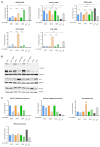
Expression of LASSO-identified genes in in vitro models of cervical disease. (A) CEBPD, KRT16, NOTCH1, SLPI, and RPS29 transcript levels (determined by qPCR) in HFK1-3 with and without HPV 16E6E7, as well as cancer cells C33A and SiHa. Samples are normalized to a housekeeping gene (36B4 or GAPDH) and for HFKs to their respective parent and cancer cells to HFK1. Error bars represent standard deviation for technical replicates (n = 3 for HFKs; n = 9 for cancer cells). Asterisks denote significant differences between HFK cells with and without HPV 16E6E7, and between either cancer cell or HFK1 as determined by Student t-Test. p value: *** = 0.0002; **** ≤ 0.0001. (B) Representative immunoblots of NOTCH1, SLPI, and RPS29 are shown. GAPDH is included as a loading control. (C) Densitometry of shown immunoblots (NOTCH1, SLPI, and RPS29). Samples normalized to GAPDH, and to the parent HFK for HFK1-3 and to HFK1 for the cancer cells. Two distinct bands were detected for NOTCH1 (300 kDa and 120 kDa).
Figure 6
Figure 6
Correlation of LASSO-identified genes with NFX1 expression in premalignant cervical lesions. The expression of each LASSO-identified genes was plotted (y-axis) against the expression of NFX1 (x-axis) in premalignant cervical lesions. LASSO-identified genes whose expression has significantly correlated with NFX1 as determined by (A) Spearman correlation analysis or (B) Pearson correlation analysis is shown. For all, R and p-values are shown. CIN1 = cervical intraepithelial neoplasia grade 1 (green), CIN2 = cervical intraepithelial neoplasia grade 2 (orange), and CIN3 = cervical intraepithelial neoplasia grade 3 (purple).
Figure 7
Figure 7
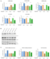
Influence of NFX1 expression on LASSO-identified gene expression. (A) CEBPD, SPRR2G, NOTCH1, LCE1B, and SLPI transcript levels (determined by qPCR) in HFK1-3 with and without exogenous NFX1-123 expression. Samples are normalized to a housekeeping gene (36B4 or GAPDH) and HFKs are normalized to their LXSN vector control for each pair. Error bars represent standard deviation for technical replicates (n = 3). Asterisks denote significant differences between HFK cells with and without exogenous NFX1-123 expression as determined by Student t-Test. p value: * = 0.03; ** = 0.002; *** = 0.0002; **** ≤ 0.0001. (B) Representative immunoblots of NOTCH1 (top) and SLPI (bottom) are shown. GAPDH is included as a loading control. (C) Densitometry of shown immunoblots (NOTCH1 and SLPI) normalized to GAPDH abundance and similarly to qPCR to each HFK LXSN vector control. Two distinct bands were detected for NOTCH1.

Similar articles

Cited by

References

    1. Van Doorslaer K., Li Z., Xirasagar S., Maes P., Kaminsky D., Liou D., Sun Q., Kaur R., Huyen Y., McBride A.A. The Papillomavirus Episteme: A major update to the papillomavirus sequence database. Nucleic Acids Res. 2017;45:D499–D506. doi: 10.1093/nar/gkw879. - DOI - PMC - PubMed
    1. Munoz N., Bosch F.X., de Sanjose S., Herrero R., Castellsague X., Shah K.V., Snijders P.J., Meijer C.J. Epidemiologic classification of human papillomavirus types associated with cervical cancer. N. Engl. J. Med. 2003;348:518–527. doi: 10.1056/NEJMoa021641. - DOI - PubMed
    1. Brianti P., De Flammineis E., Mercuri S.R. Review of HPV-related diseases and cancers. New Microbiol. 2017;40:80–85. - PubMed
    1. Giuliano A.R., Lee J.H., Fulp W., Villa L.L., Lazcano E., Papenfuss M.R., Abrahamsen M., Salmeron J., Anic G.M., Rollison D.E., et al. Incidence and clearance of genital human papillomavirus infection in men (HIM): A cohort study. Lancet. 2011;377:932–940. doi: 10.1016/S0140-6736(10)62342-2. - DOI - PMC - PubMed
    1. Kreimer A.R., Pierce Campbell C.M., Lin H.Y., Fulp W., Papenfuss M.R., Abrahamsen M., Hildesheim A., Villa L.L., Salmeron J.J., Lazcano-Ponce E., et al. Incidence and clearance of oral human papillomavirus infection in men: The HIM cohort study. Lancet. 2013;382:877–887. doi: 10.1016/S0140-6736(13)60809-0. - DOI - PMC - PubMed