Review
. 2021 Dec 9;10(24):5757.
doi: 10.3390/jcm10245757.

Spectral Photon-Counting CT Technology in Chest Imaging

Affiliations
Review

Spectral Photon-Counting CT Technology in Chest Imaging

Salim Aymeric Si-Mohamed et al. J Clin Med. .

Abstract

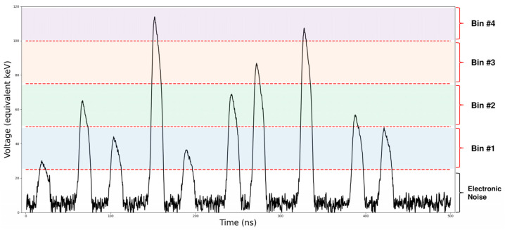
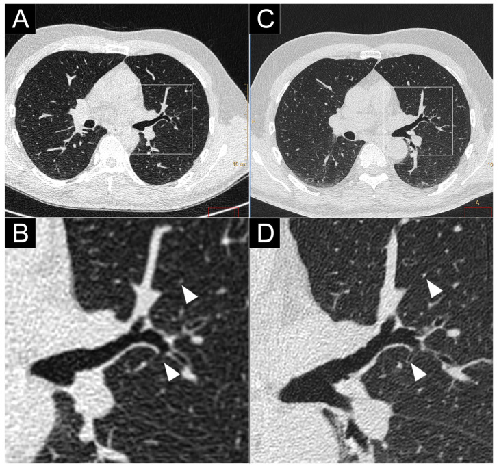
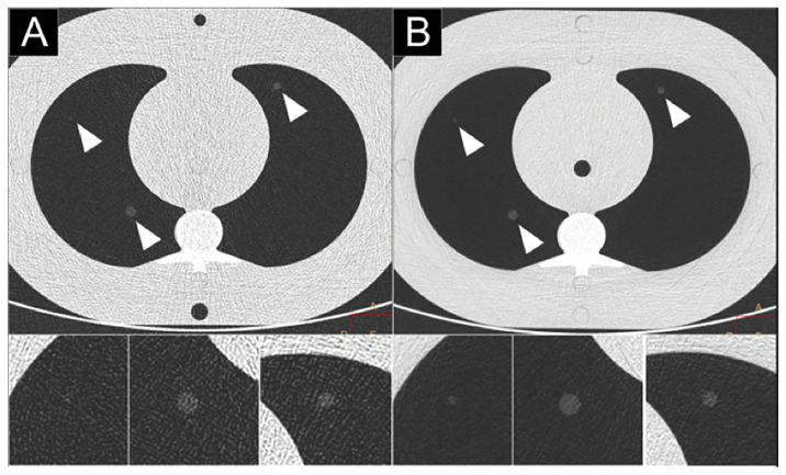
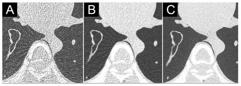
The X-ray imaging field is currently undergoing a period of rapid technological innovation in diagnostic imaging equipment. An important recent development is the advent of new X-ray detectors, i.e., photon-counting detectors (PCD), which have been introduced in recent clinical prototype systems, called PCD computed tomography (PCD-CT) or photon-counting CT (PCCT) or spectral photon-counting CT (SPCCT) systems. PCD allows a pixel up to 200 microns pixels at iso-center, which is much smaller than that can be obtained with conventional energy integrating detectors (EID). PCDs have also a higher dose efficiency than EID mainly because of electronic noise suppression. In addition, the energy-resolving capabilities of these detectors allow generating spectral basis imaging, such as the mono-energetic images or the water/iodine material images as well as the K-edge imaging of a contrast agent based on atoms of high atomic number. In recent years, studies have therefore been conducted to determine the potential of PCD-CT as an alternative to conventional CT for chest imaging.

Keywords: computed tomography; diagnostic imaging; lung; photon-counting detectors; thorax.


Conflict of interest statement

The authors declare no conflict of interest.

Figures

Figure 1
Figure 1
Schematic representation of photon detection technologies: energy-integrating (top) and photon-counting detectors (bottom).
Figure 2
Figure 2
Example of a 500 ns signal output of a photon-counting detector pixel.
Figure 3
Figure 3
Energy weighting of energy integrating detectors (EID) and photon-counting detectors (PCD). The optimal weight for differentiating calcium from water is derived from the attenuation coefficients subtraction (dashed lines).
Figure 4
Figure 4
Attenuation coefficients of contrast agents with a K-edge in the medical energy range.
Figure 5
Figure 5
Comparison of high-resolution lung imaging between a conventional CT (Brilliance 64; Philips Haifa, Israel; (A,B)), a clinical prototype PCD-CT (SPCCT; Philips; (C,D)), and in a human volunteer. Close-up views on the left pulmonary hilum found a greater overall image quality as well as better depiction and a greater number of small structures (bronchial wall, vessels; white arrows).
Figure 6
Figure 6
Comparison of high-resolution CT lung imaging of ground-glass nodules (2, 4, 6 mm) between a dual-layer EID-CT (iQon; Philips, Haifa, Israel) (A) and a clinical prototype PCD-CT (SPCCT; Philips) (B) in an anthropomorphic thorax phantom with an extension ring simulating an obese patient, using a standard dose protocol (120 kVp, 40 mAs).
Figure 7
Figure 7
Comparison of high-resolution low dose lung imaging (120 kVp, 10 mAs) between a dual-layer EID-CT (iQon, Philips, Haifa, Israel; (A): filter YB, (B): filter F) and a clinical prototype PCD-CT (Philips; (C): filter detailed 1) in an anthropomorphic phantom CT Torso CTU-41 (Kyoto Kagaku, Tokyo, Japan). Both 1024 matrix and field-of-view of 350 mm were matched. Beam hardening was greatly reduced on the PCD-CT images, greatly improving the image quality.
Figure 8
Figure 8
Comparison of high-resolution low dose lung imaging (120 kVp, 10 mAs) between a conventional dual-layer CT (iQon; Philips, Haifa, Israel; (A): filter YB) and a clinical PCD-CT (Philips; (B): filter detailed 1) in an anthropomorphic phantom CT Torso CTU-41 (Kyoto Kagaku, Tokyo, Japan). Both 1024 matrix and field-of-view of 350 mm were matched. Noise was significantly reduced on the PCD-CT images in the upper and basal lobes, greatly improving the image quality.
Figure 9
Figure 9
Comparison of the airways imaging in a 72-year-old patient with a respiratory bronchiolitis, which was on the same day as a clinical prototype PCD-CT (first row; Philips, Haifa, Israel) and a dual-layer EID-CT (second row; iQon, Philips). Sagittal views (A,B) demonstrated thickening of the bronchial wall as well as a mosaicism probably due to air trapping. Close-up views of the airspaces (C,D) showed a better sharpness and conspicuity of the bronchial wall (white arrowheads) with PCD-CT (D). Greater sharpness of the bronchial calcifications were noticed on PCD-CT images (F) compared to EID-CT images ((E); white empty arrowheads).
Figure 10
Figure 10
Comparison of the distal arterial pulmonary tree with a dual-layer EID-CT (iQon, Philips, Haifa, lsrael; (A)) and a clinical prototype PCD-CT (Philips; (B)) and) after injection of iodinated contrast agent. The improvement in quality is visible, notably of the distal vessel lumen and calipers that are depicted until the pleural space.
Figure 11
Figure 11
Spectral photon-counting CT (SPCCT) images of a phantom containing tubes with clockwise decrease concentrations of gadolinium from 15 mg/mL to 1 mg/mL of atoms using a clinical prototype (SPCCT; Philips, Haifa, Israel). Conventional image (A) and material decomposition of the K-edge image was obtained by reconstructing three material bases (B): K-edge image, (C): water image, (D): iodine image. Only the K-edge images showed the specific signal in the tubes according to the gadolinium concentrations, while water image showed signal from the plastic phantom made of water and the water in the tubes. Iodine image showed no signal accordingly to the absence of iodine.
Figure 12
Figure 12
Bicolor imaging of the lung perfusion blood volume in a rabbit after injection of a standard iodinated contrast agent and a contrast agent on a K-edge material (gold nanoparticles) using a spectral photon-counting CT (SPCCT; Philips, Haifa, Israel). Conventional image (A) showed the enhancement of the chest vessels as well as the underlying tissue, while the gold K-edge image showed only the signal of gold (B). The iodine image showed the signal of iodine as well as misclassification of the bone (C).

References

    1. Si-Mohamed S., Bar-Ness D., Sigovan M., Cormode D.P., Coulon P., Coche E., Vlassenbroek A., Normand G., Boussel L., Douek P. Review of an initial experience with an experimental spectral photon-counting computed tomography system. Nucl. Instrum. Methods Phys. Res. Sect. A Accel. Spectrometers Detect. Assoc. Equip. 2017;873:27–35. doi: 10.1016/j.nima.2017.04.014. - DOI
    1. Si-Mohamed S., Boussel L., Douek P. Clinical Applications of Spectral Photon-Counting CT. In: Taguchi K., Blevis I., Iniewski K., editors. Spectral, Photon Counting Computed Tomography: Technology and Applications. CRC Press; Boca Raton, FL, USA: 2020. pp. 97–116.
    1. Willemink M.J., Persson M., Pourmorteza A., Pelc N.J., Fleischmann D. Photon-counting CT: Technical Principles and Clinical Prospects. Radiology. 2018;289:293–312. doi: 10.1148/radiol.2018172656. - DOI - PubMed
    1. Sandfort V., Persson M., Pourmorteza A., Noël P.B., Fleischmann D., Willemink M.J. Spectral photon-counting CT in cardiovascular imaging. J. Cardiovasc. Comput. Tomogr. 2021;15:218–225. doi: 10.1016/j.jcct.2020.12.005. - DOI - PubMed
    1. Sawall S., Amato C., Klein L., Wehrse E., Maier J., Kachelrieß M. Toward molecular imaging using spectral photon-counting computed tomography? Curr. Opin. Chem. Biol. 2021;63:163–170. doi: 10.1016/j.cbpa.2021.04.002. - DOI - PubMed

Grants and funding

LinkOut - more resources