

Immediate Implant Placement in the Maxillary Aesthetic Zone: A Cone Beam Computed Tomography Study
- PMID: 34945150
- PMCID: PMC8708737
- DOI: 10.3390/jcm10245853
Immediate Implant Placement in the Maxillary Aesthetic Zone: A Cone Beam Computed Tomography Study
Abstract
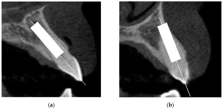
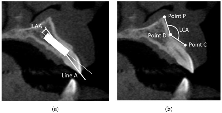
This study aimed to investigate the factors that could be associated with the risk of labial cortical bone wall perforation with immediate implant placement (IIP) in the maxillary aesthetic zone, in a cone-beam computed tomography (CBCT) virtual study. CBCT exams from 126 qualified subjects (756 teeth) were included. Implants were virtually positioned in two different positions: in the long axis of the tooth (prosthetically-driven position) and in an ideal position in relation to adjacent anatomical structures (bone-driven position). Two different implant diameters were planned for each tooth position, namely, 3.75 and 4.3 mm for central incisors and canines, and 3.0 and 3.3 mm for lateral incisors. The incidence of perforation was nearly 80% and 5% for prosthetically- and bone-driven position, respectively. Factors associated with a higher risk of cortical bone wall perforation (bone-driven position), according to logistic regression analysis, were women, wider implants, Sagittal Root Position class IV, and decrease of the labial concavity angle. Perforation of the labial cortical bone wall can be greatly minimized when the implant is placed in a bone-driven position compared to a prosthetically-driven position. It is important to preoperatively evaluate the morphological features of the implant site for risk assessment and to individualize the treatment plan.
Keywords: anterior maxilla; cone beam computed tomography; dental implant; fenestration; immediate implant placement; risk assessment; virtual treatment planning.
Conflict of interest statement
The authors declare no conflict of interest.
Figures
References
-
- Lee S.R., Jang T.S., Seo C.S., Choi I.O., Lee W.P. Hard Tissue Volume Stability Effect beyond the Bony Envelope of a Three-Dimensional Preformed Titanium Mesh with Two Different Collagen Barrier Membranes on Peri-Implant Dehiscence Defects in the Anterior Maxilla: A Randomized Clinical Trial. Materials. 2021;14:5618. doi: 10.3390/ma14195618. - DOI - PMC - PubMed
LinkOut - more resources
Full Text Sources
