Case Reports
. 2021 Dec 19;57(12):1383.
doi: 10.3390/medicina57121383.

Use of the Cover-Lifting Technique in Mandibular Cemento-Ossifying Fibroma Excision to Preserve the Inferior Alveolar Nerve

Affiliations
Case Reports

Use of the Cover-Lifting Technique in Mandibular Cemento-Ossifying Fibroma Excision to Preserve the Inferior Alveolar Nerve

Juan-You Qiu et al. Medicina (Kaunas). .

Abstract

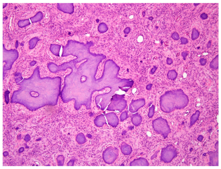
Cemento-ossifying fibroma (also known as ossifying fibroma or cementifying fibroma) is a benign osteogenic neoplasm. Pain and paresthesia are rarely associated with cemento-ossifying fibroma; thus, nerves must be preserved during excision. With the advent of computer-aided techniques, the use of virtual surgical planning and a customized template can improve the precision of resection and reconstruction, reduce operating time, and improve postoperative outcomes. In this report, we describe a case of cemento-ossifying fibroma in a female patient who underwent segmental mandibulectomy and reconstruction with an iliac bone graft. Additionally, we describe a simple and effective way to preserve the inferior alveolar nerve.

Keywords: 3D-printed cutting guide; cemento-ossifying fibromas; inferior alveolar nerve.


Conflict of interest statement

The authors declare no conflict of interest.

Figures

Figure 1
Figure 1
Pre-operative images of the jaw. (A) The panoramic radiograph, revealing a mixed (radiolucent–radiopaque) bony lesion (3 × 3 cm) in the posterior region of the left mandible region. The lesion was very close to the inferior border of the mandible. (B) Pre-operative three-dimensional image (left-sided view), showing the expansile tumor. (C) Pre-surgery plan to simulate the iliac bone graft reconstruction model.
Figure 2
Figure 2
Three-dimensional virtual surgical plan for (A) a customized mandibular cutting guide and (B) an iliac cutting guide. (C) The appearance of the inferior alveolar nerve and tumor after mandibular the cortical bone block was removed. (D) Segmental mandibulectomy with preservation of the inferior alveolar nerve.
Figure 3
Figure 3
Post-operative computed tomographic reconstructed model, showing the iliac bone graft fixed with mini-plates and mini-screws according to the pre-operative plan, and intermaxillary fixation was performed.
Figure 4
Figure 4
Histopathological micrograph, revealing curvilinear trabeculae formed by cementum-like calcifications in a fibrous stroma. The final diagnosis was cemento-ossifying fibroma.

References

    1. Ram R., Singhal A., Singhal P. Cemento-ossifying fibroma. Contemp. Clin. Dent. 2012;3:83–85. doi: 10.4103/0976-237X.94553. - DOI - PMC - PubMed
    1. Katti G., Khan M.M., Chaubey S.S., Amena M. Cemento-ossifying fibroma of the jaw. BMJ Case Rep. 2016;2016:bcr2015214327. doi: 10.1136/bcr-2015-214327. - DOI - PMC - PubMed
    1. Neville B.W., Damm D.D., Allen C.M., Bouquot J.E. Oral and Maxillofacial Pathology. 3rd ed. Saunders; Philadelphia, PA, USA: 2008.
    1. Mainville G.N., Turgeon D.P., Kauzman A. Diagnosis and management of benign fibro-osseous lesions of the jaws: A current review for the dental clinician. Oral Dis. 2017;23:440–450. doi: 10.1111/odi.12531. - DOI - PubMed
    1. Huang D., Chen M., He D., Yang C., Yuan J., Bai G., Wang Y., Wei W., Chen Z. Preservation of the inferior alveolar neurovascular bundle in the osteotomy of benign lesions of the mandible using a digital template. Br. J. Oral Maxillofac. Surg. 2015;53:637–641. doi: 10.1016/j.bjoms.2015.04.013. - DOI - PubMed

Publication types

LinkOut - more resources