

Buccal Bone Thickness in Anterior and Posterior Teeth-A Systematic Review
- PMID: 34946389
- PMCID: PMC8700878
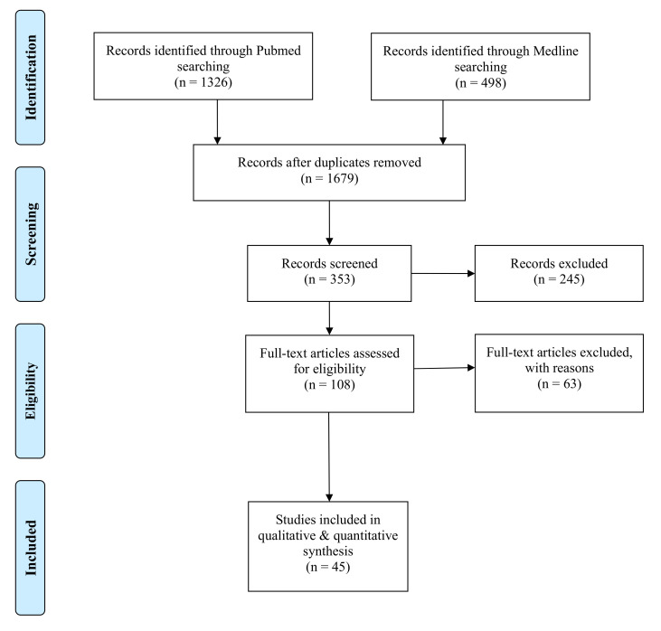
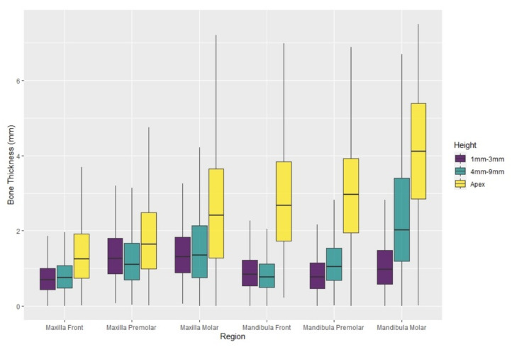
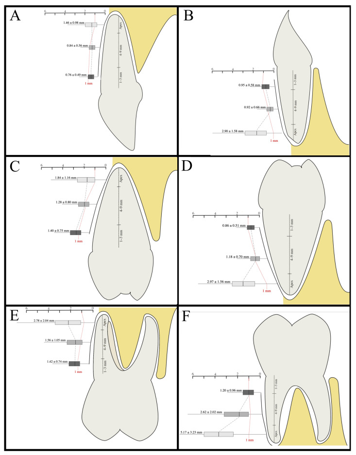
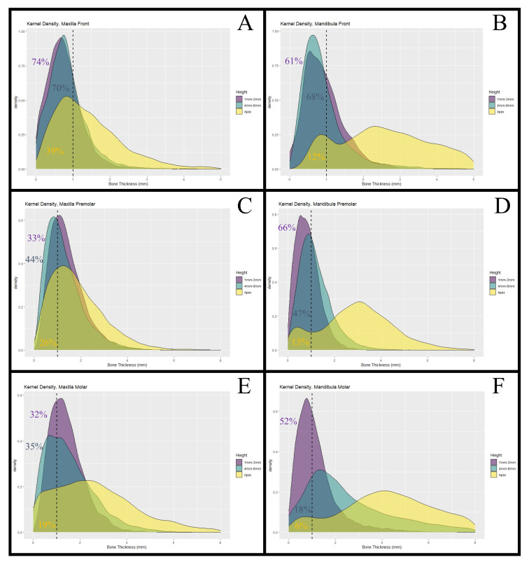
- DOI: 10.3390/healthcare9121663
Buccal Bone Thickness in Anterior and Posterior Teeth-A Systematic Review
Abstract
(1) Background: Immediate dental implant placement has been a subject of great interest over the last decade. Here, information regarding the anatomy and bone thickness of the jaw prior to dental implant placement is crucial to increase the surgery's success and the patient's safety. The clinical premises for this approach have been controversially discussed. One of those heavily discussed premises is a buccal bone thickness of at least 1 mm thickness. This meta-analysis aims to systematically review buccal bone thickness (BBT) in healthy patients. Thus, the feasibility of immediate dental implant placement in daily practice can be assessed. (2) Methods: A search in the electronic databases was performed to identify articles reporting on BBT that was measured by computed tomography in adults. (3) Results: We were able to find 45 studies, including 4324 patients with 25,452 analyzed teeth. The analysis showed a BBT at the alveolar crest of 0.76 ± 0.49 mm in the maxillary frontal and of 1.42 ± 0.74 mm in the maxillary posterior region. In the mandible, the average measured values were similar to those in the maxilla (front: 0.95 ± 0.58 mm; posterior: 1.20 ± 0.96 mm). In the maxillary frontal region 74.4% and in the mandibular frontal region 61.2% of the crestal buccal bones showed widths <1 mm. (4) Conclusions: In more than 60% of the cases, the BBT at the alveolar crest is <1 mm in maxillary and mandibular frontal regions. This anatomic data supports careful pre-surgical assessment, planning of a buccal graft, and critical selection of indication for immediate implant placement, especially in the maxillary and mandibular frontal and premolar region.
Keywords: alveolar bone; buccal bone thickness; dental implant; dental implant loading; immediate; tomography.
Conflict of interest statement
The authors declare no conflict of interest.
Figures
References
-
- Hammerle C.H., Chen S.T., Wilson T.G., Jr. Consensus statements and recommended clinical procedures regarding the placement of implants in extraction sockets. Int. J. Oral Maxillofac. Implant. 2004;19:26–28. - PubMed
Publication types
LinkOut - more resources
Full Text Sources
