Case Reports
. 2021 Dec 13;12(12):1980.
doi: 10.3390/genes12121980.

New Mutations in HFE2 and TFR2 Genes Causing Non HFE-Related Hereditary Hemochromatosis

Affiliations
Case Reports

New Mutations in HFE2 and TFR2 Genes Causing Non HFE-Related Hereditary Hemochromatosis

Gonzalo Hernández et al. Genes (Basel). .

Abstract

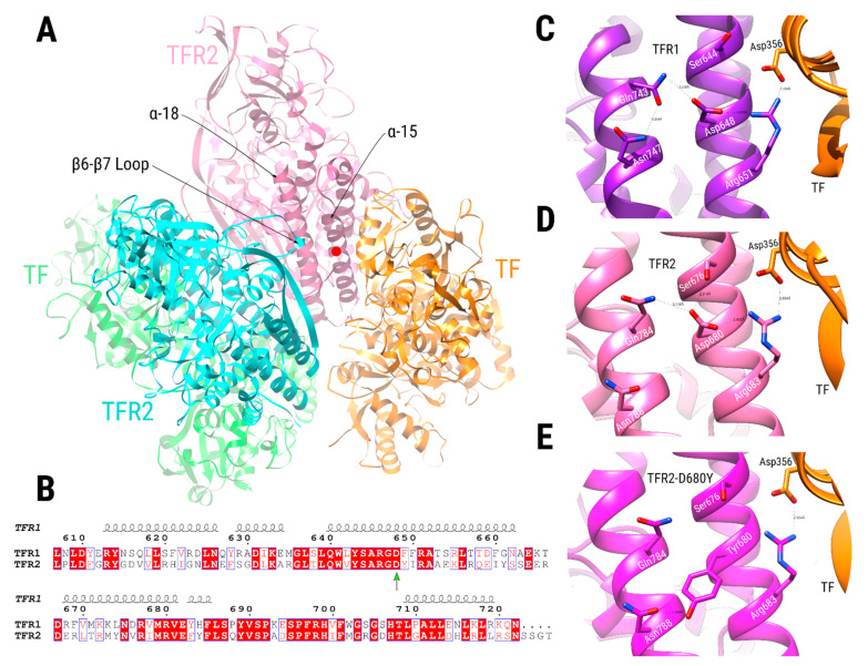
Hereditary hemochromatosis (HH) is an iron metabolism disease clinically characterized by excessive iron deposition in parenchymal organs such as liver, heart, pancreas, and joints. It is caused by mutations in at least five different genes. HFE hemochromatosis is the most common type of hemochromatosis, while non-HFE related hemochromatosis are rare cases. Here, we describe six new patients of non-HFE related HH from five different families. Two families (Family 1 and 2) have novel nonsense mutations in the HFE2 gene have novel nonsense mutations (p.Arg63Ter and Asp36ThrfsTer96). Three families have mutations in the TFR2 gene, one case has one previously unreported mutation (Family A-p.Asp680Tyr) and two cases have known pathogenic mutations (Family B and D-p.Trp781Ter and p.Gln672Ter respectively). Clinical, biochemical, and genetic data are discussed in all these cases. These rare cases of non-HFE related hereditary hemochromatosis highlight the importance of an earlier molecular diagnosis in a specialized center to prevent serious clinical complications.

Keywords: HFE related hereditary hemochromatosis; HFE2 gene; TFR2 gene; homozygous; iron overload; missense; non-HFE related hereditary hemochromatosis; nonsense.


Conflict of interest statement

C.T. is CEO of BloodGenetics, S.L. and M.S. are co-founders of BloodGenetics.

Figures

Figure A1
Figure A1
Liver biopsy of a patient with HJV-related hemochromatosis. (a). The specimen contains several pseudo-lobules and shows massive iron overload in Rappaport zone 1 and decreasing hemosiderin gradient from Rappaport zone 1 to 3 (Perls’ Prussian blue stain, 2X magnification). (b) A magnification (20×) of the previous image focusing a portal tract and part of the hepatic lobules showing massive hemosiderin granules in hepatocytes, biliocytes, macrophages and portal vein endothelium. Deugnier’s score: Total Iron Score 51 (Hepatic Iron Score 12,9,9; Sinusoidal Iron Score 3,3,3; Portal Iron score 4,4,4).
Figure A2
Figure A2
Sequence similarity network for TFR1 and TFR2 orthologous proteins. Three hundred fifty-two sequences were used, where each node represents an orthologous sequence to the human TFR1 (red nodes) or TFR2 (green nodes) proteins. Edges are drawn between two nodes if their sequence similarity is higher than 0.59. Node border colors indicate amino acid identities of positions aligned with the human TFR2 position 680. Sequences with aspartate at this position have thin black borders. Sequences with an identity different from aspartate appear with thick blue borders. Finally, sequences with no residue aligned at this position appear with yellow node borders. Nodes representing the human TFR1 and TFR2 sequences have labels indicating their protein name. The right bottom table indicates the percentage of sequences bearing a particular amino acid identity (or lack of it: ‘-’) at the position aligned with the human TFR2 position 680.
Figure A3
Figure A3
Global view of the binding mode between TFR2 (pink) and TF (orange) proteins. Black dots encircle all domains, and their corresponding names are indicated. The region where TFR2 position 680 is located is shown with a red circle.
Figure 1
Figure 1
Model for the hepcidin expression and its alterations in hereditary hemochromatosis (HH). Left, the coordinated signaling of the TFR1, TFR2, HFE, HJV and BMP receptors activated the SMAD complex that promotes HAMP expression. The hepcidin produced is then released to the blood stream and will stimulate the FPN degradation in the duodenum epithelial cells, limiting iron absorption. Right, in an individual affected by HH, mutations in any of the proteins marked with a red cross result in a reduction in the serum hepcidin levels. This leads to an uncontrolled iron absorption by the duodenum that will result in an iron accumulation in the tissues.
Figure 2
Figure 2
Localization of the reported pathogenic mutations in the HJV precursor. The new mutations reported in this work are boxed. The black region in the N-terminal represents the signal peptide while the white box at the C-terminal represents a portion that will be removed in the maturation process, exposing the Asp400 residue (in red) necessary for the GPI anchor of the HJV protein to the membrane.
Figure 3
Figure 3
Localization of the reported mutations in the TFR2 protein. The new mutations reported are boxed. Dimeric, Transferrin Receptor-like dimerization region; TM, transmembrane domain; PA, Protease-Associated domain. Image adapted from Joshi et al. [23].
Figure 4
Figure 4
Non-HFE hereditary hemochromatosis families: families and mutations. The probands are indicated with an arrow. Black symbols denote affected individuals and half-filled black symbols unaffected carriers. Individuals studied at the molecular level are indicated with the # symbol. (a) Pedigrees of the two non-HFE related HH families carrying HFE2 mutations. (b) Pedigrees of the three non-HFE related HH families carrying TFR2 mutations.
Figure 5
Figure 5
Comparative modeling of the TFR2 dimer complexed with TF. (A) TFR2 chains are shown in cyan and pink, while TF chains are shown in green and orange. Dimeric interactions between TFR2 chains, close to Asp680 position (red circle at the α-15 helix, pink chain), are made between residues in the α-18 helix of one TFR2 chain (pink chain) and the loop located between the β-6 and β-7 strands of the other (cyan chain). (B) Sequence alignment of the TFR1 and TFR2 proteins near the TFR2 680 position (green arrow). Numbering is according to the TFR1 sequence, and the secondary structure depiction is based on the TFR1 structure (PDB ID 1CX8). The alignment coloring was produced by the ESPript 3.0 web server [40], where position with red background means absolute sequence conservation. (CE) Structural context of positions TFR1-Asp648 (C, purple), TFR2-Asp680 (D, pink), and TFR2-Asp680Tyr (E, magenta). Residue names of TFR1/2 appear in white, while residues in TF are in black. Significant interactions are shown with dashed lines.

References

    1. Hentze M.W., Muckenthaler M.U., Galy B., Camaschella C. Two to tango: Regulation of Mammalian iron metabolism. Cell. 2010;142:24–38. doi: 10.1016/j.cell.2010.06.028. - DOI - PubMed
    1. Roetto A., Papanikolaou G., Politou M., Alberti F., Girelli D., Christakis J., Loukopoulos D., Camaschella C. Mutant antimicrobial peptide hepcidin is associated with severe juvenile hemochromatosis. Nat. Genet. 2003;33:21–22. doi: 10.1038/ng1053. - DOI - PubMed
    1. Kawabata H., Yang R., Hirama T., Vuong P.T., Kawano S., Gombart A.F., Koeffler H.P. Molecular cloning of transferrin receptor 2. A new member of the transferrin receptor-like family. J. Biol. Chem. 1999;274:20826–20832. doi: 10.1074/jbc.274.30.20826. - DOI - PubMed
    1. Nai A., Lidonnici M.R., Rausa M., Mandelli G., Pagani A., Silvestri L., Ferrari G., Camaschella C. The second transferrin receptor regulates red blood cell production in mice. Blood. 2015;125:1170–1179. doi: 10.1182/blood-2014-08-596254. - DOI - PMC - PubMed
    1. Silvestri L., Nai A., Dulja A., Pagani A. Hepcidin and the BMP-SMAD pathway: An unexpected liaison. Vitam. Horm. 2019;110:71–99. - PubMed

Publication types

LinkOut - more resources